
Can you explain to me what is actually retropatentorial structures?
retropatentorial structures anatomy meaning
"Retropatentorial" - Clarifying the Term
| Term | Meaning |
|---|---|
| Retropetrosal | Behind (posterior to) the petrous part of the temporal bone |
| Infratentorial / Subtentorial | Below the tentorium cerebelli (the dural fold separating the cerebrum from the cerebellum) |
| Posterior cranial fossa | The bony compartment containing all these structures |
The Posterior Cranial Fossa (Infratentorial Space)
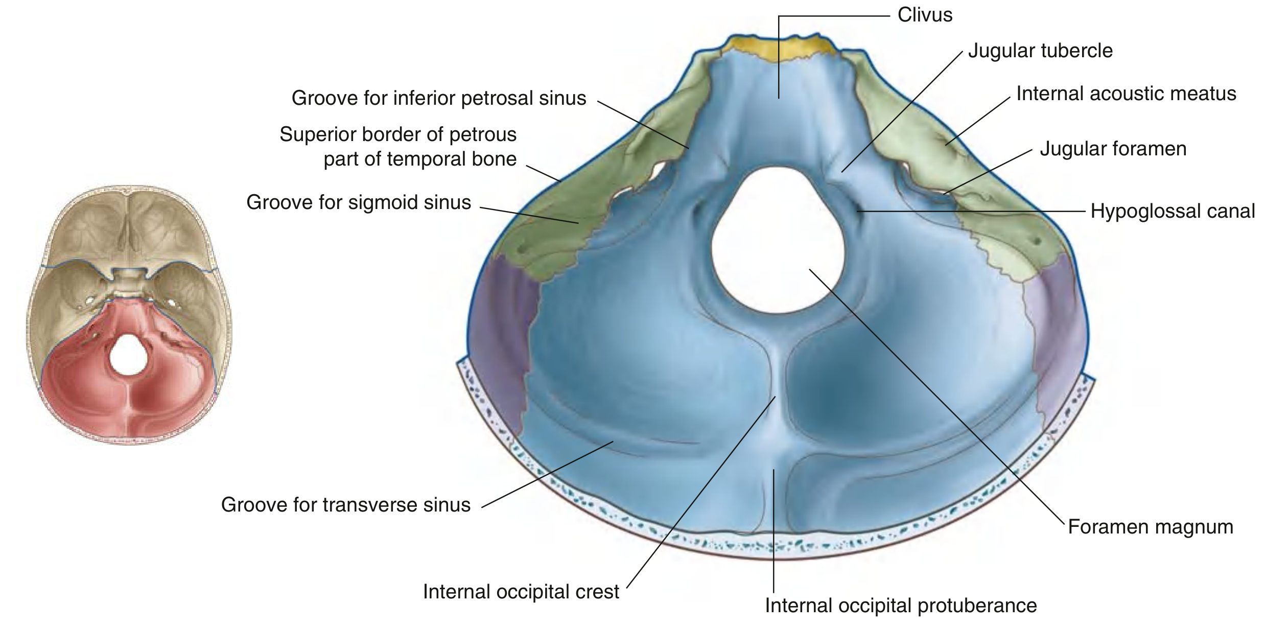
Bony Boundaries
- Anteriorly (midline): The dorsum sellae and the clivus - a sloped piece of bone running from the foramen magnum upward, formed by the sphenoid body and the basilar occipital bone
- Anteriorly (lateral): The superior border of the petrous part of the temporal bone (this is the "petrosal" landmark)
- Posteriorly: The squamous occipital bone (up to the level of the transverse groove) and laterally the petromastoid part of the temporal bone
Neural Contents (What's Inside)
-
Brainstem - three parts:
- Midbrain (mesencephalon)
- Pons
- Medulla oblongata
-
Cerebellum - the two cerebellar hemispheres sit in the concave bony depressions on either side of the internal occipital crest
-
4th ventricle - the CSF-filled space between the brainstem and cerebellum
Cranial Nerves and Foramina
| Foramen | Contents |
|---|---|
| Internal acoustic meatus | CN VII (facial), CN VIII (vestibulocochlear), labyrinthine artery |
| Jugular foramen | CN IX (glossopharyngeal), CN X (vagus), CN XI (accessory), sigmoid + inferior petrosal sinuses → internal jugular vein |
| Hypoglossal canal | CN XII (hypoglossal) |
| Foramen magnum | Spinal cord/brainstem junction, vertebral arteries, meninges, spinal roots of CN XI |
Venous Sinuses
- Transverse sinuses (run laterally from the internal occipital protuberance)
- Sigmoid sinuses (curve inferiorly, draining into the jugular foramina)
- Inferior petrosal sinus (runs between the basilar occipital bone and petrous temporal bone)
Clinical Relevance
- Posterior fossa tumors (e.g., medulloblastoma in children, cerebellar metastases in adults) can compress the 4th ventricle → obstructive hydrocephalus
- Posterior fossa malformations include Dandy-Walker malformation (cystic enlargement of 4th ventricle + cerebellar vermis hypoplasia) and Chiari malformations (herniation of posterior fossa structures through the foramen magnum)
- Syringomyelia workup requires imaging of posterior fossa structures to assess for Chiari malformation
- In neurosurgery, the retropetrosal approach is a specific surgical corridor that goes behind the petrous bone to reach tumors like acoustic neuromas or petroclival meningiomas
Retroperitonial
retroperitoneal space anatomy compartments diagram
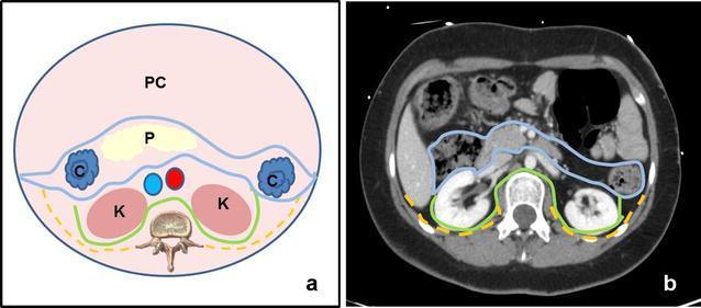
This dual-panel educational material illustrates the anatomy of the retroperitoneal space using a comparative approach. Panel (a) is a schematic anatomical diagram in axial cross-section. It delineates the three main retroperitoneal compartments: the anterior pararenal space (outlined in blue), containing the pancreas (P) and ascending/descending colon (C); the perirenal space (outlined in green), housing the kidneys (K), adrenal glands, and perirenal fat; and the posterior pararenal space (outlined in yellow dashed lines), located between the posterior renal fascia and the transversalis fascia. The peritoneal cavity (PC) is shown anteriorly. Panel (b) presents a corresponding axial CT scan with intravenous contrast, applying the same color-coded boundary lines to clinical imaging. The CT demonstrates the contrast-enhanced kidneys within the perirenal space and the relative positions of the pancreas and bowel. This comparison aids in understanding fascial planes and the compartmentalization of potential retroperitoneal pathology, such as hematomas or abscesses, which are often confined by these anatomical boundaries.
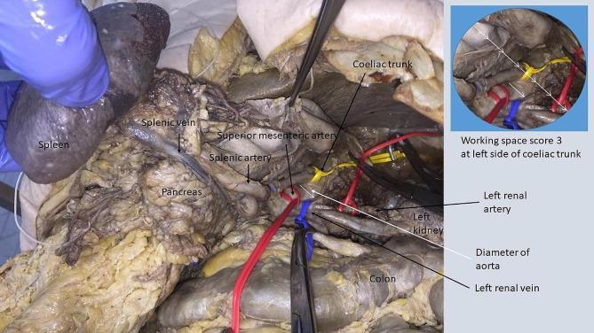
An intraoperative clinical photograph and annotated diagram demonstrating the mobilization of the spleen and pancreas to expose retroperitoneal vasculature during a surgical procedure. The main image shows the spleen being retracted superiorly by a gloved hand, revealing the underlying pancreas. Key vascular structures are labeled, including the splenic vein and artery, superior mesenteric artery (SMA), coeliac trunk, left renal artery, and left renal vein. Several vessels are isolated with colored vessel loops (red, blue, and yellow) and manipulated with surgical forceps. An inset image in the upper right corner provides a quantified assessment of the 'working space' at the left side of the coeliac trunk. The diagram illustrates a 'working space score 3,' defined as a space approximately three times the diameter of the abdominal aorta. This educational content focuses on retroperitoneal anatomy, surgical exposure techniques for tumor resection, and the quantification of surgical access in the left lateral anatomic region.
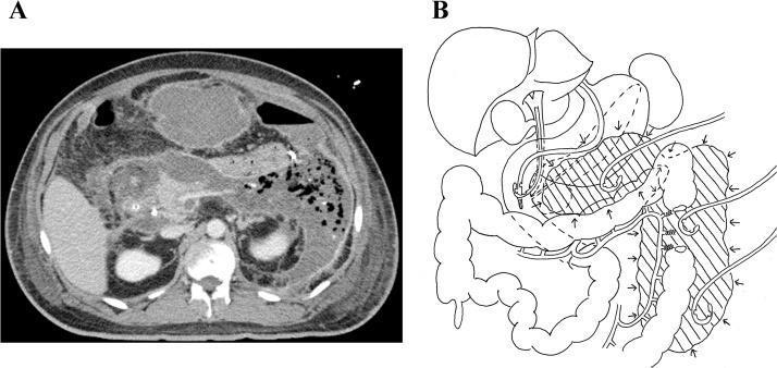
This composite educational graphic details the diagnosis and management of a complex retroperitoneal abscess. Panel A is an axial contrast-enhanced CT scan of the abdomen demonstrating a large, heterogeneous, low-attenuation collection in the retroperitoneal space. The collection is predominantly located in the left hemi-abdomen, involving the peripancreatic region and extending laterally toward the splenic flexure and descending colon. Notable features include internal debris and gas bubbles (mottled air sign), highly suggestive of an infected necrotic process or abscess secondary to severe pancreatitis. Panel B is a corresponding anatomical line diagram illustrating the ultrasound-guided drainage strategy. It depicts three percutaneous pigtail catheters placed to target distinct fluid compartments: the pre-pancreatic space, the area around the splenic flexure, and the paracolic gutter along the descending colon. The diagram clarifies the spatial relationship between the abscess (represented by hatched shading) and the major abdominal organs, including the stomach, liver, pancreas, and colon, highlighting the multidisciplinary approach to managing deep-seated abdominal sepsis.
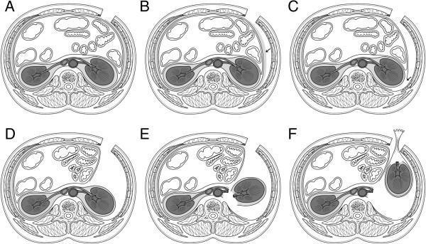
This medical diagram consists of six axial schematic frames (A–F) illustrating the surgical progression of a Gasless Radical Nephrectomy via Reduced Port Surgery (RASI) in the supine position. Frame A shows the baseline anatomy with a subcostal skin incision. Frame B depicts the excision of fatty tissue on the lateroconal fascia (LCF). Frame C demonstrates the longitudinal incision of the LCF to expose Gerota’s fascia. Frame D illustrates the medial retraction of the peritoneum to gain access to the retroperitoneal space. Frame E shows the dissection of the renal hilum, including the renal artery and vein, and the mobilization of the kidney from its attachments. Finally, frame F displays the extraction of the renal specimen through the surgical site using a specialized retrieval device. Key anatomical structures visible throughout the series include the abdominal wall muscles (external oblique, internal oblique, and transversus abdominis), the aorta, inferior vena cava, kidneys, and surrounding peritoneal contents. This educational graphic focuses on the surgical pathway and tissue layers involved in retroperitoneal urologic procedures.
The Retroperitoneum - Complete Anatomy
What is it?
| Boundary | Structure |
|---|---|
| Anterior | Posterior parietal peritoneum |
| Posterior | Iliopsoas and lumbar muscles |
| Superior | Diaphragm |
| Inferior | Levator ani muscles (pelvic floor) |
The Three Compartments
1. Anterior Pararenal Space
- Bounded anteriorly by the posterior parietal peritoneum and posteriorly by the anterior lamina of Gerota's fascia
- Contains the secondarily retroperitoneal organs - those that started as intraperitoneal structures and migrated posteriorly during embryogenesis:
- Ascending and descending colon
- Duodenum (2nd and 3rd parts)
- Pancreas (head, neck, body)
2. Perirenal Space (Perinephric Space)
- Enclosed within Gerota's fascia (renal fascia), which has an anterior and posterior lamina merging laterally as the lateroconal fascia
- Contains the primarily retroperitoneal organs:
- Kidneys
- Adrenal glands
- Proximal ureters
- Gonadal vessels (testicular/ovarian)
- Perirenal fat
- Abdominal aorta and inferior vena cava
- The perirenal space has a cone shape open inferiorly into the extraperitoneal pelvis, meaning fluid can track downward
3. Posterior Pararenal Space
- Bounded anteriorly by the posterior lamina of Gerota's fascia and posteriorly by the transversalis fascia
- Contains only fat (no organs)
- Continuous with the preperitoneal fat of the anterior abdominal wall
Retroperitoneal Structures at a Glance
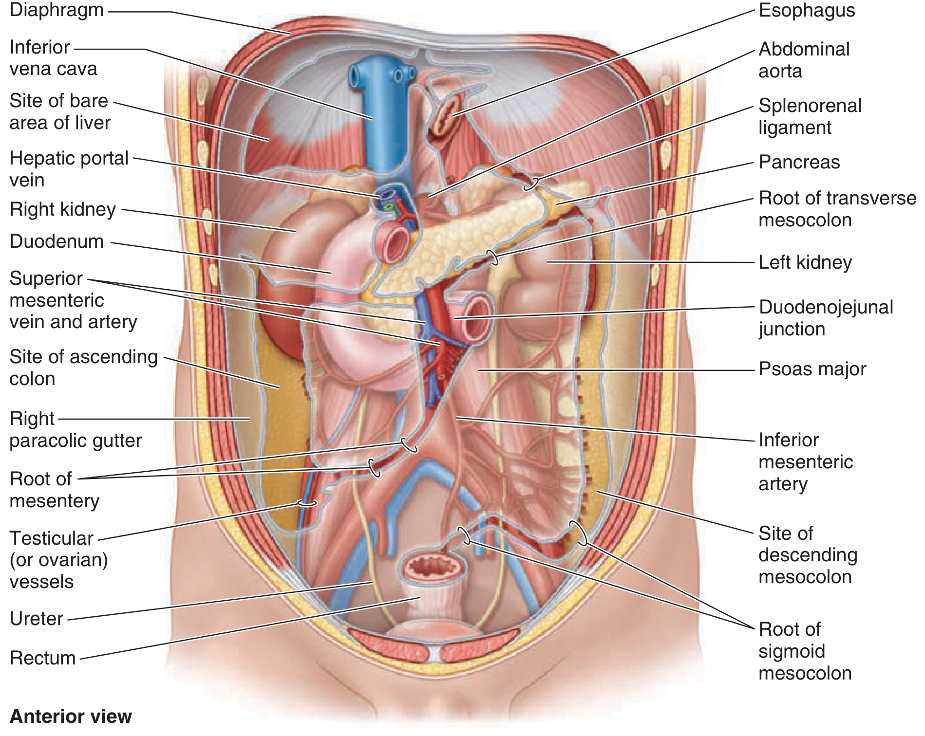
Primary vs. Secondary Retroperitoneal Structures
| Type | Definition | Structures |
|---|---|---|
| Primary | Always retroperitoneal throughout embryonic development | Kidneys, adrenal glands, ureters, abdominal aorta, inferior vena cava, rectum |
| Secondary | Started as intraperitoneal, migrated posteriorly during development | Duodenum (D2-D4), pancreas (head/neck/body), ascending colon, descending colon |
Trauma Zones (Surgical Relevance)
| Zone | Location | Action |
|---|---|---|
| Zone 1 (Central) | Midline - major vessels (aorta, IVC) | Always explore after obtaining proximal/distal vascular control |
| Zone 2 (Lateral/Flank) | Perinephric area | Explore only if expanding, pulsatile, or penetrating injury present |
| Zone 3 (Pelvic) | Pelvic retroperitoneum | Avoid opening; manage with packing or angioembolization |
Clinical Conditions of the Retroperitoneum
-
Retroperitoneal infections/abscesses - can arise from perforated retrocecal appendix, diverticulitis, pancreatitis, or duodenal perforation. May present insidiously with back/flank pain and fever. Diagnosed best by CT with IV contrast. Treated with drainage ± IV antibiotics.
-
Retroperitoneal fibrosis (Ormond's disease) - rare condition where a grey/white fibrous plaque develops in the low lumbar region, spreading to encase the ureters, aorta, and common iliac vessels. Usually idiopathic but can be caused by lymphoma, carcinoid, drugs, or inflammatory aortic aneurysm.
-
Retroperitoneal hematoma - commonly from trauma, aortic aneurysm rupture, or anticoagulation. Given the compliance of the anterior boundary, collections tend to expand anteriorly toward the peritoneal cavity.
-
Retroperitoneal tumors - primary tumors include soft tissue sarcomas (liposarcoma most common), lymphomas, and germ cell tumors. Metastases can come from kidney, adrenal, colon, and other retroperitoneal organs.
-
Perinephric collections - urine (urinoma), blood (hematoma), or pus (perinephric abscess) can accumulate within the perirenal space and track inferiorly through the open cone of Gerota's fascia into the pelvis.
Can you show me 3D picture of this?
A detailed 3D anatomical illustration of the retroperitoneal space in the human abdomen, showing a cross-sectional and slightly angled perspective. Label and clearly show the three compartments: anterior pararenal space (containing pancreas, ascending colon, descending colon, duodenum), perirenal space enclosed by Gerota's fascia (containing both kidneys, adrenal glands, ureters, abdominal aorta, inferior vena cava), and posterior pararenal space (containing only fat). Show the posterior parietal peritoneum as the anterior boundary, the transversalis fascia posteriorly, and the iliopsoas muscles. Use different translucent colors for each compartment. Include labels for all major structures. Medical illustration style, highly detailed and educational.
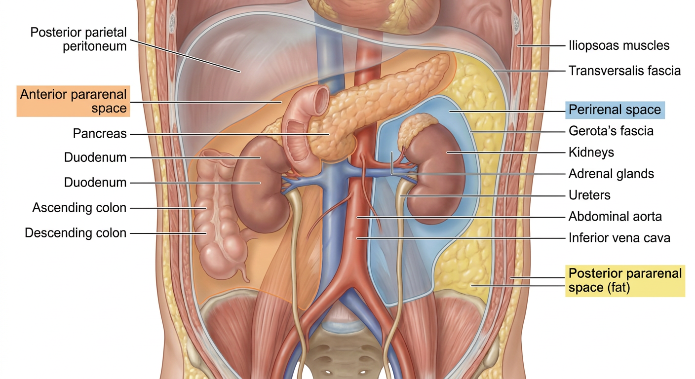
- Anterior pararenal space - the frontmost layer behind the peritoneum, housing the pancreas, duodenum, and ascending/descending colon
- Perirenal space (Gerota's fascia) - the middle zone enclosing both kidneys, adrenal glands, ureters, aorta, and IVC
- Posterior pararenal space - the deepest fat-only layer between Gerota's fascia and the transversalis fascia, with the iliopsoas muscles forming the posterior wall