
Ventricles of brain anatomy detailed explanation in simple way with images
Reading File
Now let me search the medical library for ventricles of brain content, along with images.
Finding Sources
Finding Sources
Excellent! I found great resources. Let me now read the detailed content from Neuroanatomy and Color Atlas books, plus grab the images.
Reading File
Reading File
Reading File
Now let me read more of the Neuroanatomy textbook content on ventricles, and fetch the key images in parallel.
Reading File
Loading Image
Loading Image
Loading Image
Loading Image
I now have excellent images and rich textbook content. Let me fetch one more key image showing the sagittal view with all 4 ventricles, then also get the Guyton physiology diagram.
Loading Image
Loading Image
I now have all the content and images I need for a thorough, well-illustrated answer. Let me compile everything.
Ventricles of the Brain - Detailed Anatomy (Simple Explanation)
The ventricles are a system of four fluid-filled cavities (hollow spaces) inside the brain. Think of them as a network of interconnected "rooms" filled with cerebrospinal fluid (CSF) - a clear liquid that cushions and protects the brain. The entire cranial cavity holds about 1600-1700 mL total, of which ~150 mL is CSF at any given time.
Overview - The Ventricular System
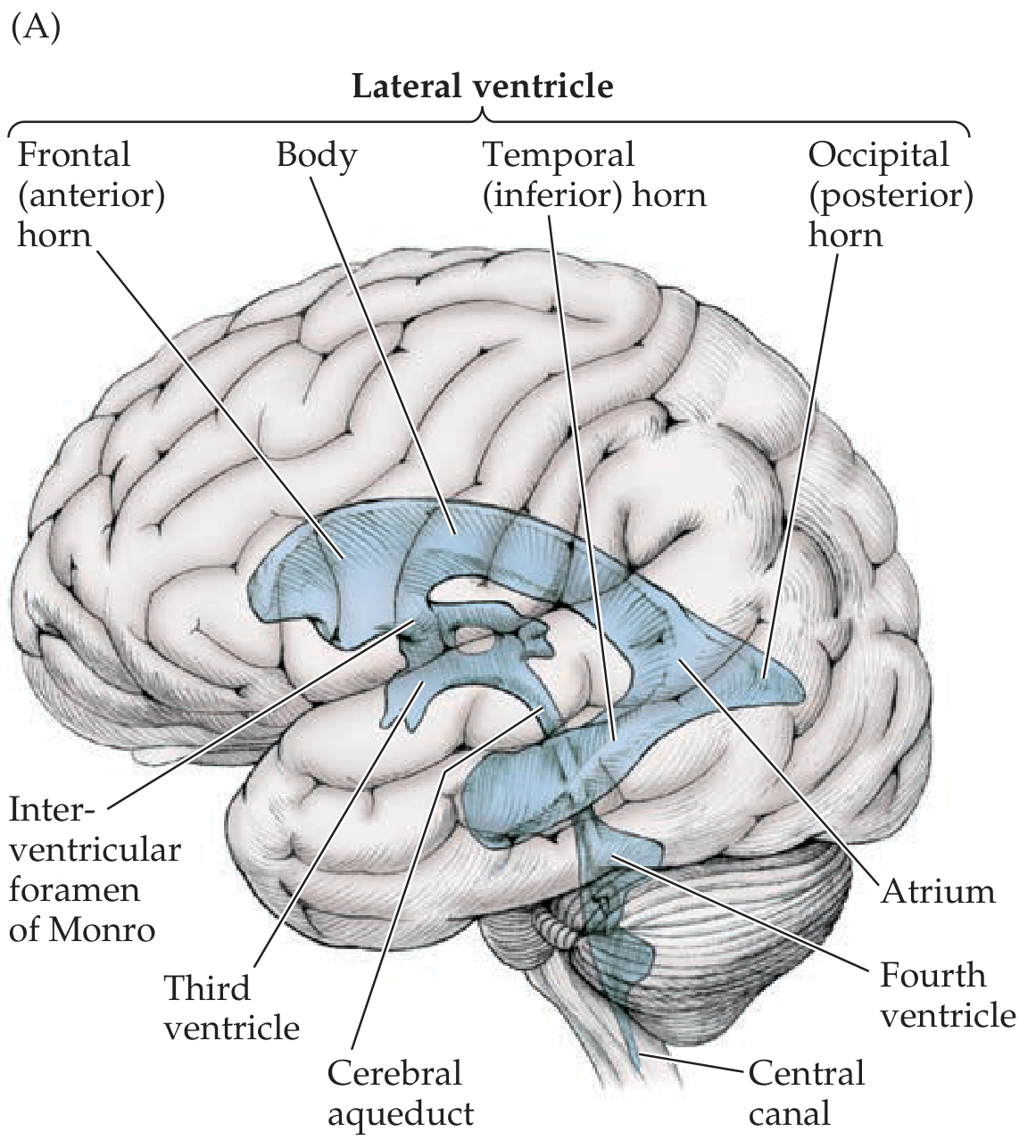
Fig. 5.11A - All four ventricles as seen from the lateral (side) surface of the brain - Neuroanatomy through Clinical Cases, 3rd Ed.
1. The Lateral Ventricles (Left & Right)
These are the two largest ventricles, one inside each cerebral hemisphere. They were formerly called the 1st and 2nd ventricles. Each lateral ventricle is shaped like a C (or a curved ram's horn), following the natural curve of the surrounding brain structures.
Each lateral ventricle has four parts:
| Part | Also Called | Location |
|---|---|---|
| Frontal horn | Anterior horn | Extends forward into the frontal lobe; starts anterior to the foramen of Monro |
| Body | - | Runs through the frontal and parietal lobes, posterior to the foramen of Monro |
| Atrium (Trigone) | - | A junction point where the body, occipital horn, and temporal horn all meet |
| Occipital horn | Posterior horn | Extends backward into the occipital lobe |
| Temporal horn | Inferior horn | Extends downward and forward into the temporal lobe |
Simple memory tip: The lateral ventricle is like a "C"-shaped hallway with rooms branching off at the back - one going to the vision area (occipital), one going down to the memory area (temporal).
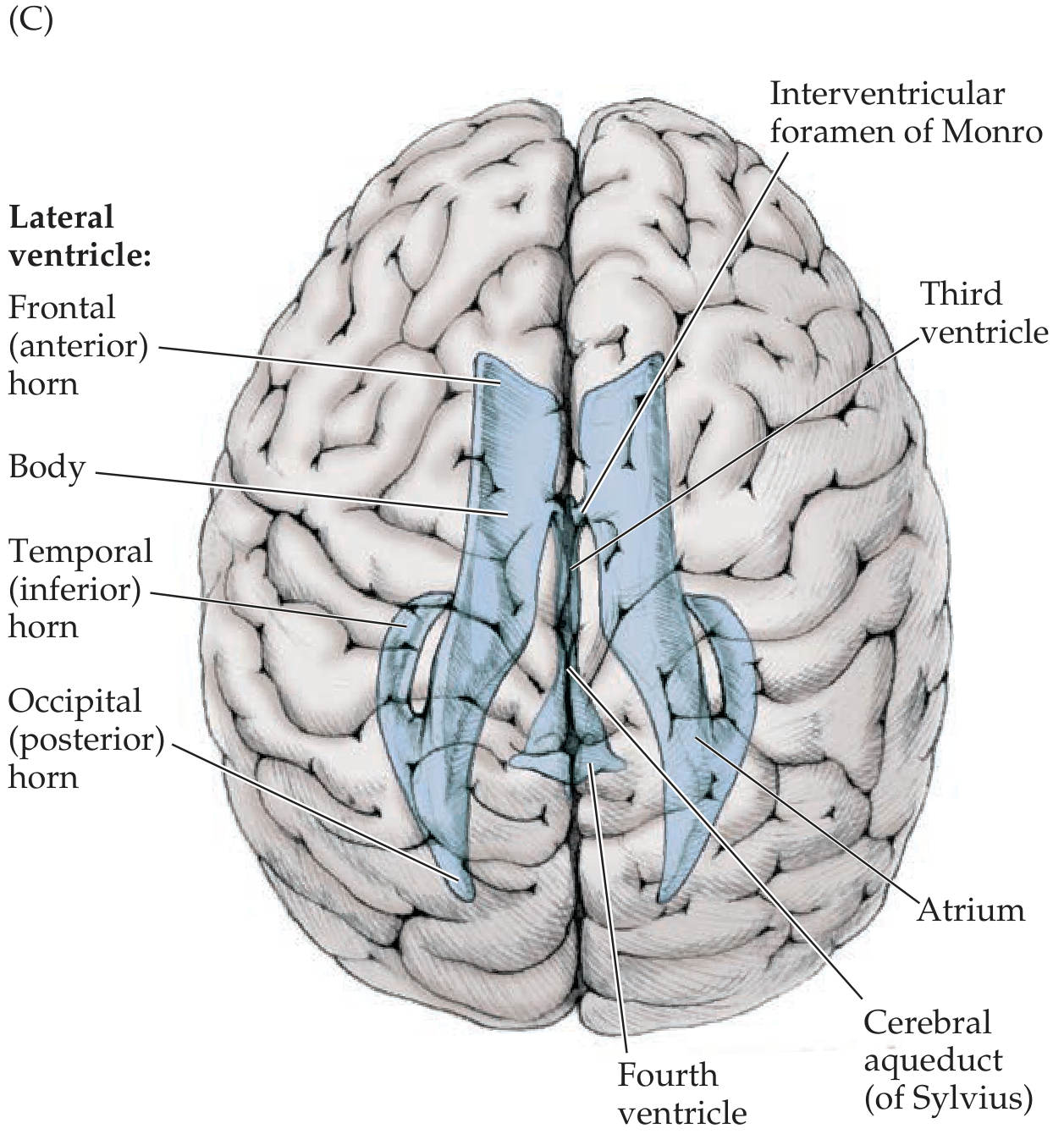
Fig. 5.11C - View from above (superior) showing both lateral ventricles symmetrically - Neuroanatomy through Clinical Cases, 3rd Ed.
2. The Third Ventricle
The third ventricle is a narrow, slit-like space sitting right in the middle of the brain, between the two halves of the thalamus and hypothalamus (which form its walls). It is much smaller than the lateral ventricles.
Key features:
- Communicates with the lateral ventricles above via the interventricular foramen of Monro (one on each side)
- Has several small recesses (pockets) projecting into surrounding structures:
- Supraoptic recess - above the optic chiasm
- Infundibular recess - going down toward the pituitary stalk
- Pineal recess - toward the pineal gland
- Suprapineal recess - above the pineal gland
- Connected below/behind to the fourth ventricle via the cerebral aqueduct
Simple way to think of it: The third ventricle is like a narrow corridor sitting in the very center of the brain, connecting the upper rooms (lateral ventricles) to the lower room (fourth ventricle).
3. The Cerebral Aqueduct (Aqueduct of Sylvius)
This is not a ventricle itself but a narrow tube (the smallest passage in the whole system) connecting the third and fourth ventricles. It travels through the midbrain. Because it is so narrow, it is the most common site of obstruction that causes hydrocephalus.
4. The Fourth Ventricle
The fourth ventricle sits at the back of the brain, between the cerebellum (roof) and the pons + medulla (floor). It is shaped roughly like a tent or diamond.
Key features:
- Roof: formed by the cerebellum
- Floor: formed by the pons and medulla (this area is called the rhomboid fossa)
- Opens into the subarachnoid space (the space around the outside of the brain) through three openings:
- Foramen of Magendie (1 median opening in the posterior roof)
- Foramina of Luschka (2 lateral openings, one on each side)
- Continues downward as the central canal of the spinal cord
Complete 3D Structure - All Ventricles Together
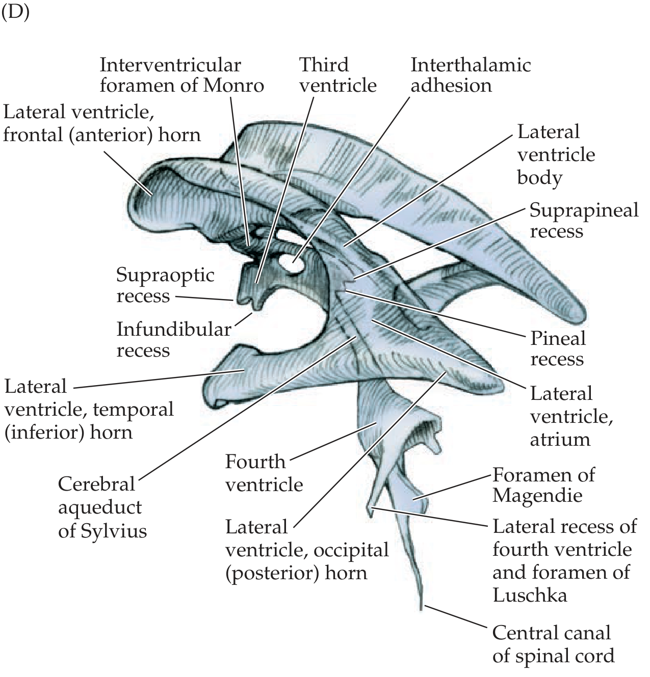
Fig. 5.11D - Full 3D detail showing all ventricles, horns, recesses, and connections - Neuroanatomy through Clinical Cases, 3rd Ed.
CSF Flow Path - The Journey of Cerebrospinal Fluid
CSF is produced by the choroid plexus (a special network of cells inside the ventricles, especially the lateral ventricles). It then flows in a set path:
Lateral ventricles → Foramen of Monro → Third ventricle → Cerebral aqueduct → Fourth ventricle → Foramina of Magendie & Luschka → Subarachnoid space → Absorbed by arachnoid granulations into venous blood (superior sagittal sinus)
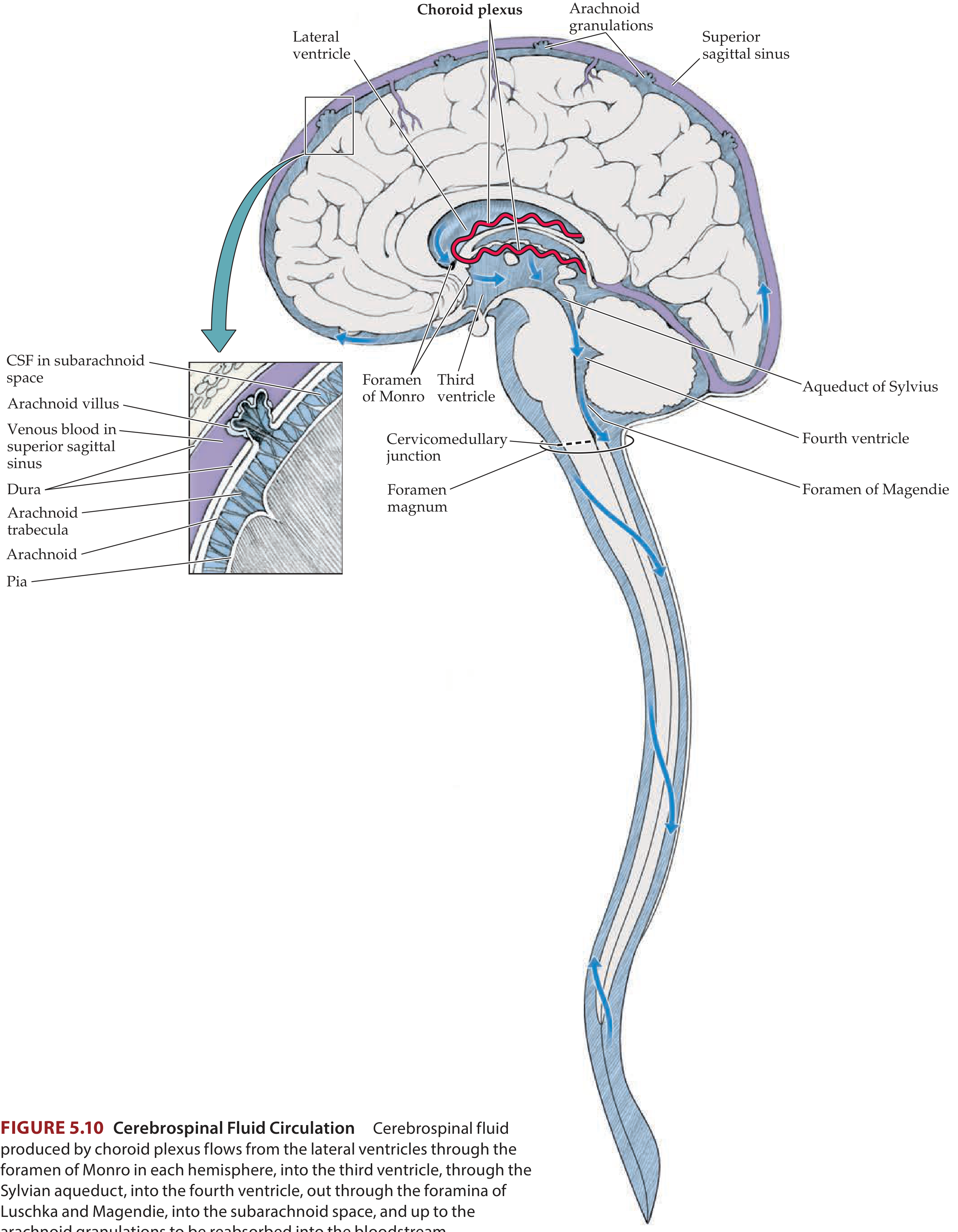
Fig. 5.10 - Complete CSF circulation pathway - Neuroanatomy through Clinical Cases, 3rd Ed.
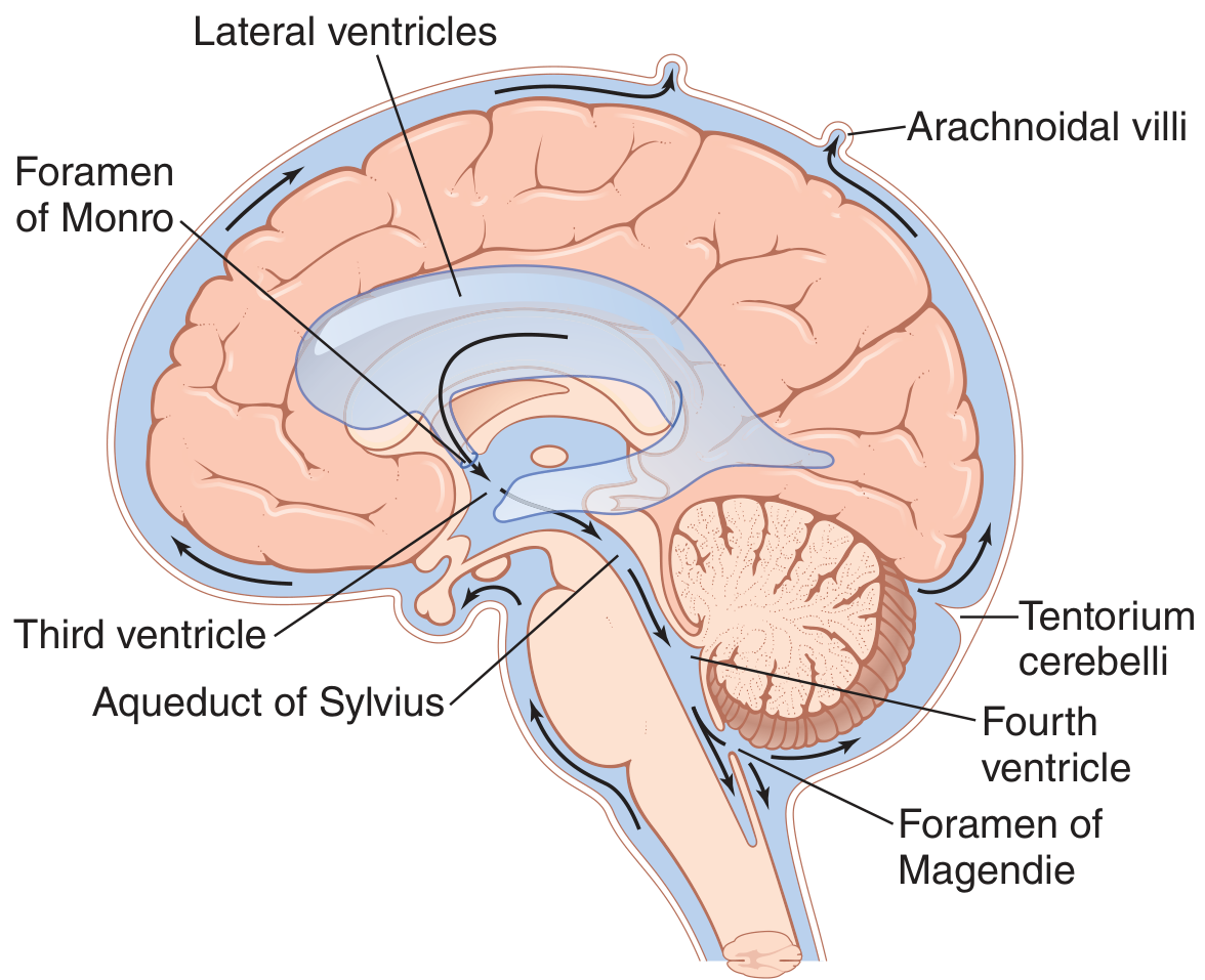
Fig. 62.6 - CSF pathway and ventricle anatomy - Guyton & Hall Textbook of Medical Physiology
Quick Summary Table
| Ventricle | Location | Walls/Boundaries | Connects To |
|---|---|---|---|
| Lateral (×2) | Inside each cerebral hemisphere | Frontal, parietal, temporal, occipital lobes | 3rd ventricle via foramen of Monro |
| Third | Between thalamus & hypothalamus | Thalamus (lateral walls), hypothalamus (floor) | Lateral ventricles above; cerebral aqueduct below |
| Cerebral Aqueduct | Through midbrain | Midbrain walls | 3rd and 4th ventricles |
| Fourth | Between cerebellum + pons/medulla | Cerebellum (roof), pons & medulla (floor) | Subarachnoid space via foramina of Magendie & Luschka; spinal canal |
Why Are Ventricles Important Clinically?
- Hydrocephalus: If any part of this system gets blocked (especially the narrow cerebral aqueduct), CSF builds up, ventricles enlarge, and brain pressure rises. This can cause headache, vomiting, and brain damage if untreated.
- Intraventricular hemorrhage: Bleeding into the ventricles (e.g., in premature babies or trauma)
- Choroid plexus tumors: Can cause excess CSF production
- The ventricles are clearly visible on CT and MRI scans and are a key landmark for neuroimaging interpretation
Sources: Neuroanatomy through Clinical Cases, 3rd Ed. | Guyton & Hall Textbook of Medical Physiology | Color Atlas of Human Anatomy, Vol. 3
This is a shared conversation. Sign in to Orris to start your own chat.