
How to write lacrimal apparatus in exam while covering all topics to write in exam in 4 mins
Reading File
The lacrimal apparatus includes the lacrimal gland (tear production) and the drainage system (puncta, canaliculi, lacrimal sac, nasolacrimal duct). Let me pull the full content from the textbooks now.
Searching sources...
Finding Sources
Finding Sources
Reading File
Finding Sources
Reading File
Finding Sources
Finding Sources
Reading File
Loading Image
Finding Sources
I now have all the information needed to write a comprehensive, exam-structured answer. Here is the complete exam-ready write-up:
Lacrimal Apparatus - Exam Answer (4 Minutes)
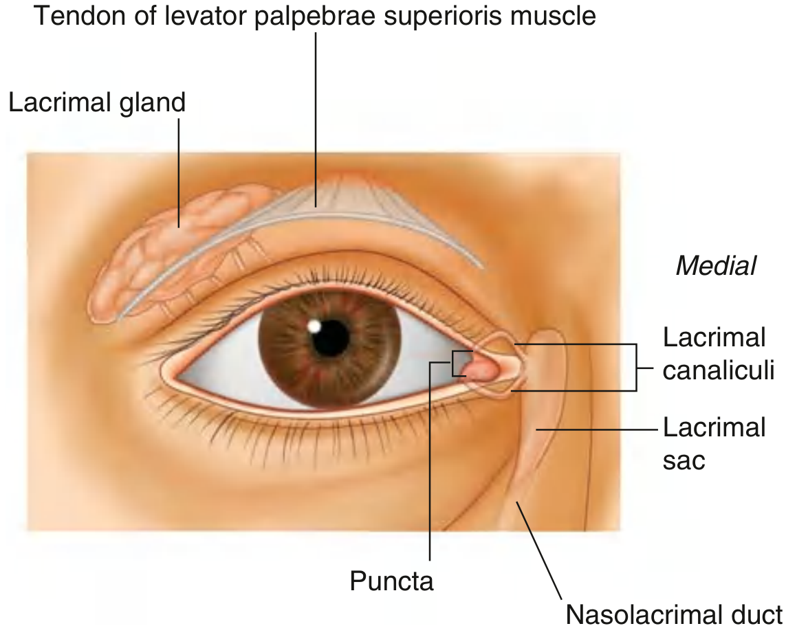
Definition
The lacrimal apparatus is the system involved in the production, movement, and drainage of tears (lacrimal fluid) from the surface of the eyeball. It consists of two parts:
- Secretory part - Lacrimal gland + its ducts
- Drainage part - Lacrimal puncta → Canaliculi → Lacrimal sac → Nasolacrimal duct
PART 1: LACRIMAL GLAND (Secretory)
Location: Superolateral region of the orbit, in the lacrimal fossa of the frontal bone.
Divided into 2 parts by the tendon of levator palpebrae superioris:
| Part | Location |
|---|---|
| Orbital (larger) | In the lacrimal fossa, frontal bone |
| Palpebral (smaller) | Inferior to levator tendon, in the superolateral eyelid |
Ducts: Numerous ducts open into the lateral part of the superior conjunctival fornix.
PART 2: DRAINAGE PATHWAY (Medial to Lateral Flow)
Tears flow lateral → medial with each blink, accumulating in the lacrimal lake at the medial canthus, then drain as:
Lacrimal Lake
      ↓
Lacrimal Puncta (one per eyelid, upper & lower)
      ↓
Lacrimal Canaliculi (superior & inferior)
      ↓
Lacrimal Sac (in the lacrimal fossa, between anterior & posterior lacrimal crests)
      ↓
Nasolacrimal Duct
      ↓
Inferior Meatus of Nose (under inferior turbinate, via Hasner's valve)
Key points to mention:
- The lacrimal sac lies between the anterior and posterior lacrimal crests, posterior to the medial palpebral ligament
- The lacrimal part of orbicularis oculi (Horner's muscle) dilates the lacrimal sac during blinking, actively pumping tears
- The nasolacrimal duct runs downward, laterally, and posteriorly; opens via Hasner's valve into the inferior meatus ~1 cm posterior to the anterior nasal spine
PART 3: INNERVATION OF LACRIMAL GLAND
| Type | Nerve | Pathway |
|---|---|---|
| Sensory | Lacrimal branch of ophthalmic nerve [V1] | Direct |
| Secretomotor (Parasympathetic) | Facial nerve [VII] → Greater petrosal nerve → Nerve of pterygoid canal → Pterygopalatine ganglion → Post-ganglionic fibers → Zygomatic nerve [V2] → Zygomaticotemporal nerve → Lacrimal nerve → Lacrimal gland | Relay in pterygopalatine ganglion |
| Sympathetic | Superior cervical ganglion → Deep petrosal nerve → Nerve of pterygoid canal → (same path as parasympathetic) | No relay (post-ganglionic) |
PART 4: BLOOD SUPPLY
- Arterial supply: Branches of the ophthalmic artery
- Venous drainage: Ophthalmic veins
PART 5: CLINICAL NOTES (for extra marks)
| Condition | Structure affected | Key feature |
|---|---|---|
| Dacryoadenitis | Lacrimal gland | Painful swelling superolaterally |
| Dacryocystitis | Lacrimal sac | Painful medial canthal swelling; caused by nasolacrimal duct obstruction; S. aureus commonest in adults; neonatal form due to non-canalization of Hasner's valve |
| Epiphora | Drainage failure | Overflow of tears due to obstruction |
| Congenital NLD obstruction | Hasner's valve | Commonest cause of watery eye in neonates; resolves spontaneously by 6-12 months |
Quick Exam Memory Trick
"P-C-S-N" = Punctum → Canaliculus → Sac → Nasolacrimal duct
Parasympathetic relay: VII → Greater petrosal → Pterygoid canal → Pterygopalatine ganglion → V2 → Zygomatic → Lacrimal nerve
Writing Sequence for 4 Minutes
- Definition + components (30 sec) - Write 2 lines
- Draw diagram (1 min) - Simple flow diagram of drainage + gland location
- Lacrimal gland - 2 parts + ducts (30 sec)
- Drainage pathway with Hasner's valve (45 sec)
- Innervation table - Sensory/Para/Sympathetic (45 sec)
- Blood supply (15 sec)
- 1-2 clinical points (15 sec)
Sources: Gray's Anatomy for Students; Scott-Brown's Otorhinolaryngology; Goodman & Gilman's
This is a shared conversation. Sign in to Orris to start your own chat.