
This is the report. What does it signify and what’s the treatment?
python3 -c " import pdfplumber with pdfplumber.open('/tmp/workspace/attachments/5f987d73-626f-4a49-9a3a-6df7884008a1/Mrs_ISHIKA_22_F_report.pdf') as pdf: for i, page in enumerate(pdf.pages): print(f'--- Page {i+1} ---') print(page.extract_text()) "
brain granuloma MRI cortical young adult
neurocysticercosis granuloma MRI treatment
PMID: 41484678
brain granuloma MRI T2 hypointense cortical parietal peripheral enhancement
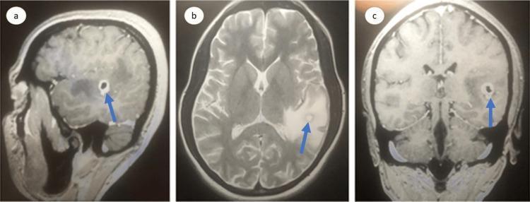
This diagnostic imaging composite displays Magnetic Resonance Imaging (MRI) of the brain in three planes: sagittal (a), axial (b), and coronal (c). The images reveal a single, well-defined, rounded ring-enhancing lesion located in the left parietal region. In the T2-weighted axial view (b), the lesion is hyperintense with a hypointense rim and is surrounded by extensive hyperintense signal indicating vasogenic edema, which results in partial effacement of the adjacent cortical sulci and subarachnoid space. Contrast-enhanced sections (a and c) demonstrate moderate, discrete peripheral rim enhancement with a darker central core. Blue arrows in each panel point to the lesion and its associated inflammatory changes. This presentation is characteristic of a neuroinflammatory process or an infectious granuloma, such as neurocysticercosis or a cerebral abscess. The imaging is critical for medical students and clinicians to study intracranial mass effects, the breakdown of the blood-brain barrier, and differential diagnosis of ring-enhancing lesions.
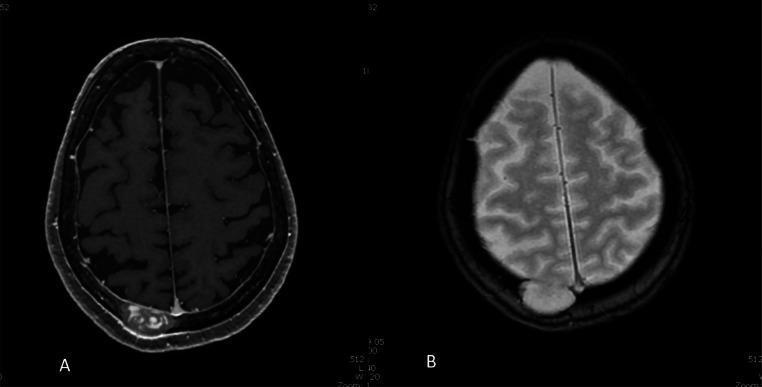
This diagnostic image consists of two axial MRI slices of the head, labeled A and B, illustrating a prominent osteolytic lesion of the right posterior parietal skull vault. Image A is a T1-weighted sequence showing a heterogeneous, predominantly hypointense lesion measuring 34 x 17 mm in the transverse plane. There is visible peripheral enhancement along the inner table, suggesting dural or pachymeningeal involvement. Image B is a T2-weighted sequence where the lesion appears markedly hyperintense relative to the brain parenchyma. The mass demonstrates full-thickness erosion of the skull vault with focal inward bulging into the epidural space. Notably, there is no evidence of underlying brain parenchymal edema or cortical signal abnormality, indicating the lesion remains extradural without pial passage. These images are characteristic of calvarial tumors or inflammatory processes (such as eosinophilic granuloma or metastatic disease) requiring multiplanar evaluation to assess dural integrity and intracranial extension.
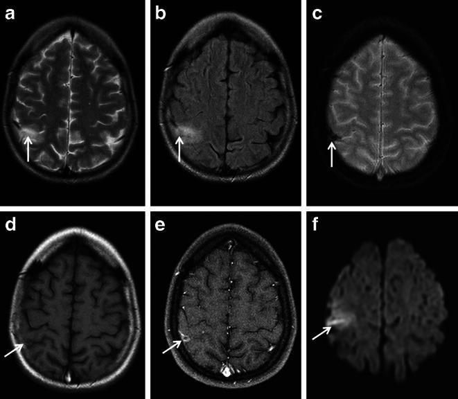
This composite diagnostic image displays six axial MRI brain sequences (a-f) of a 27-year-old female, illustrating the neuroradiological features of cortical vein thrombosis in the right postcentral parietal region. Sequence (a) T2-weighted and (b) FLAIR images demonstrate a hyperintense cortical lesion (arrows). The (c) T2*-weighted image reveals significant signal loss and a 'blooming' artifact localized to a central cortical vein, indicative of paramagnetic blood products such as deoxyhemoglobin or hemosiderin within a thrombus. The (d) pre-contrast T1-weighted image shows a subtle isointense to hypointense lesion, while (e) the post-contrast T1-weighted image exhibits circumscribed peripheral enhancement, suggesting blood-brain barrier disruption. Finally, (f) the Diffusion-Weighted Imaging (DWI) shows restricted diffusion (arrow), confirming an acute thrombus. These findings serve as a critical differential diagnostic tool to distinguish cortical vein thrombosis from cortical superficial siderosis (cSS). The presence of the tubular blooming effect and intravasal diffusion restriction are key pedagogical markers for identifying venous outflow disorders in clinical practice.
MRI Report Summary - Mrs. Ishika, 22F
Finding 1: Normal Pituitary - No Microadenoma
- Check a serum prolactin level (as advised in the report)
- If prolactin is mildly elevated (< 2-3x upper limit), causes such as medications (antipsychotics, metoclopramide), hypothyroidism, or physiological stress should be excluded
- If prolactin is significantly elevated and a microadenoma is still suspected clinically despite a negative MRI, a repeat high-resolution study or dedicated pituitary protocol at 3T may be considered
- If prolactin is normal, the headache has no pituitary origin
Finding 2: Tiny Left Parietal Granulomatous Lesion (The More Important Finding)
"A T2/FLAIR hypointense nodular focus of size 4 x 3 mm along the cortical aspect of the left parietal lobe with subtle adjoining gliosis. On post-contrast T1 FS: subtle peripheral enhancement."
| Feature | Neurocysticercosis (NCC) | Tuberculoma |
|---|---|---|
| Cause | Taenia solium larvae | Mycobacterium tuberculosis |
| T2 signal | Hypointense (calcified/colloidal) | Hypointense (central) |
| Enhancement | Peripheral ring/nodular | Ring or nodular |
| Gliosis | Present | Present |
| Size | Usually small (<2 cm) | Variable |
| Common in India | Very common | Very common |
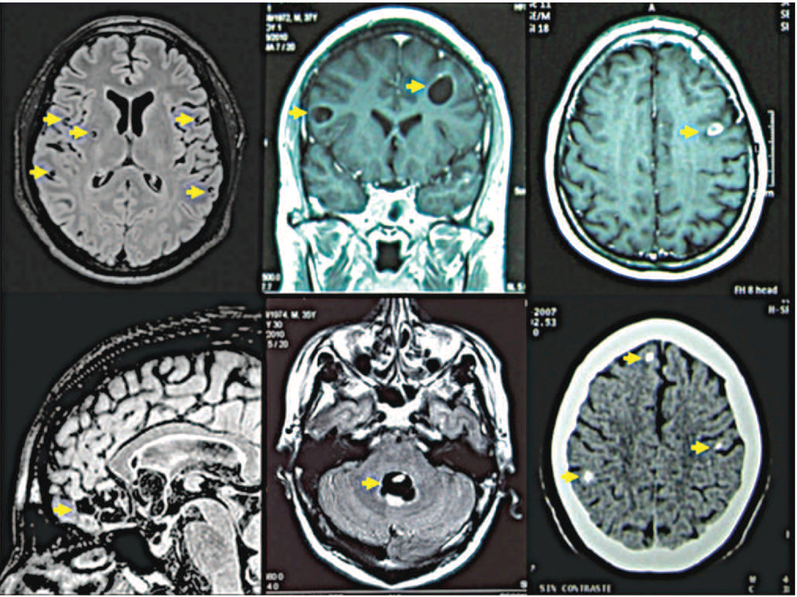
Recommended Workup
- Dedicated CE-MRI Brain with thin sections, DWI, and ideally MR spectroscopy - to characterize the lesion better and look for additional lesions
- Serology: NCC serology (ELISA for T. solium antibodies), Mantoux/TST, IGRA (for TB)
- Chest X-ray - to look for pulmonary TB
- CSF analysis (if clinically indicated) - for cells, protein, ADA, and Gram stain/culture
- Ophthalmology review - to rule out ocular cysticercosis or TB
Treatment Approach
If Neurocysticercosis (single lesion, parenchymal):
- Albendazole 15 mg/kg/day (max 800 mg/day) for 8-28 days - monotherapy for 1-2 cysts
- Dexamethasone 0.1-0.4 mg/kg/day or prednisone 60 mg/day concurrently - mandatory to prevent inflammatory flare
- Anti-seizure medication if seizures occur (can be tapered at 6 months if lesion resolves and patient is seizure-free)
- Antiparasitic treatment is never an emergency - stabilize the patient first
- (Bradley and Daroff's Neurology in Clinical Practice, Treatment Table 79.7)
If Tuberculoma:
- Standard anti-tuberculous therapy (ATT): HRZE for 2 months, then HR for 7-10 months (total 9-12 months for CNS TB)
- Corticosteroids (dexamethasone) to reduce inflammation, especially if edema is significant
- Serial MRI monitoring for response
If the lesion is calcified (no active enhancement on dedicated MRI):
- No antiparasitic treatment is needed
- Treat symptoms (anti-seizure drugs if seizures occur)
Summary for the Patient
| What it means | |
|---|---|
| Pituitary: Normal | No adenoma found; check serum prolactin blood test |
| Left parietal lesion | Small 4x3 mm granuloma - likely a parasite (tapeworm cyst) or TB focus |
| Urgent next step | Dedicated brain MRI + blood/CSF tests to confirm which one |
| Treatment | Anthelmintic or anti-TB drugs + steroids, depending on diagnosis |