
Nerve supply of wrist
Reading File
Now let me search the medical library for wrist nerve supply:
Finding Sources
Finding Sources
Finding Sources
Finding Sources
Reading File
Reading File
The Gray's Anatomy section on the wrist joint doesn't directly list innervation. Let me search more specifically:
Finding Sources
Finding Sources
Reading File
Finding Sources
Reading File
Finding Sources
Reading File
I now have enough information from authoritative sources. Let me compile a thorough answer.
Nerve Supply of the Wrist
The wrist (radiocarpal joint and surrounding structures) receives contributions from all three major nerves of the forearm: the median, ulnar, and radial nerves, along with their deep branches.
1. Median Nerve
-
The median nerve crosses the volar (anterior) aspect of the wrist within the carpal tunnel, just radial and deep to the palmaris longus tendon.
-
Its terminal branch, the anterior interosseous nerve (AIN), carries articular (sensory) fibers to the radiocarpal, radioulnar, intercarpal, and carpometacarpal joints on the volar side.
-
Injury to the terminal branch of the AIN can cause persistent, dull, aching volar wrist pain.
-
The AIN has no cutaneous representation - it is a purely articular/deep sensory branch at the wrist level.
-
Localization in Clinical Neurology, 8e, p. 98
2. Ulnar Nerve
-
The ulnar nerve at the wrist is contained within Guyon's canal, between the pisiform and the hook of the hamate.
-
The dorsal sensory branch of the ulnar nerve crosses the dorsum of the wrist near the ulnar styloid, supplying the dorsal ulnar skin of the hand.
-
Provides articular branches to the ulnar side of the wrist joint.
-
Rosen's Emergency Medicine, p. 3687
3. Radial Nerve - Posterior Interosseous Nerve (PIN)
-
The superficial branch of the radial nerve crosses the dorsum of the wrist near the radial styloid, supplying dorsal skin on the radial side.
-
The posterior interosseous nerve (PIN) - the deep continuation of the radial nerve - gives articular branches to the joint capsule of the wrist and the four radial metacarpophalangeal joints.
-
Injury to the PIN at the wrist spares wrist extension (the extensor carpi radialis longus is supplied more proximally) but affects finger extension.
-
THIEME Atlas of Anatomy, p. 4831-4833
Summary Table
| Nerve | Route at Wrist | Articular Supply | Cutaneous Area |
|---|---|---|---|
| Median - AIN | Carpal tunnel (volar) | Radiocarpal, radioulnar, intercarpal, CMC joints (volar) | None (purely articular) |
| Ulnar | Guyon's canal | Ulnar wrist joint | Dorsal and palmar ulnar hand/fingers |
| Radial (superficial branch) | Dorsum, near radial styloid | - | Dorsal radial hand/thumb web |
| Radial (PIN / deep branch) | Dorsal forearm | Wrist joint capsule + radial 4 MCP joints | None |
Muscles Acting on the Wrist and Their Nerve Supply
| Movement | Muscle | Nerve |
|---|---|---|
| Flexion | Flexor carpi radialis | Median (C6, C7) |
| Flexion | Flexor carpi ulnaris | Ulnar (C7, C8) |
| Flexion | Palmaris longus | Median (C8, T1) |
| Extension | Extensor carpi radialis longus | Radial (C6, C7) |
| Extension | Extensor carpi radialis brevis | PIN/Radial (C7, C8) |
| Extension | Extensor carpi ulnaris | PIN/Radial (C7, C8) |
| Radial deviation | APL, EPB, EPL, ECRL | Radial (C7, C8) |
| Ulnar deviation | FCU, ECU | Ulnar + PIN |
Clinical Correlations
- Carpal tunnel syndrome: Compression of the median nerve in the carpal tunnel - sensory loss in radial 3.5 digits, thenar wasting.
- Guyon's canal syndrome: Ulnar nerve compression - weakness of intrinsic hand muscles, sensory loss in ulnar 1.5 digits.
- Wrist drop: Proximal radial nerve injury paralyzing all wrist and finger extensors (innervated by PIN); sensory loss is limited to the dorsal radial web space.
- PIN syndrome: Isolated deep branch injury - finger drop without wrist drop (ECRL spared), no sensory loss.
- Wrist denervation (surgical): Involves sectioning of both the posterior interosseous nerve and the anterior interosseous nerve near the wrist joint, used for chronic wrist pain relief. - Campbell's Operative Orthopaedics 15th Ed, 2026
Muscles of fore arm
The forearm muscles are divided into anterior (flexor) and posterior (extensor) compartments. The anterior compartment has 3 layers (superficial, intermediate, deep) innervated mainly by the median nerve, and the posterior compartment has superficial and deep groups innervated by the radial nerve (PIN). Let me get the full detailed picture from the medical library.
Searching sources...
Finding Sources
Finding Sources
Reading File
Reading File
Reading File
Finding Sources
Reading File
Reading File
Reading File
Now let me get the posterior compartment and radial group details:
Finding Sources
Finding Sources
Reading File
Reading File
Reading File
Reading File
I now have comprehensive data from Gray's Anatomy for Students. Let me also fetch the forearm muscle diagram images for visual clarity:
Loading Image
Loading Image
Excellent - I have all the data needed for a comprehensive, well-illustrated answer.
Muscles of the Forearm
The forearm has two compartments separated by the interosseous membrane and intermuscular septa:
- Anterior (flexor) compartment - 3 layers, mainly median nerve
- Posterior (extensor) compartment - 2 layers, entirely radial nerve (PIN)
There is also a lateral (radial) group bridging both compartments.
ANTERIOR COMPARTMENT (Flexor)
"All muscles in the anterior compartment of the forearm are innervated by the median nerve, except for the flexor carpi ulnaris and the medial half of the flexor digitorum profundus, which are innervated by the ulnar nerve."
- Gray's Anatomy for Students, p. 897
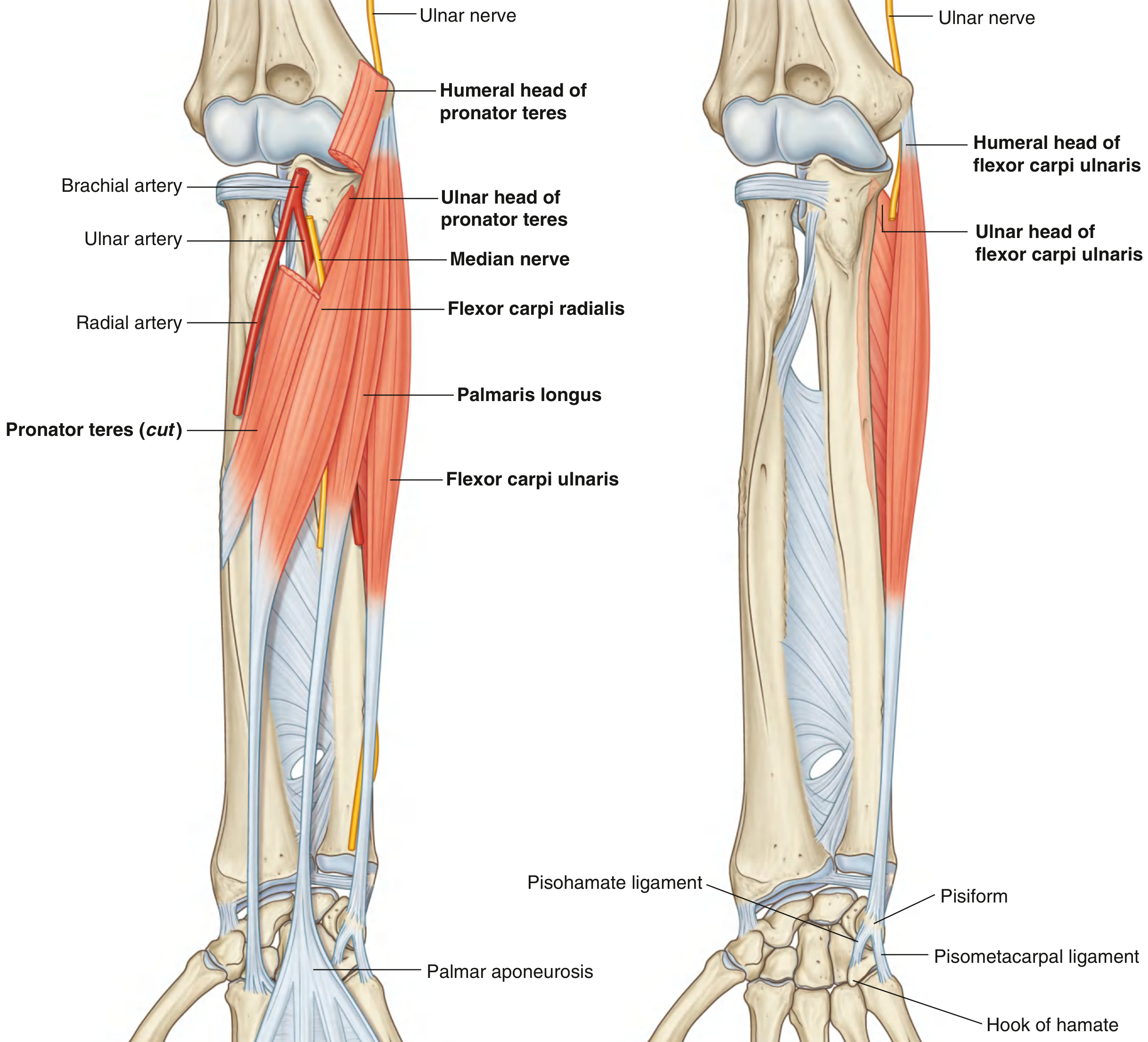
Layer 1 - Superficial (4 muscles, all from medial epicondyle)
| Muscle | Origin | Insertion | Nerve | Action |
|---|---|---|---|---|
| Pronator teres | Medial epicondyle (humeral head) + coronoid process of ulna (ulnar head) | Lateral midshaft of radius | Median (C6, C7) | Pronation; medial boundary of cubital fossa |
| Flexor carpi radialis | Medial epicondyle | Base of metacarpals II & III | Median (C6, C7) | Flexes + abducts (radial deviates) wrist |
| Palmaris longus | Medial epicondyle | Palmar aponeurosis | Median (C7, C8) | Wrist flexion; tightens palmar aponeurosis; absent in ~15% |
| Flexor carpi ulnaris | Medial epicondyle (humeral head) + olecranon/posterior ulna (ulnar head) | Pisiform → hamate + base of metacarpal V | Ulnar (C7, C8, T1) | Flexes + adducts (ulnar deviates) wrist |
Memory aid: "Please Find Professor Faulkner" (Pronator teres, FCR, Palmaris longus, FCU)
Layer 2 - Intermediate (1 muscle)
| Muscle | Origin | Insertion | Nerve | Action |
|---|---|---|---|---|
| Flexor digitorum superficialis (FDS) | Humero-ulnar head (medial epicondyle + coronoid process) + radial head (anterior oblique line of radius) | Margins of middle phalanges of index, middle, ring, little fingers | Median (C8, T1) | Flexes MCP + PIP joints of fingers; accessory wrist flexor |
Note: The median nerve and ulnar artery pass deep to FDS between its two heads. FDS tendons split near the proximal phalanx to allow FDP tendons to pass through (Camper's chiasma).
Layer 3 - Deep (3 muscles)
| Muscle | Origin | Insertion | Nerve | Action |
|---|---|---|---|---|
| Flexor digitorum profundus (FDP) | Anterior + medial surfaces of ulna + interosseous membrane | Distal phalanges of index, middle, ring, little fingers | Lateral half (index, middle): Median - AIN (C8, T1); Medial half (ring, little): Ulnar (C8, T1) | Flexes DIP, PIP, MCP joints; accessory wrist flexion |
| Flexor pollicis longus (FPL) | Anterior surface of radius + interosseous membrane | Base of distal phalanx of thumb | Median - AIN (C7, C8) | Flexes IP + MCP joints of thumb |
| Pronator quadratus | Linear ridge on distal anterior ulna | Distal anterior radius | Median - AIN (C7, C8) | Pronation (primary pronator) |
POSTERIOR COMPARTMENT (Extensor)
"All muscles in the posterior compartment of the forearm are innervated by the radial nerve."
- Gray's Anatomy for Students, p. 906
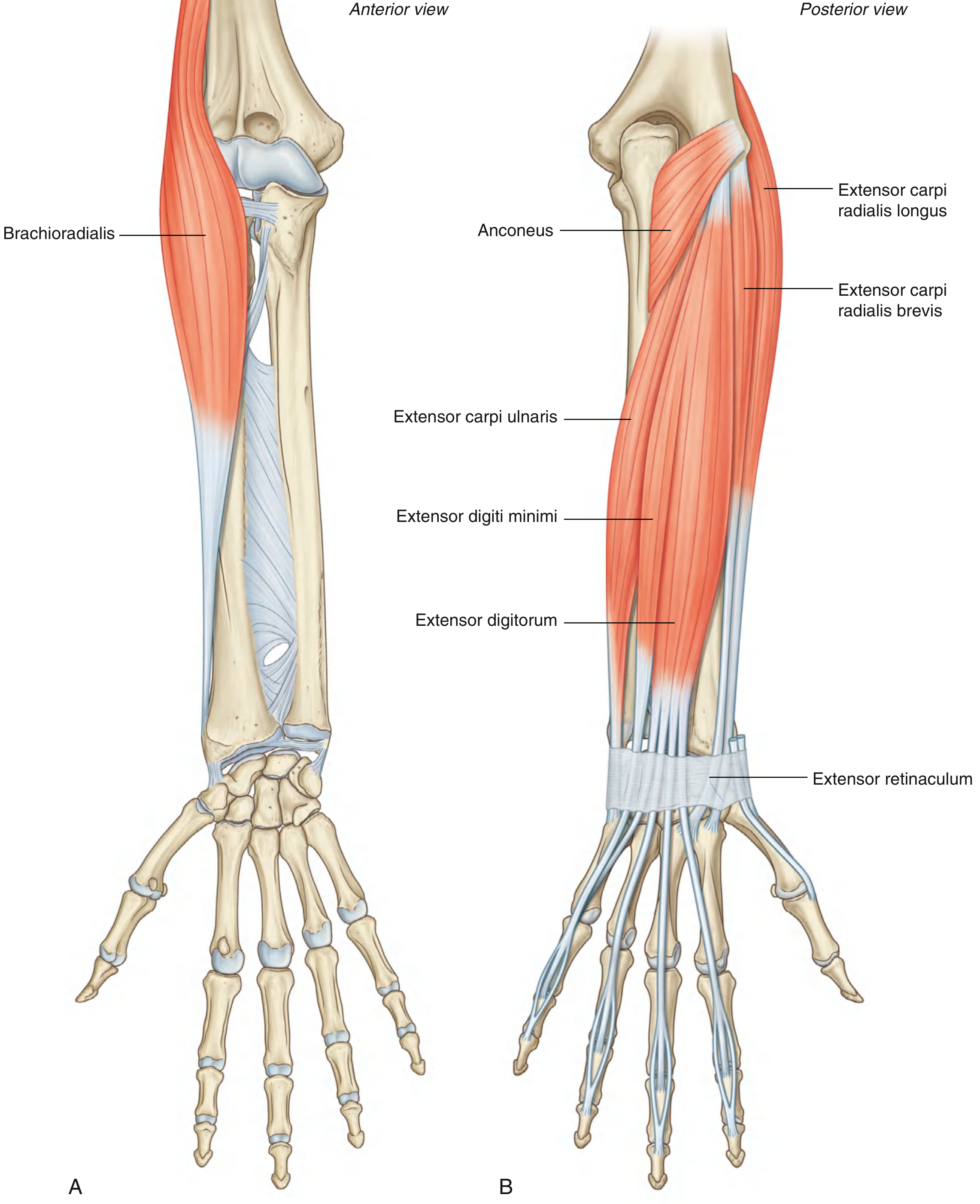
Superficial Layer (7 muscles, common origin: lateral epicondyle / supraepicondylar ridge)
| Muscle | Origin | Insertion | Nerve | Action |
|---|---|---|---|---|
| Brachioradialis | Proximal lateral supraepicondylar ridge | Lateral distal radius (above radial styloid) | Radial (C5, C6) - before bifurcation | Flexes elbow (when forearm midproned); forms lateral border of cubital fossa |
| Extensor carpi radialis longus (ECRL) | Distal lateral supraepicondylar ridge | Base of metacarpal II | Radial (C6, C7) - before bifurcation | Extends + abducts wrist |
| Extensor carpi radialis brevis (ECRB) | Lateral epicondyle | Base of metacarpals II & III | Deep branch radial/PIN (C7, C8) | Extends + abducts wrist |
| Extensor digitorum (ED) | Lateral epicondyle | Extensor hoods → middle + distal phalanges of index, middle, ring, little | PIN (C7, C8) | Extends MCP, PIP, DIP joints; accessory wrist extension |
| Extensor digiti minimi (EDM) | Lateral epicondyle | Extensor hood of little finger | PIN (C7, C8) | Extends little finger |
| Extensor carpi ulnaris (ECU) | Lateral epicondyle + posterior ulna | Base of metacarpal V | PIN (C7, C8) | Extends + adducts (ulnar deviates) wrist |
| Anconeus | Lateral epicondyle | Lateral olecranon + posterior ulna | Radial (C7, C8) | Accessory elbow extension; stabilizes elbow |
Deep Layer (5 muscles, origin: posterior radius/ulna + interosseous membrane)
| Muscle | Origin | Insertion | Nerve | Action |
|---|---|---|---|---|
| Supinator | Lateral epicondyle + anular ligament + supinator crest of ulna | Lateral proximal radius (above oblique line) | PIN (C6, C7) | Supination (the deep branch of radial nerve passes through its two heads to become PIN) |
| Abductor pollicis longus (APL) | Posterior surfaces of radius + ulna + interosseous membrane | Base of metacarpal I | PIN (C7, C8) | Abducts thumb; radial deviation of wrist |
| Extensor pollicis brevis (EPB) | Posterior surface of radius (distal to APL) | Base of proximal phalanx of thumb | PIN (C7, C8) | Extends MCP + CMC joints of thumb; lateral border of anatomical snuffbox |
| Extensor pollicis longus (EPL) | Posterior surface of ulna (distal to APL) | Base of distal phalanx of thumb | PIN (C7, C8) | Extends all thumb joints; medial border of anatomical snuffbox |
| Extensor indicis (EI) | Posterior ulna (distal to EPL) | Extensor hood of index finger | PIN (C7, C8) | Independent index finger extension |
Note on the anatomical snuffbox: APL + EPB form the lateral border; EPL forms the medial border. The scaphoid and radial artery lie in its floor.
LATERAL (RADIAL) GROUP - Summary
The brachioradialis, ECRL, and ECRB form the "radial group" - visible as the lateral muscle mass of the forearm. Brachioradialis is unique: it is in the posterior compartment yet acts as an elbow flexor, not an extensor.
Nerve Supply - Quick Reference
| Nerve | Muscles Supplied |
|---|---|
| Median nerve | Pronator teres, FCR, Palmaris longus, FDS, FPL, Lateral FDP (index + middle) |
| Median - AIN | FDP (lateral half), FPL, Pronator quadratus |
| Ulnar nerve | FCU, Medial FDP (ring + little) |
| Radial nerve (before bifurcation) | Brachioradialis, ECRL |
| Radial - deep branch | ECRB, Supinator (before becoming PIN) |
| Posterior interosseous nerve (PIN) | ED, EDM, ECU, APL, EPB, EPL, EI, Anconeus |
Key Clinical Points
- Median nerve injury at elbow: Loss of pronation (PT, PQ), weak wrist flexion (FCR), loss of FDS, FPL, lateral FDP - "hand of benediction" when making a fist
- AIN syndrome: Loss of FPL + lateral FDP only - inability to make "OK sign" (pinch posture); no sensory loss
- Ulnar nerve injury: Loss of FCU + medial FDP (weak ring/little finger DIP flexion); "claw hand" with intrinsic muscle loss
- Radial nerve / PIN injury (posterior interosseous): Finger drop (cannot extend at MCP joints); wrist drop only if radial nerve injured proximal to ECRL origin; no sensory loss with pure PIN injury
- Tennis elbow (lateral epicondylitis): Degeneration at origin of ECRB at the lateral epicondyle
Sources: Gray's Anatomy for Students, pp. 897-911; THIEME Atlas of General Anatomy and Musculoskeletal System, pp. 2348-2384
This is a shared conversation. Sign in to Orris to start your own chat.