
Explain about vitamin d and biochemistry in it in an easy way
Reading File
Now let me search the medical library for Vitamin D biochemistry content in parallel.
Finding Sources
Finding Sources
Good results. Let me now read the key sections from Robbins (best biochemistry source) and the Tietz lab medicine book, plus grab the diagram image.
Reading File
Reading File
Reading File
Excellent content found. Let me now read the rest of Robbins for functions/deficiency effects, and fetch the metabolism diagram.
Reading File
Loading Image
Loading Image
I now have everything needed for a complete, well-illustrated explanation. Here is a thorough, easy-to-understand breakdown of Vitamin D and its biochemistry.
Vitamin D - A Simple, Complete Guide
What is Vitamin D?
Vitamin D is a fat-soluble vitamin that your body actually uses more like a hormone than a regular vitamin. Its biggest job is keeping calcium and phosphorus at the right levels in your blood - which is essential for strong bones, working muscles, and nerve transmission.
It comes in two main forms:
- Vitamin D2 (Ergocalciferol) - found in some fungi and added to foods
- Vitamin D3 (Cholecalciferol) - made in your skin, and found in oily fish, dairy, and egg yolk
Where Does Vitamin D Come From?
Think of Vitamin D as having two doors:
| Source | What Happens |
|---|---|
| ☀️ Sunlight (UVB, 290-315 nm) | ~90% of your needs - UV light hits skin and converts 7-dehydrocholesterol into Vitamin D3 |
| 🍽️ Diet | ~10% - fish, dairy, fortified foods, supplements; absorbed via the gut |
Note: People with darker skin produce less Vitamin D because melanin absorbs UVB light before it can work on the skin precursor. People at higher latitudes also get less UVB in winter.
The Biochemistry: Step-by-Step Activation
Vitamin D is inactive when it enters the body - it needs two chemical modifications (called hydroxylations) to become active. Think of it as a rough gem that needs two rounds of polishing.
Here is the full chemical journey:
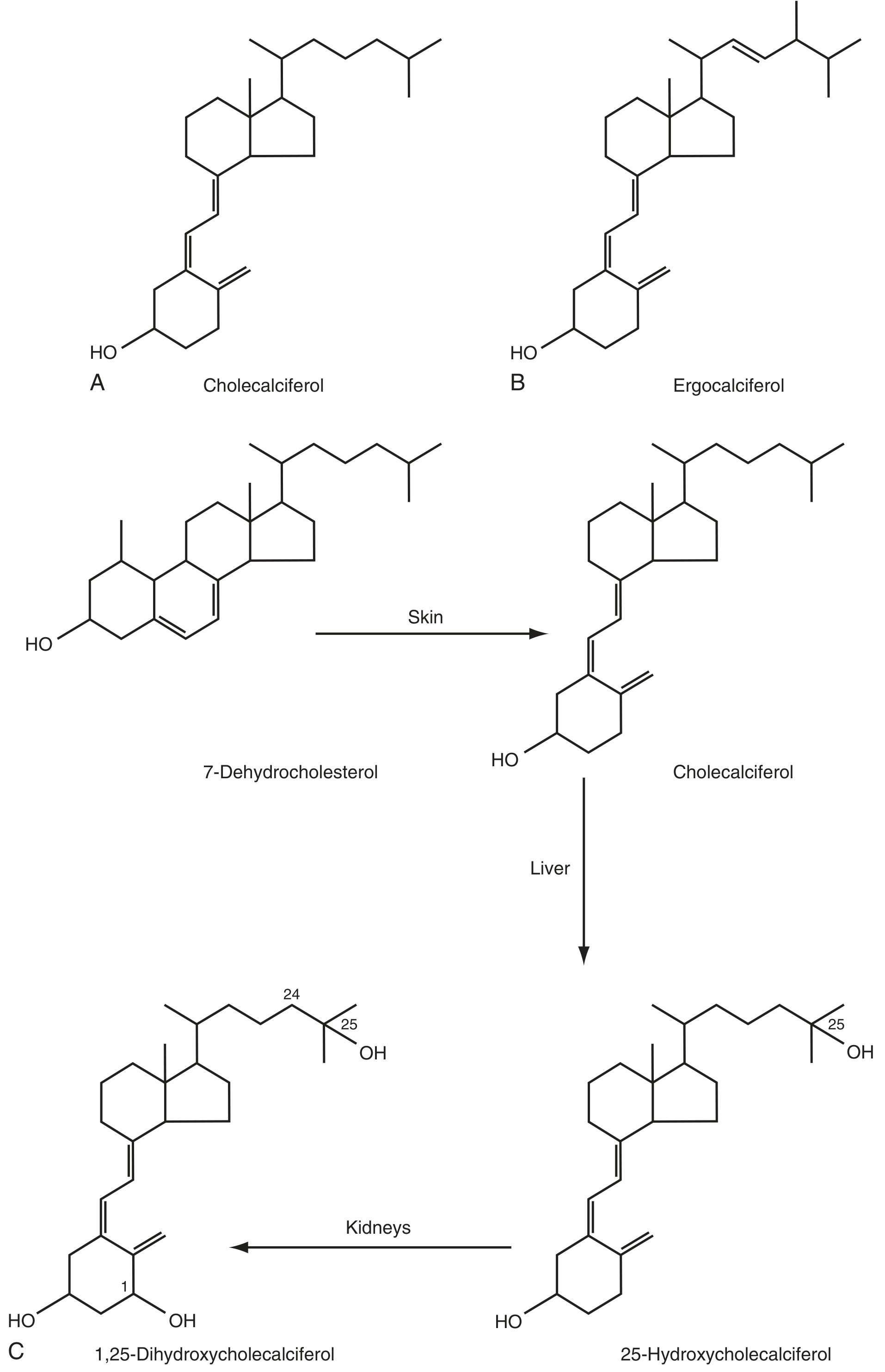
Step 1 - In the Skin (or Gut)
UVB radiation converts 7-dehydrocholesterol in the skin into Vitamin D3 (cholecalciferol). Dietary Vitamin D is absorbed in the gut with dietary fats.
Step 2 - Transport to the Liver
Vitamin D from both sources binds to a carrier protein called Vitamin D Binding Protein (DBP), an alpha-1 globulin in the blood, and is transported to the liver.
Step 3 - In the Liver (First Hydroxylation)
The enzyme 25-hydroxylase (CYP27A1) adds an -OH group at position 25, producing:
25-hydroxyvitamin D (also called calcidiol or 25-OH-D)
This is the main storage form that circulates in the blood. When doctors test your Vitamin D level, this is what they measure. It has little biological activity on its own.
Step 4 - In the Kidney (Second, Final Hydroxylation)
The enzyme 1α-hydroxylase adds another -OH group at position 1, producing:
1,25-dihydroxyvitamin D (also called calcitriol or 1,25-(OH)₂D₃)
This is the fully active, hormonal form of Vitamin D - and it is extremely potent.
The Whole Picture in One Diagram
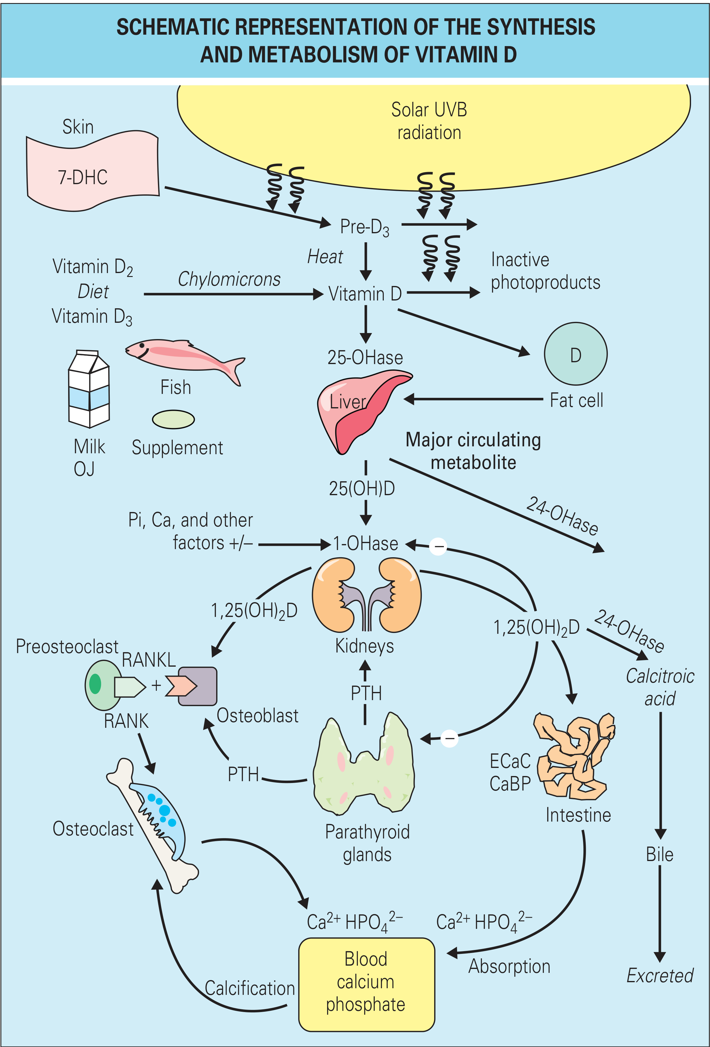
What Controls the Kidney Activation Step?
The kidney's 1α-hydroxylase is tightly regulated - your body only activates as much Vitamin D as it needs:
| Signal | Effect on 1α-Hydroxylase |
|---|---|
| Low blood calcium (hypocalcemia) → triggers PTH | Stimulates (turns it ON) |
| Low blood phosphate (hypophosphatemia) | Stimulates (turns it ON) |
| High 1,25-(OH)₂D already present | Inhibits (negative feedback - turns it OFF) |
| FGF-23 (a bone hormone) | Inhibits (turns it OFF) |
This is a beautiful self-regulating loop - the body only makes active Vitamin D when it actually needs calcium or phosphate.
How Does Active Vitamin D Work in the Body?
Calcitriol (1,25-dihydroxyvitamin D) acts like a steroid hormone - it enters cells and binds to the Vitamin D Receptor (VDR), a nuclear receptor. The VDR then partners with another receptor (RXR) to form a complex that switches specific genes ON or OFF.
Main Actions:
1. Intestine - Increases Calcium Absorption
- Activates the gene for TRPV6, a calcium transport channel in gut cells
- Also stimulates synthesis of calbindins - proteins that carry calcium across intestinal cells
- Stimulates phosphate absorption too
2. Kidney - Saves Calcium
- Increases expression of TRPV5 in kidney tubules, so less calcium is lost in urine
3. Bone - Promotes Mineralization
- Stimulates osteoblasts to make osteocalcin, a protein involved in depositing calcium into bone
- Together with PTH, increases RANKL on osteoblasts → activates osteoclasts → releases calcium from bone when needed
4. Parathyroid Glands - Feedback
- High calcitriol suppresses PTH production (negative feedback loop)
5. Immune System and Other Tissues
- The Vitamin D receptor is found in almost every cell in the body
- Has anti-proliferative and immunomodulatory effects - active research area
The PTH-Vitamin D Loop (Easy Summary)
Here is how the system works as a team:
Blood Ca²⁺ drops
     ↓
Parathyroid glands sense it
     ↓
PTH is released
     ↓
PTH → activates kidney 1α-hydroxylase → more 1,25(OH)₂D
     ↓
1,25(OH)₂D → intestine absorbs more Ca + kidney keeps more Ca + bone releases Ca
     ↓
Blood Ca²⁺ rises back to normal
     ↓
High Ca + high 1,25(OH)₂D → PTH secretion is suppressed
What Happens When Vitamin D is Deficient?
When there is not enough Vitamin D, calcium absorption from the gut falls. The body compensates through secondary hyperparathyroidism - PTH rises and pulls calcium out of bone. The consequence:
| Age Group | Disease | What Happens |
|---|---|---|
| Children (before growth plates close) | Rickets | Soft, deformed bones; bowed legs; enlarged wrists; pot belly |
| Adults | Osteomalacia | Soft bones, bone pain, muscle weakness, stress fractures |
| Any age (acute) | Hypocalcemic Tetany | Muscle cramps, spasms (uncommon because PTH usually compensates) |
Quick Biochemistry Summary Table
| Form | Name | Where Made | Activity |
|---|---|---|---|
| Vitamin D3 | Cholecalciferol | Skin (UVB) | Inactive |
| 25-OH-D | Calcidiol | Liver | Storage form (inactive) |
| 1,25-(OH)₂D | Calcitriol | Kidney | Active hormone |
| 24,25-(OH)₂D | Calcitroic acid | Kidney/intestine | Inactive - excreted via bile |
Key Takeaways in Plain Language
- Sunlight makes most of your Vitamin D - about 90% is produced in your skin using UVB rays
- Vitamin D needs two upgrades before it works - one in the liver, one in the kidney
- The active form is calcitriol - it acts like a hormone, not just a vitamin
- Its main job is calcium control - keeping bones strong and muscles working
- Low calcium triggers more Vitamin D activation - through PTH stimulation of the kidney enzyme
- Deficiency causes rickets in kids, osteomalacia in adults
- Vitamin D also affects immunity - the receptor exists in nearly every cell, not just bones
Sources: Robbins, Cotran & Kumar - Pathologic Basis of Disease, p. 417-420 | Tietz Textbook of Laboratory Medicine, 7th Edition, p. 1682 | Rheumatology, 2-Volume Set (2022, Elsevier), p. 1859
👍
Glad it was helpful! 😊 Feel free to ask anytime you want to explore another topic — whether it's more biochemistry, clinical medicine, or anything else!
This is a shared conversation. Sign in to Orris to start your own chat.