Why vitamin b12 deficiency leads to neural defects

Reading File
Finding Sources
Finding Sources
Finding Sources
Reading File
Reading File
Reading File
Reading File
Searching PubMed

"vitamin B12 deficiency" AND "subacute combined degeneration"

Searching Images

subacute combined degeneration spinal cord posterior columns vitamin B12 deficiency
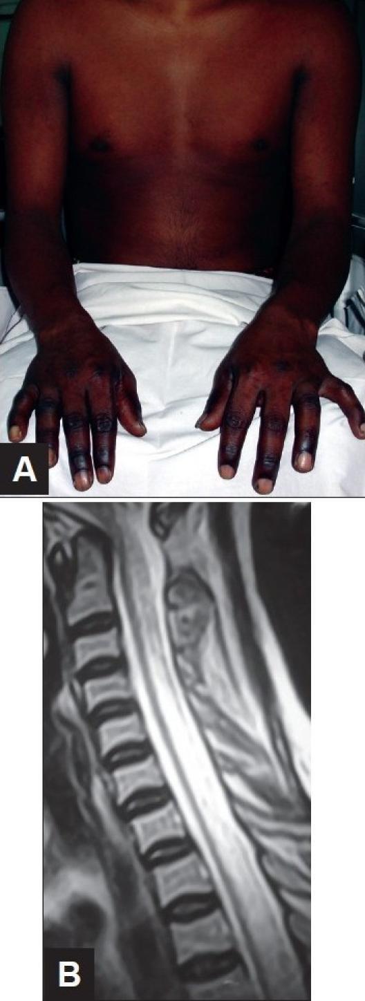
This composite figure demonstrates clinical and diagnostic findings associated with Vitamin B12 deficiency (Subacute Combined Degeneration of the spinal cord).
This composite figure demonstrates clinical and diagnostic findings associated with Vitamin B12 deficiency (Subacute Combined Degeneration of the spinal cord).

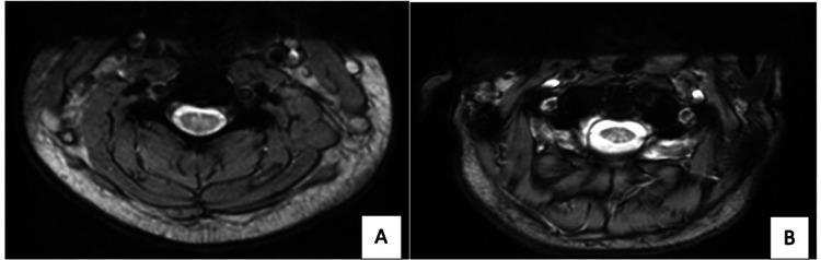
Two axial T2-weighted magnetic resonance imaging (MRI) scans of the spine (A: cervical region, B: thoracic region). The primary diagnostic feature is a symmetric, inverted V-shaped hyperintense signal within the dorsal columns of the spinal cord. This radiological finding, often referred to as the 'inverted V sign,' is highly characteristic of subacute combined degeneration of the spinal cord. The images demonstrate significant contrast between the high-signal pathology in the posterior cord and the normal intermediate signal of the surrounding cord parenchyma. Surrounding anatomical structures include the vertebral bodies (low signal bone), paraspinal muscles, and subcutaneous fat. The educational focus is on identifying neurologic sequelae of Vitamin B12 deficiency or nitrous oxide toxicity, illustrating the specific localization of degeneration in the dorsal columns responsible for proprioception and vibratory sense loss.
Two axial T2-weighted magnetic resonance imaging (MRI) scans of the spine (A: cervical region, B: thoracic region). The primary diagnostic feature is a symmetric, inverted V-shaped hyperintense signal within the dorsal columns of the spinal cord. This radiological finding, often referred to as the 'inverted V sign,' is highly characteristic of subacute combined degeneration of the spinal cord. The images demonstrate significant contrast between the high-signal pathology in the posterior cord and the normal intermediate signal of the surrounding cord parenchyma. Surrounding anatomical structures include the vertebral bodies (low signal bone), paraspinal muscles, and subcutaneous fat. The educational focus is on identifying neurologic sequelae of Vitamin B12 deficiency or nitrous oxide toxicity, illustrating the specific localization of degeneration in the dorsal columns responsible for proprioception and vibratory sense loss.

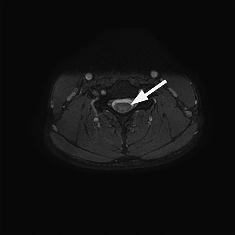
An axial T2-weighted Magnetic Resonance Imaging (MRI) scan of the cervical spine at the C5 vertebral level. The image demonstrates a well-defined spinal cord situated centrally within the spinal canal. A prominent white arrow points to a localized area of increased T2 signal intensity (hyperintensity) within the dorsal (posterior) columns. This radiographic finding is characteristic of subacute combined degeneration of the spinal cord, often associated with vitamin B12 deficiency or nitrous oxide toxicity. The surrounding anatomical structures, including the vertebral body, dark-appearing cerebrospinal fluid (CSF), and paraspinal soft tissues, are visible in the cross-sectional view. The hyperintensity specifically affects the posterior white matter tracts, illustrating the selective pathology typically seen in demyelinating processes of the dorsal columns.
An axial T2-weighted Magnetic Resonance Imaging (MRI) scan of the cervical spine at the C5 vertebral level. The image demonstrates a well-defined spinal cord situated centrally within the spinal canal. A prominent white arrow points to a localized area of increased T2 signal intensity (hyperintensity) within the dorsal (posterior) columns. This radiographic finding is characteristic of subacute combined degeneration of the spinal cord, often associated with vitamin B12 deficiency or nitrous oxide toxicity. The surrounding anatomical structures, including the vertebral body, dark-appearing cerebrospinal fluid (CSF), and paraspinal soft tissues, are visible in the cross-sectional view. The hyperintensity specifically affects the posterior white matter tracts, illustrating the selective pathology typically seen in demyelinating processes of the dorsal columns.

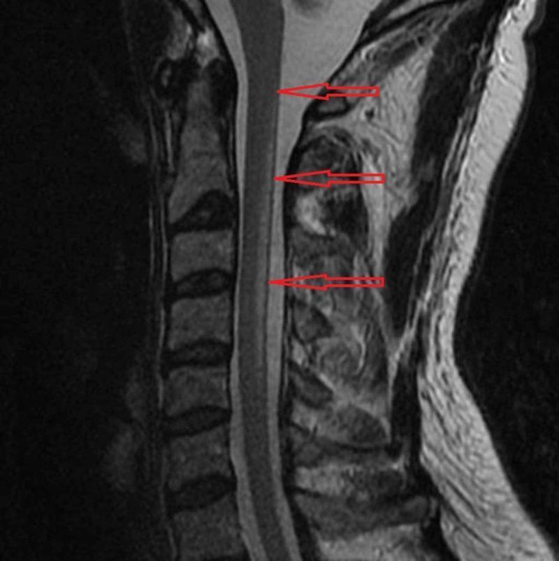
A sagittal T2-weighted magnetic resonance imaging (MRI) scan of the cervical spine. The diagnostic image demonstrates abnormal hyperintense (high) signal intensity within the posterior aspect of the spinal cord, specifically localized to the dorsal columns. This longitudinal signal abnormality is marked by three red horizontal arrows and extends from the C2 vertebral level down to the C5 level. The spinal cord maintains its normal caliber without significant edema or expansion. These radiographic findings are characteristic of Subacute Combined Degeneration (SCD) of the cord, typically associated with Vitamin B12 deficiency or nitrous oxide toxicity. The image serves as an educational example of metabolic myelopathy, illustrating the selective involvement of the posterior columns in the cervical region for medical students and neurology residents.

A sagittal T2-weighted magnetic resonance imaging (MRI) scan of the cervical spine. The diagnostic image demonstrates abnormal hyperintense (high) signal intensity within the posterior aspect of the spinal cord, specifically localized to the dorsal columns. This longitudinal signal abnormality is marked by three red horizontal arrows and extends from the C2 vertebral level down to the C5 level. The spinal cord maintains its normal caliber without significant edema or expansion. These radiographic findings are characteristic of Subacute Combined Degeneration (SCD) of the cord, typically associated with Vitamin B12 deficiency or nitrous oxide toxicity. The image serves as an educational example of metabolic myelopathy, illustrating the selective involvement of the posterior columns in the cervical region for medical students and neurology residents.

I have enough to give a complete mechanistic answer. Here it is:

Why vitamin B12 deficiency causes neurological damage

Vitamin B12 (cobalamin) is a cofactor for only two human enzymatic reactions, and both feed into pathways that maintain the nervous system. Loss of either explains the neurological injury.

1. Methionine synthase reaction (homocysteine → methionine)

Methylcobalamin is required by methionine synthase to convert homocysteine to methionine. Methionine is then converted to S-adenosylmethionine (SAM), the universal methyl donor used for methylation of myelin basic protein, phospholipids, neurotransmitters, and DNA/histones in the nervous system.
  • The nervous system has no alternative (betaine-based) route to regenerate methionine the way the liver does, so it is entirely dependent on the B12-methionine synthase pathway.
  • When B12 is deficient, homocysteine cannot be remethylated, SAM falls, and hypomethylation occurs in neural tissue. This destabilizes myelin and impairs normal neuronal function - "Basic Medical Biochemistry - A Clinical Approach - 6e" describes this as one of the two central mechanisms of B12 neurotoxicity.

2. Methylmalonyl-CoA mutase reaction (propionate metabolism)

The second B12-dependent enzyme, methylmalonyl-CoA mutase, converts methylmalonyl-CoA to succinyl-CoA (which enters the Krebs cycle). Without B12:
  • Methylmalonyl-CoA and its precursor propionyl-CoA accumulate.
  • Propionyl-CoA abnormally displaces succinyl-CoA, the normal primer for fatty acid synthesis, causing odd-chain and branched-chain fatty acids to be incorporated into membrane phospholipids instead of normal even-chain fatty acids.
  • These abnormal fatty acids are laid down in the myelin sheath, destabilizing it and impairing its ability to insulate axons and support normal action potential conduction (Adams and Victor's Principles of Neurology, 12th Ed; Basic Medical Biochemistry, 6e).
Both mechanisms converge on the same structural target: myelin. Pathologically, this produces the classic lesion, subacute combined degeneration (SCD) of the spinal cord:
"The earliest histologic event is swelling of myelin sheaths, characterized by the formation of intramyelinic vacuoles and separation of myelin lamellae... The changes begin in the posterior columns of the lower cervical and upper thoracic segments of the cord and spread... representing a myelinopathy." - Adams and Victor's Principles of Neurology, 12th Ed, p. 1177
"Subacute combined degeneration of the spinal cord is caused by deficiency of vitamin B12... The lesions result from a defect in myelin formation... On microscopic examination, there is swelling of myelin layers, producing vacuoles in the affected tracts; with time, axons degenerate as well." - Robbins, Cotran & Kumar Pathologic Basis of Disease, p. 694

Why specific tracts are affected

The posterior columns (proprioception, vibration) and lateral corticospinal tracts (motor) of the spinal cord are hit hardest, explaining the classic triad: symmetric paresthesias/numbness starting in the feet, loss of vibration/position sense and ataxia (posterior column), followed by spastic weakness and hyperreflexia (corticospinal tract involvement). Peripheral nerves, optic nerves, and cerebral white matter can also be affected, producing optic neuropathy, cognitive changes ("megaloblastic madness," depression, dementia), and rarely psychosis.
Inverted-V sign of subacute combined degeneration on MRI
Axial T2 MRI showing the characteristic "inverted V sign" - symmetric hyperintensity in the dorsal columns, reflecting the myelin damage described above.

Important nuance

Because neurons are post-mitotic and don't divide, the DNA-synthesis defect that causes megaloblastic anemia (via the folate "methyl-trap") does not explain the neurological damage - which is why some patients (especially if folic acid is given without B12) can have normal blood counts yet still develop or even worsen neurological disease. This is also why treating pernicious anemia with folic acid alone is a recognized clinical error: it corrects the anemia but does nothing for, and may unmask, the myelin pathology.

A related but distinct issue - fetal neural tube defects

Separately, maternal B12 deficiency (independent of folate status) is increasingly recognized as a risk factor for fetal neural tube defects, likely via the same homocysteine/SAM-methylation pathway affecting neural tube closure during early embryogenesis. A recent systematic review/meta-analysis addresses this:
  • Nie L, et al. "Beyond Folate: The Emerging Role of Maternal Vitamin B12 in Neural Tube Development." Nutrients 2025 (PMID: 40573151)
If your question was about neural tube defects in pregnancy rather than the classic adult neuropathy (subacute combined degeneration), let me know and I can go deeper into that literature specifically.

Why vitamin b12 deficiency leads to neural defects for 2 mark question

For a 2-mark exam answer, keep it short and mechanistic:
Vitamin B12 deficiency → neurological defects (2-mark answer):
Vitamin B12 is a cofactor for two key reactions:
  1. Methionine synthase reaction: B12 is needed to convert homocysteine → methionine → S-adenosylmethionine (SAM), the methyl donor required for myelin synthesis. Deficiency causes hypomethylation and defective myelin formation.
  2. Methylmalonyl-CoA mutase reaction: B12 deficiency causes accumulation of methylmalonyl-CoA and propionyl-CoA, leading to incorporation of abnormal odd-chain fatty acids into myelin, destabilizing it.
Both mechanisms damage myelin in the posterior and lateral columns of the spinal cord, causing subacute combined degeneration - presenting as paresthesia, loss of vibration/position sense, and spastic weakness.
One-line version if space is very limited: "B12 deficiency impairs methionine synthase and methylmalonyl-CoA mutase activity, causing defective myelin synthesis and abnormal fatty acid incorporation into myelin, leading to demyelination of the spinal cord (subacute combined degeneration) and peripheral nerves."

Why vitamin b12 deficiency leads to neural defects for 2 mark question anatomy question not biochemistry

Vitamin B12 deficiency → neural defects (Anatomy answer, 2 marks):
B12 deficiency causes degeneration of the posterior (dorsal) columns and lateral corticospinal tracts of the spinal cord - a condition called Subacute Combined Degeneration (SCD).
  • Posterior columns (fasciculus gracilis/cuneatus) carry proprioception, vibration, and fine touch → deficiency causes loss of position/vibration sense and sensory ataxia.
  • Lateral corticospinal tracts carry descending motor fibers → deficiency causes spastic weakness, hyperreflexia, and extensor plantar (Babinski) response.
Anatomically, the lesion begins in the lower cervical and upper thoracic cord and spreads up and down, and may also involve peripheral nerves and optic nerves, producing a mixed picture of sensory loss, spastic paraparesis, and absent ankle jerks with extensor plantars ("combined" upper and lower motor neuron + sensory signs).
One-line version: "B12 deficiency causes demyelination of the posterior columns and lateral corticospinal tracts of the spinal cord (subacute combined degeneration), leading to loss of vibration/position sense and spastic weakness of the limbs."
This is a shared conversation. Sign in to Orris to start your own chat.