
Mechanism of urine formation
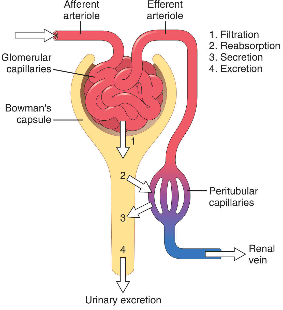
The kidneys form urine through three main processes: glomerular filtration, tubular reabsorption, and tubular secretion. About 180 L of plasma is filtered daily, but only ~1.5 L is excreted as urine, meaning roughly 99% of the filtrate is reabsorbed.
Searching sources...
Reading File
Finding Sources
Searching Images
mechanism of urine formation nephron glomerular filtration reabsorption secretion diagram
Reading File
Finding Sources
Reading File
Reading File
Reading File
Loading Image
Now let me get more detail on glomerular filtration specifically, then tubular handling:
Finding Sources
Finding Sources
Reading File
Reading File
Finding Sources
Finding Sources
Reading File
Reading File
I now have comprehensive material to write a thorough, well-structured answer. Let me also fetch the countercurrent diagram:
Loading Image
I now have everything needed for a comprehensive, well-illustrated answer.
Mechanism of Urine Formation
Urine formation is the result of three sequential, interrelated renal processes: glomerular filtration, tubular reabsorption, and tubular secretion. The net result is summarized as:
Urinary Excretion = Glomerular Filtration - Tubular Reabsorption + Tubular Secretion
(Guyton and Hall Textbook of Medical Physiology)
1. Glomerular Filtration
What is filtered
Urine formation begins when a large volume of fluid - virtually protein-free - is filtered from the glomerular capillaries into Bowman's capsule. Most plasma substances (water, electrolytes, glucose, amino acids, urea, creatinine) pass freely; plasma proteins and substances bound to them are retained. The filtrate in Bowman's capsule therefore has almost the same concentration as plasma.
Driving force - Starling forces
Glomerular filtration is governed by the Starling equation:
GFR = K_f [(P_GC - P_BS) - π_GC]
| Force | Value | Effect on filtration |
|---|---|---|
| P_GC - glomerular capillary hydrostatic pressure | ~45 mm Hg | Favors filtration |
| P_BS - Bowman's space hydrostatic pressure | ~10 mm Hg | Opposes filtration |
| π_GC - glomerular capillary oncotic pressure | ~28 mm Hg (rises along capillary) | Opposes filtration |
| Net ultrafiltration pressure | ~10 mm Hg | Favors filtration |
- K_f (filtration coefficient) for glomerular capillaries is 100-fold that of systemic capillaries because of their enormous surface area and high intrinsic water permeability - this allows 180 L of filtrate to be produced every day.
- The oncotic pressure progressively rises along the capillary length as fluid is filtered out; eventually it reaches filtration equilibrium and filtration stops.
- (Costanzo Physiology 7th Edition)
GFR in numbers
- Normal GFR: ~125 mL/min = ~180 L/day of filtrate formed
- Only ~1.5 L is excreted as urine - meaning ~99% is reabsorbed
2. Tubular Reabsorption
As the filtrate flows through the nephron tubules, essential substances are recovered back into the peritubular capillary blood. Reabsorption is quantitatively large and highly selective - unlike filtration, which is nonselective.
Segment-by-segment reabsorption
Proximal Convoluted Tubule (PCT)
- Reabsorbs ~65% of filtered Na+, water, and Cl-; 100% of glucose and amino acids; most bicarbonate
- The luminal surface has an extensive brush border (increases surface area), and cells are packed with mitochondria to power active transport
- Mechanism: Na+-K+-ATPase on the basolateral side maintains low intracellular Na+; this drives Na+ entry from the lumen via:
- Co-transport with glucose and amino acids (SGLT in early PCT)
- Counter-transport with H+ (NHE - reabsorbs Na+, secretes H+, removing HCO3-)
- Passive Cl- diffusion in the late PCT (higher luminal Cl- concentration drives it)
- Water follows by osmosis through aquaporin channels
- (Guyton and Hall Textbook of Medical Physiology)
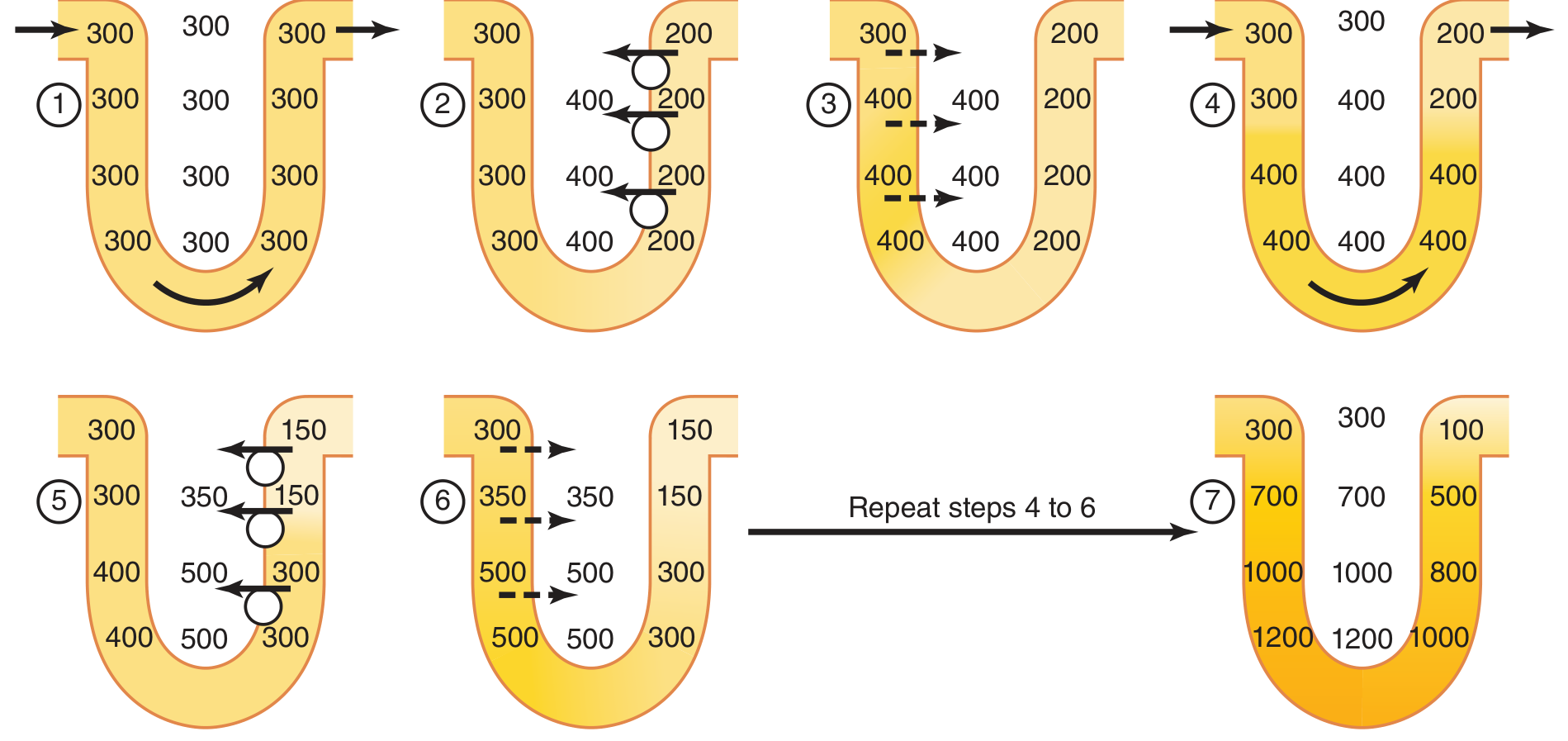
Loop of Henle - Countercurrent Multiplier
This is responsible for producing the hyperosmotic renal medullary interstitium that allows urine concentration.
| Segment | Water permeability | Solute transport |
|---|---|---|
| Descending thin limb | High (water exits by osmosis) | Low active transport |
| Ascending thin limb | Low | Passive NaCl efflux |
| Thick ascending limb (TAL) | Impermeable to water | Active Na-K-2Cl (NKCC2) cotransport OUT |
The thick ascending limb actively pumps NaCl into the interstitium but water cannot follow (impermeable). This creates a 200 mOsm/L gradient at each horizontal level. As new fluid enters from the proximal tubule, the concentrated fluid from the descending limb flows up the ascending limb - and the process repeats, multiplying the concentration gradient. The end result is an interstitial osmolarity gradient from ~300 mOsm/L at the cortex to ~1200-1400 mOsm/L deep in the medulla.
(Guyton and Hall Textbook of Medical Physiology)
Distal Convoluted Tubule (DCT)
- Fluid leaving the loop of Henle enters the DCT at only ~100-140 mOsm/L (dilute)
- The early DCT, like the TAL, actively reabsorbs NaCl (via NCC cotransporter) but is impermeable to water - further diluting tubular fluid
- Aldosterone (acting on principal cells) increases Na+ reabsorption and K+ secretion in the late DCT and collecting duct
- PTH promotes Ca2+ reabsorption here
Collecting Duct (Cortical and Medullary)
- The final arbiter of urine concentration
- ADH (vasopressin) inserts aquaporin-2 (AQP2) channels into the luminal membrane, making the duct water-permeable
- With high ADH: water is osmotically drawn out into the hyperosmotic medullary interstitium → concentrated urine (~1200 mOsm/L)
- Without ADH: water stays in the tubule → dilute urine (~50-100 mOsm/L)
- Urea recycling in the inner medullary collecting duct also contributes significantly to medullary hyperosmolarity
3. Tubular Secretion
Secretion moves substances from peritubular capillary blood into the tubular lumen - this adds to what the filtrate already contains.
Key secreted substances:
| Substance | Primary site | Significance |
|---|---|---|
| H+ | PCT, intercalated cells of collecting duct | Acid-base balance |
| K+ | Principal cells of collecting duct | Regulated by aldosterone; major route of K+ excretion |
| NH4+/NH3 | PCT and collecting duct | Buffer in acidosis |
| Organic acids & bases | PCT | Drug/toxin elimination (e.g., PAH, penicillin, uric acid) |
| Creatinine | Small amount in PCT | Near-complete excretion (mainly filtered) |
Secretion is especially important for K+ and H+ - their urinary excretion rates can actually exceed their filtered loads, proving net secretion has occurred. (Costanzo Physiology 7th Edition)
Summary Table: Fate of Key Filtered Substances
| Substance | Filtered/day | Excreted/day | % Reabsorbed | Key mechanism |
|---|---|---|---|---|
| Water | 180 L | 1.5 L | 99% | Osmosis (AQP), ADH control |
| Na+ | 25,200 mEq | 100 mEq | 99.6% | Na-K-ATPase, aldosterone |
| Glucose | 180 g | 0 g | 100% | SGLT1/2 (PCT) |
| HCO3- | ~4320 mEq | ~2 mEq | ~99.9% | Linked to H+ secretion |
| Urea | 56 g | 28 g | ~50% | Passive (PCT, inner medulla) |
| Creatinine | ~1.8 g | ~1.8 g | ~0% | Marker of GFR |
| K+ | ~800 mEq | ~100 mEq | ~87% | Active reabsorption + secretion |
Key Regulatory Hormones
| Hormone | Site of action | Effect |
|---|---|---|
| ADH (vasopressin) | Collecting duct | ↑ water reabsorption (AQP2 insertion) |
| Aldosterone | Late DCT, collecting duct | ↑ Na+ reabsorption, ↑ K+ secretion |
| ANP/BNP | Glomerulus, collecting duct | ↑ GFR, ↓ Na+ reabsorption |
| PTH | DCT | ↑ Ca2+ reabsorption, ↓ phosphate reabsorption |
| Angiotensin II | PCT, afferent/efferent arterioles | ↑ Na+ reabsorption, vasoconstriction |
Why Filter 180 L and Reabsorb 99%?
This seemingly wasteful strategy has two key advantages:
- Rapid clearance of metabolic waste products (urea, creatinine, uric acid) that are poorly reabsorbed - a high GFR ensures they are quickly removed
- Precise regulation - fine control over excretion is achieved by adjusting reabsorption, even for substances filtered in large amounts
About 10% of the body's resting energy goes to powering this reabsorption machinery.
(Guyton and Hall Textbook of Medical Physiology, p. 330)
This is a shared conversation. Sign in to Orris to start your own chat.