
Do you know about pubic symphosys
Reading File
Now let me search the medical library for pubic symphysis content.
Finding Sources
Finding Sources
Good results. Let me now read the most relevant sections - Gray's Anatomy for the joint description, and the Imaging Anatomy atlas for a structured overview.
Reading File
Reading File
Reading File
Let me also read the osteitis pubis section and fetch a relevant image.
Reading File
Loading Image
Loading Image
I now have excellent textbook content and images to give a thorough answer. Here it is:
Pubic Symphysis
What it is
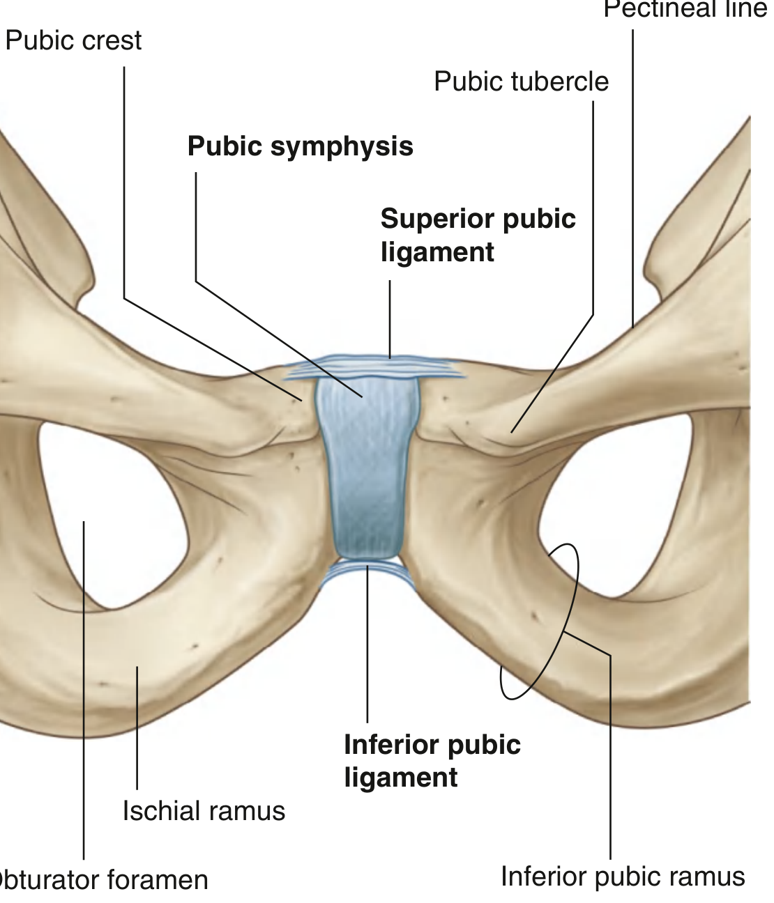
The pubic symphysis is a secondary cartilaginous joint (symphysis) located at the midline of the anterior pelvis, joining the two pubic bones. It is classified as a syndesmotic joint with a fibrocartilage disk - called the interpubic disk - interposed between the hyaline cartilage-lined articular surfaces of the two pubic bones.
- Articular surface mean length: 30-35 mm
- Articular surface mean width: 10-12 mm
- A small non-synovial cavity may exist within the disk
(Imaging Anatomy: Bones, Joints, Vessels and Nerves)
Location and Pelvic Role
The pubic symphysis forms the anterior midline boundary of the pelvic outlet. The diamond-shaped pelvic outlet is bounded:
- Anteriorly - by the pubic symphysis
- Laterally - by the ischial tuberosities
- Posteriorly - by the coccyx and sacrotuberous ligaments
(Gray's Anatomy for Students)
Ligaments
Four ligaments reinforce and stabilize the joint:
| Ligament | Location / Description |
|---|---|
| Superior pubic ligament | Attached superiorly to the pubic crest, extending laterally to the pubic tubercles |
| Inferior pubic ligament (arcuate pubic ligament) | Forms an arch spanning the inferior pubic rami; a small fat-filled space between it and the perineal membrane contains the deep dorsal vein of the penis or clitoris |
| Anterior pubic ligament | Connects the pubic bones anteriorly; its superficial fibers interweave with the tendons of rectus abdominis, oblique abdominal muscles, and pyramidalis |
| Posterior pubic ligament | Very small |
Movement
The joint allows a small amount of translation and rotation, but movement here always requires simultaneous movement at the sacroiliac joint.
During pregnancy, the hormone relaxin induces structural changes in the fibrocartilaginous disk - increasing symphyseal width and mobility to facilitate childbirth. This can sometimes progress to painful symphysis pubis dysfunction (SPD).
Age-Related Changes
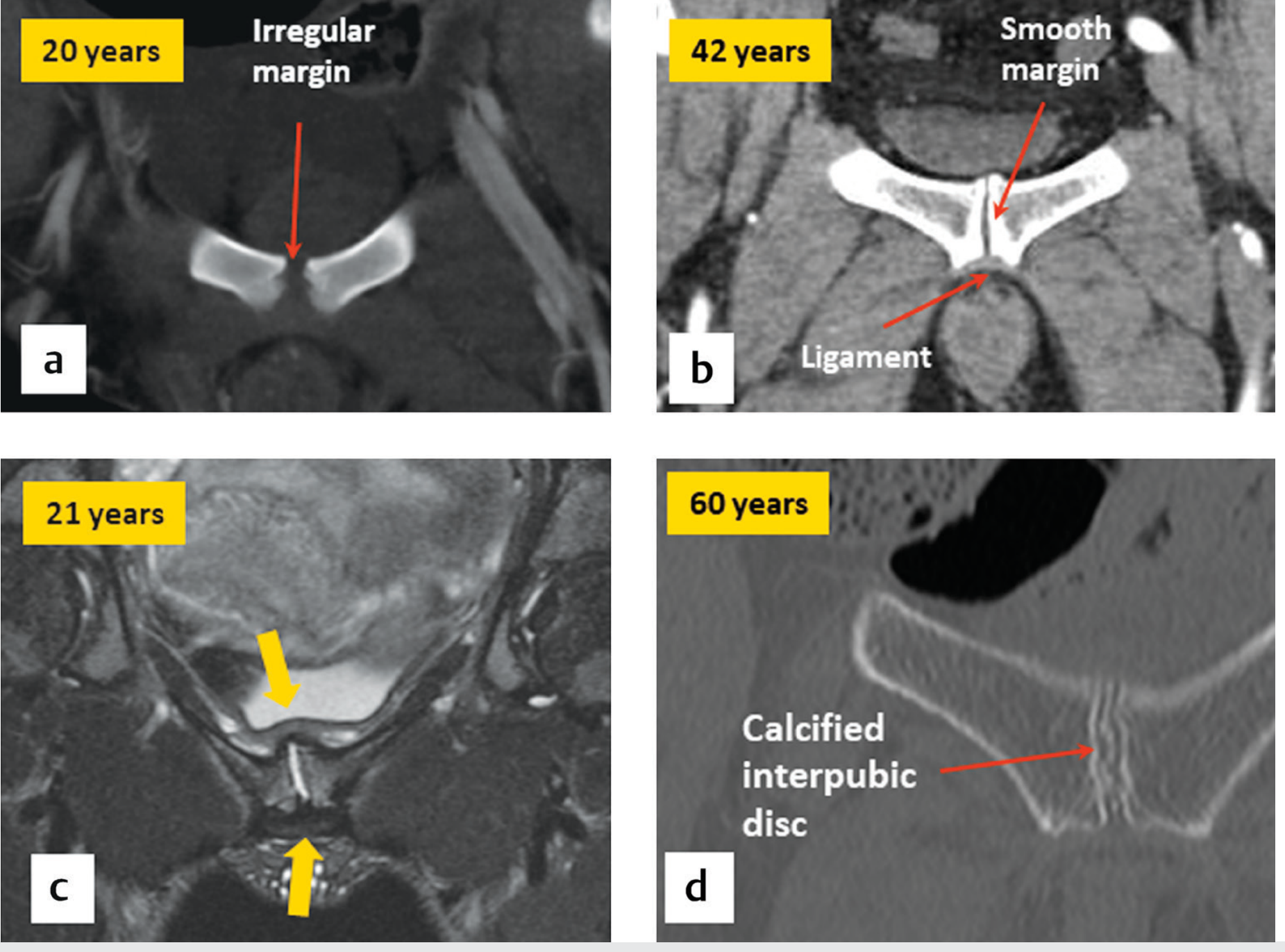
- Young adults (<30 yrs): Subchondral bony surface is irregular
- ~30 years: Surface becomes smooth and straighter
- ~60 years: Degenerative changes appear - joint narrowing, subchondral sclerosis, and irregularity become common findings
The interpubic disk can also become calcified due to degeneration or calcium pyrophosphate crystal deposition (CPPD) - the pubic symphysis is actually a common site for CPPD disease.
(Imaging Anatomy: Bones, Joints, Vessels and Nerves)
Muscle Attachments
Two major muscle/tendon groups attach here:
- Rectus abdominis - passes over the pubic bones anteriorly, inserting onto the anterior inferior aspect
- Adductor muscles - share a common aponeurosis with the rectus abdominis at the pubic bone
This common aponeurosis is clinically significant (see below).
Clinical Conditions
1. Osteitis Pubis
Caused by abnormal stresses applied to the symphysis. Features include:
- Subchondral sclerosis
- Symphyseal irregularity and bone resorption
- MRI: marrow edema and disruption of the rectus abdominis-adductor aponeurosis
2. Athletic Pubalgia ("Sports Hernia")
Results from avulsion of the common rectus abdominis-adductor aponeurosis from the bone and capsule. Most commonly seen in older adolescent or young adult male athletes.
3. Traumatic Diastasis
Disruption/widening of the symphysis, typically from high-energy pelvic trauma (e.g., open-book pelvic fractures). May require surgical fixation with a plate (ORIF of symphysis pubis).
4. Symphysis Pubis Dysfunction (SPD) in Pregnancy
Relaxin-mediated excessive widening causing pelvic instability and pain during pregnancy.
Sources: Imaging Anatomy: Bones, Joints, Vessels and Nerves | Gray's Anatomy for Students | Rockwood and Green's Fractures in Adults, 10th ed. 2025
This is a shared conversation. Sign in to Orris to start your own chat.