
Interstitial lung disease Classification and HRCT imaging pattern
HRCT patterns interstitial lung disease UIP NSIP honeycombing ground glass
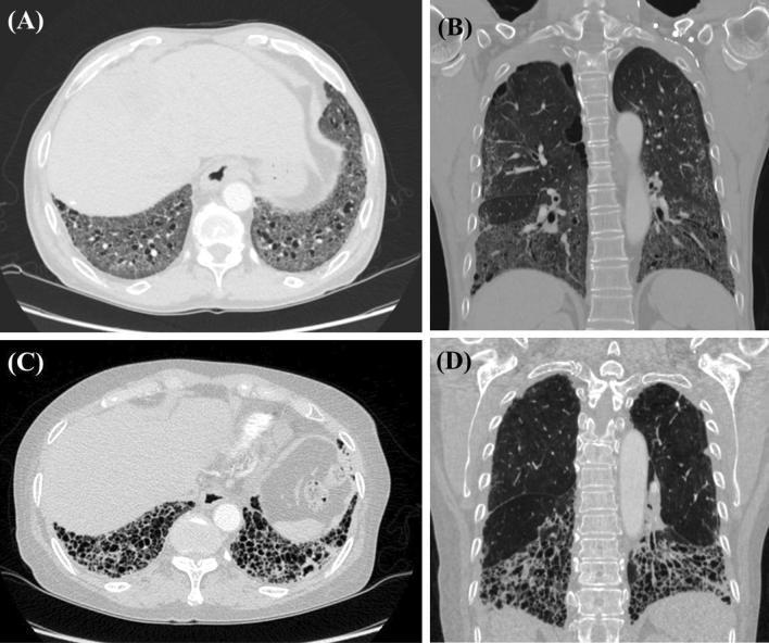
High-resolution computed tomography (HRCT) comparison of interstitial lung disease (ILD) patterns in systemic sclerosis (SSc). Figures A (axial) and B (coronal) display a Non-Specific Interstitial Pneumonia (NSIP) pattern characterized by bilateral, diffuse ground-glass opacities and mild traction bronchiectasis with a distinct basal predominance. Figures C (axial) and D (coronal) demonstrate a Usual Interstitial Pneumonia (UIP) pattern. This pattern is marked by extensive subpleural honeycombing (clusters of cystic airspaces), exuberant traction bronchiectasis, and severe architectural distortion predominantly involving the lower lung zones. These diagnostic images serve to illustrate the distinct radiologic morphology between NSIP and UIP patterns in patients with connective tissue disease-associated ILD, highlighting key features such as distribution, presence of honeycombing, and the degree of fibrotic changes essential for clinical classification and management.
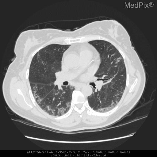
**Imaging Modality:** High-resolution computed tomography (HRCT) of the chest, axial section.
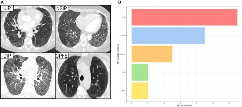
This composite educational resource consists of a diagnostic imaging panel (A) and a comparison bar chart (B) illustrating interstitial lung disease (ILD) patterns in patients with Sjögren's syndrome. Panel A displays axial High-Resolution Computed Tomography (HRCT) slices of four distinct ILD patterns: Usual Interstitial Pneumonia (UIP) characterized by peripheral reticulation and honeycombing; Non-Specific Interstitial Pneumonia (NSIP) showing more uniform ground-glass opacities; Desquamative Interstitial Pneumonia (DIP) with diffuse ground-glass attenuation; and Combined Pulmonary Fibrosis and Emphysema (CPFE) demonstrating upper-lobe emphysematous lucencies alongside fibrotic changes. Panel B is a horizontal bar chart quantifying the prevalence of these CT disease patterns among the study cohort. The chart indicates that UIP is the most frequent pattern, followed by NSIP, unspecific changes (Unspez), and lastly DIP and CPFE, which show equal, lower prevalence. This visual aid is intended for medical education regarding the radiologic classification and epidemiological distribution of pulmonary manifestations in systemic autoimmune diseases.
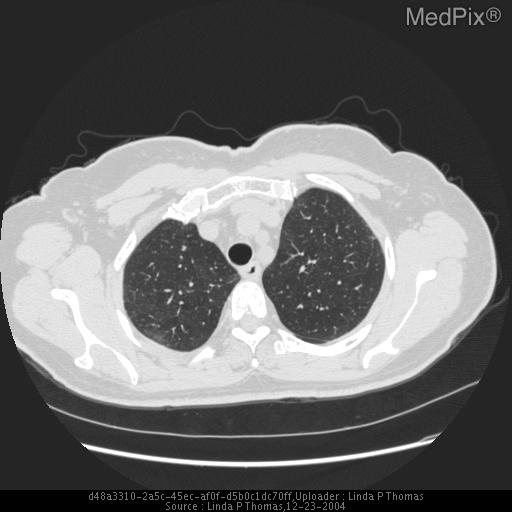
**Imaging Modality:** High-resolution computed tomography (HRCT) of the chest, axial plane.
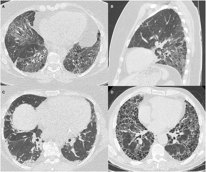
This composite of High-Resolution Computed Tomography (HRCT) images illustrates diverse patterns of Interstitial Lung Disease (ILD) associated with Antisynthetase Syndrome (ASyS). Panel A (axial) and B (sagittal) depict fibrotic Non-Specific Interstitial Pneumonitis (NSIP), characterized by bilateral, patchy ground-glass opacities, fine reticulation, and traction airway dilatation, with a clear lower zone predominance visible in the sagittal reconstruction. Panel C displays an Organizing Pneumonitis (OP) pattern, showing patchy, peripheral, and basal-predominant lung consolidation. Panel D demonstrates a Usual Interstitial Pneumonitis (UIP) pattern, featuring advanced fibrotic changes including subpleural and basal-predominant reticulation and extensive honeycombing. These images serve as an educational comparison of radiological manifestations in connective tissue disease-related ILD (CTD-ILD), highlighting key diagnostic features such as ground-glass opacification, architectural distortion, and honeycombing across different pathological patterns in patients with anti-Jo1 antibodies.
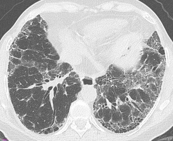
This transverse High-Resolution Computed Tomography (HRCT) axial scan of the thorax demonstrates a 'definite Usual Interstitial Pneumonia (UIP)' pattern, a hallmark of Idiopathic Pulmonary Fibrosis (IPF). The image shows bilateral, predominantly subpleural and basal lung involvement. Key diagnostic features include honeycombing—manifesting as clustered cystic airspaces with thick walls—which is particularly evident in the subpleural region of the left lung. Associated findings include a prominent reticular pattern, traction bronchiectasis (dilatation of bronchi within the fibrotic areas), and mild ground-glass opacities. The distribution is peripheral and basal-predominant, which is characteristic of the ATS/ERS/JRS/ALAT guidelines for diagnosing UIP. This imaging is crucial for medical students and radiologists to distinguish between different patterns of interstitial lung disease (ILD), specifically identifying the architectural distortion and honeycombing required for a definite diagnosis of UIP versus non-specific interstitial pneumonia (NSIP).
UIP pattern IPF honeycombing traction bronchiectasis basal subpleural HRCT
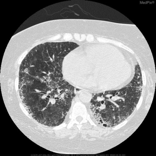
**Imaging Modality:** High-Resolution Computed Tomography (HRCT) of the chest, axial section, lung window.
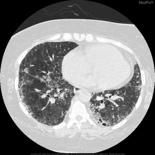
**Imaging Modality:** High-Resolution Computed Tomography (HRCT) of the chest.
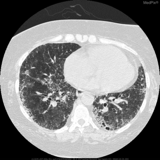
**Imaging Modality:** High-resolution computed tomography (HRCT) of the chest, axial section.
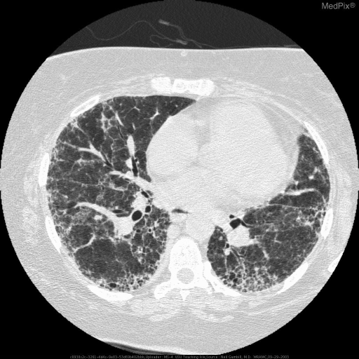
**Imaging Modality:** High-resolution computed tomography (HRCT) of the chest, axial section.
sarcoidosis HRCT perilymphatic nodules upper lobe
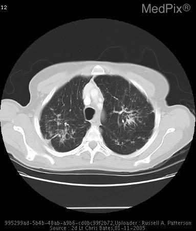
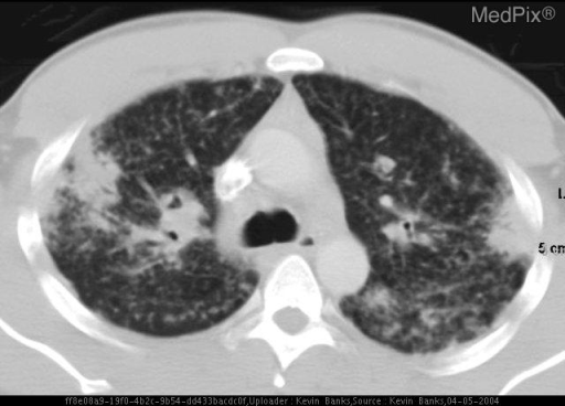
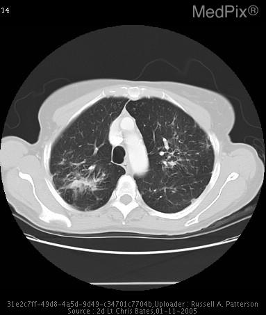
Interstitial Lung Disease (ILD): Classification and HRCT Patterns
1. Classification of ILD
A. Idiopathic Interstitial Pneumonias (IIPs)
| Group | Entities |
|---|---|
| Chronic Fibrosing | Idiopathic Pulmonary Fibrosis (IPF), Nonspecific Interstitial Pneumonia (NSIP) |
| Smoking-Related | Respiratory Bronchiolitis-ILD (RB-ILD), Desquamative Interstitial Pneumonia (DIP) |
| Acute/Subacute | Cryptogenic Organizing Pneumonia (COP), Acute Interstitial Pneumonia (AIP), Lymphoid Interstitial Pneumonia (LIP) |
| Rare IIPs | Acute Fibrinous & Organizing Pneumonia (AFOP), Pleuroparenchymal Fibroelastosis (PPFE), Broncholocentric IIP |
B. CTD-Associated ILD
- Progressive systemic sclerosis (most common cause of ILD among CTDs)
- Rheumatoid arthritis
- Systemic lupus erythematosus
- Dermatomyositis / Polymyositis
- Sjogren syndrome
- Mixed connective tissue disease
- Ankylosing spondylitis
C. Hypersensitivity Pneumonitis (HP)
- Occupational/environmental: Farmer's lung, Bird fancier's lung
- Iatrogenic
D. Drug-Induced ILD
- Amiodarone, methotrexate, nitrofurantoin, bleomycin, checkpoint inhibitors, etc.
E. Alveolar Filling Disorders
- Goodpasture syndrome, Pulmonary alveolar proteinosis, Chronic eosinophilic pneumonia
F. Pulmonary Vasculitis-Associated ILD
- Granulomatosis with polyangiitis (GPA), Eosinophilic granulomatosis with polyangiitis (EGPA)
G. Other Specific ILDs
- Sarcoidosis, Langerhans cell histiocytosis (LCH), Lymphangioleiomyomatosis (LAM), COVID-19-associated
H. Inherited ILDs
- Familial IPF, Tuberous sclerosis, Neurofibromatosis, Gaucher disease, Hermansky-Pudlak syndrome
2. HRCT Patterns - The Core Diagnostic Tool
Key HRCT Findings Defined
| Finding | Description |
|---|---|
| Honeycombing | Clustered cystic airspaces (3-10 mm), thick walls, subpleural - hallmark of irreversible fibrosis |
| Traction bronchiectasis | Irregular dilatation of bronchi/bronchioles pulled open by surrounding fibrosis |
| Ground-glass opacity (GGO) | Hazy increased attenuation; does not obscure vessels - implies active/inflammatory disease |
| Reticulation | Network of fine lines - represents fibrosis of interlobular septa |
| Consolidation | Airspace filling that obscures underlying vessels |
| Cysts | Well-defined, thin-walled airspaces |
| Centrilobular nodules | Nodules centered around the lobular artery - respiratory bronchiolitis pattern |
| Perilymphatic nodules | Along bronchovascular bundles, septa, pleura - sarcoidosis |
3. HRCT Patterns by Specific ILD
A. UIP Pattern (Usual Interstitial Pneumonia) - IPF / Idiopathic Pulmonary Fibrosis
| HRCT Category | Distribution | Key Features |
|---|---|---|
| Typical UIP | Subpleural, bibasal, peripheral; often heterogeneous | Honeycombing ± traction bronchiectasis; "propeller blade" spread to anterior upper lobes on sagittal |
| Probable UIP | Subpleural, bibasal | Reticular pattern + peripheral traction bronchiectasis; NO honeycombing |
| Indeterminate for UIP | Subpleural and basal | Subtle reticulation ± mild GGO; may have peribronchovascular distribution with relative subpleural sparing |
| Alternative diagnosis | Peribronchovascular, perilymphatic, upper/mid lung | Cysts, marked mosaic attenuation, profuse micronodules, consolidation |
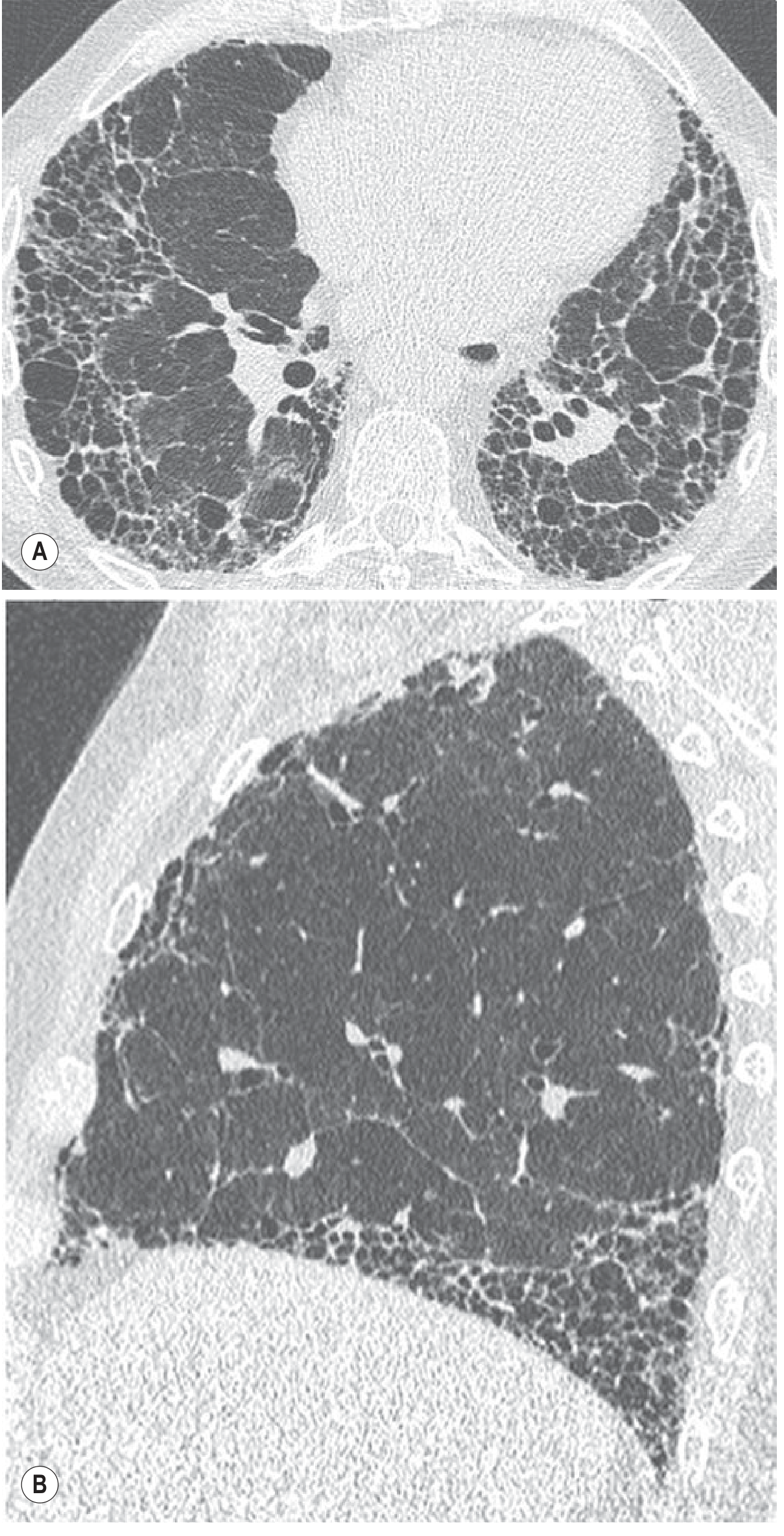
B. NSIP Pattern (Nonspecific Interstitial Pneumonia)
- Distribution: Peripheral, peribronchovascular; basal, symmetric, bilateral
- Key features: Diffuse subpleural GGO and reticular opacities; volume loss and traction bronchiectasis in lower zones
- Hallmark: Subpleural sparing - a relative sparing of the immediate subpleural zone, which helps distinguish NSIP from UIP
- Honeycombing is occasionally seen in fibrotic NSIP but is usually minor
- Associated with CTD (especially SSc, polymyositis), IPAF
C. COP Pattern (Cryptogenic Organizing Pneumonia)
- Distribution: Subpleural or peribronchovascular
- Key features: Patchy bilateral consolidation or nodules; may have "reversed halo sign" (ring of consolidation around GGO)
- Fleeting/migratory infiltrates are characteristic
D. AIP Pattern (Acute Interstitial Pneumonia / Diffuse Alveolar Damage)
- Distribution: Diffuse bilateral
- Key features: Extensive GGO and consolidation often with lobular sparing ("geographic" pattern); late traction bronchiectasis as organizing phase sets in
- Clinical correlate: Rapidly progressive, Hamman-Rich syndrome
E. DIP Pattern (Desquamative Interstitial Pneumonia)
- Distribution: Peripheral, lower lung zone predominance
- Key features: Diffuse ground-glass attenuation ± reticulation and small cysts; minimal honeycombing
- Almost exclusively in smokers
F. RB-ILD Pattern (Respiratory Bronchiolitis-ILD)
- Distribution: Diffuse, patchy
- Key features: Bronchial wall thickening, poorly defined centrilobular nodules (pigmented macrophages in RBs), patchy GGO
- Smoking-related; often reversible with cessation
G. LIP Pattern (Lymphoid Interstitial Pneumonia)
- Distribution: Diffuse
- Key features: GGO, septal thickening, bronchovascular wall thickening, and characteristic thin-walled cysts (lung cysts in a background of GGO is the classic combination)
- Associated with Sjogren syndrome, HIV, dysproteinemias
H. PPFE Pattern (Pleuroparenchymal Fibroelastosis)
- Distribution: Upper lobe predominant
- Key features: Dense subpleural consolidation with traction bronchiectasis, volume loss, and pleural thickening at upper lobes - the opposite of UIP
4. HRCT Patterns in Non-IIP ILDs
| ILD | Distribution | Characteristic HRCT |
|---|---|---|
| Sarcoidosis | Upper/mid-lung; perilymphatic | Perilymphatic micronodules along bronchovascular bundles, septa and pleura; hilar/mediastinal adenopathy; "galaxy sign" |
| Hypersensitivity Pneumonitis (HP) | Mid/lower lung; centrilobular | Subacute: centrilobular poorly-defined nodules + GGO + mosaic attenuation; Chronic/fibrotic: upper lobe fibrosis, UIP-like or NSIP-like |
| LCH | Upper lobe | Cysts (bizarre shapes) + nodules; spares costophrenic angles |
| LAM | Diffuse; uniform distribution | Thin-walled round cysts uniformly distributed, no nodules; chylothorax |
| Asbestosis | Lower lobe, subpleural | Subpleural lines, fine reticulation progressing to coarse reticulation and honeycombing identical to UIP; pleural plaques |
| CTD-ILD (SSc) | Bibasal | NSIP pattern most common (GGO + reticulation); UIP pattern possible |
5. Summary Comparison Table - IIPs on HRCT
| IIP | HRCT Distribution | Dominant Pattern | Honeycombing | GGO |
|---|---|---|---|---|
| IPF/UIP | Peripheral, subpleural, bibasal | Reticulation, traction bronchiectasis | Prominent | Mild |
| NSIP | Peripheral, peribronchovascular, bibasal, symmetric | GGO + reticulation | Rare/minimal | Prominent |
| COP | Subpleural/peribronchovascular | Consolidation | Absent | Variable |
| AIP | Diffuse bilateral | GGO + consolidation, lobular sparing | Absent | Dominant |
| DIP | Peripheral, lower zone | GGO | Minimal | Dominant |
| RB-ILD | Diffuse/patchy | Centrilobular nodules + GGO | Absent | Patchy |
| LIP | Diffuse | GGO + thin-walled cysts | Absent | Prominent |
| PPFE | Upper lobe | Dense subpleural consolidation | Absent | Absent |
6. Radiographic Distribution Clues
| Distribution | Think of |
|---|---|
| Upper lobe predominance | Sarcoidosis, silicosis, LCH, HP (chronic), ankylosing spondylitis, PPFE |
| Lower lobe predominance | IPF, asbestosis, NSIP, CTD-ILD |
| Peripheral/subpleural | IPF/UIP, NSIP, COP |
| Peribronchovascular/perilymphatic | Sarcoidosis, lymphangitic carcinomatosis |
| Centrilobular nodules | HP, RB-ILD, respiratory bronchiolitis |
| Hilar/mediastinal adenopathy | Sarcoidosis, berylliosis, silicosis, LIP |
| Cysts | LCH (irregular), LAM (round, uniform), LIP (few cysts + GGO) |
Create a comparison poster of UIP vs NSIP patterns with key discriminating features
Medical educational comparison poster titled "UIP vs NSIP: Key Discriminating Features" for pulmonology/radiology. Two-column layout side by side. Left column header: "UIP (Usual Interstitial Pneumonia)" with subtitle "IPF - Idiopathic Pulmonary Fibrosis". Right column header: "NSIP (Non-Specific Interstitial Pneumonia)". Each column contains clearly organized sections: DEMOGRAPHICS row: UIP - "Age >60, Male predominance, Smokers (2/3 cases)" | NSIP - "Age 40-50s, Female predominance, Non-smokers, CTD-associated" HRCT DISTRIBUTION row: UIP - "Subpleural, bibasal, peripheral, heterogeneous" | NSIP - "Peripheral + peribronchovascular, bibasal, symmetric" DOMINANT HRCT FEATURES section with bullet points: UIP side: "✓ Honeycombing (hallmark), ✓ Coarse reticulation, ✓ Traction bronchiectasis, ✓ 'Propeller blade' sagittal distribution, ✗ GGO - NOT dominant" NSIP side: "✓ Ground-glass opacity (dominant), ✓ Fine reticulation, ✓ Traction bronchiectasis (fibrotic), ✓ Subpleural SPARING (key clue!), ✗ Honeycombing - rare/minimal" HONEYCOMBING row: UIP - "PROMINENT - diagnostic" (red tick icon) | NSIP - "ABSENT or minimal" (red cross icon) SUBPLEURAL SPARING row: UIP - "ABSENT - extends to pleura" | NSIP - "PRESENT - hallmark feature" (highlighted in yellow/gold) TEMPORAL PATTERN row: UIP - "Temporal heterogeneity (areas at different stages of fibrosis)" | NSIP - "Temporal homogeneity (uniform appearance)" HISTOLOGY row: UIP - "Fibroblastic foci, dense fibrosis, normal areas alternating - spatially & temporally heterogeneous" | NSIP - "Uniform interstitial thickening, cellular or fibrotic, temporally homogeneous" PROGNOSIS row: UIP - "Poor - median survival 3-5 years, progressive" (red background) | NSIP - "Better - cellular NSIP responds to steroids, fibrotic NSIP variable" (green background) DIAGNOSIS CONFIDENCE row: UIP - "Typical UIP on HRCT = no biopsy needed" | NSIP - "Often requires biopsy or CTD workup" At the bottom: a KEY DIFFERENTIATOR callout box: "The single most important feature: Honeycombing = UIP | Subpleural sparing + GGO without honeycombing = NSIP" Color scheme: Deep navy blue headers, UIP column in warm red/orange tones, NSIP column in cool blue/teal tones. Clean medical infographic style with clear typography, white background, professional layout suitable for clinical teaching.
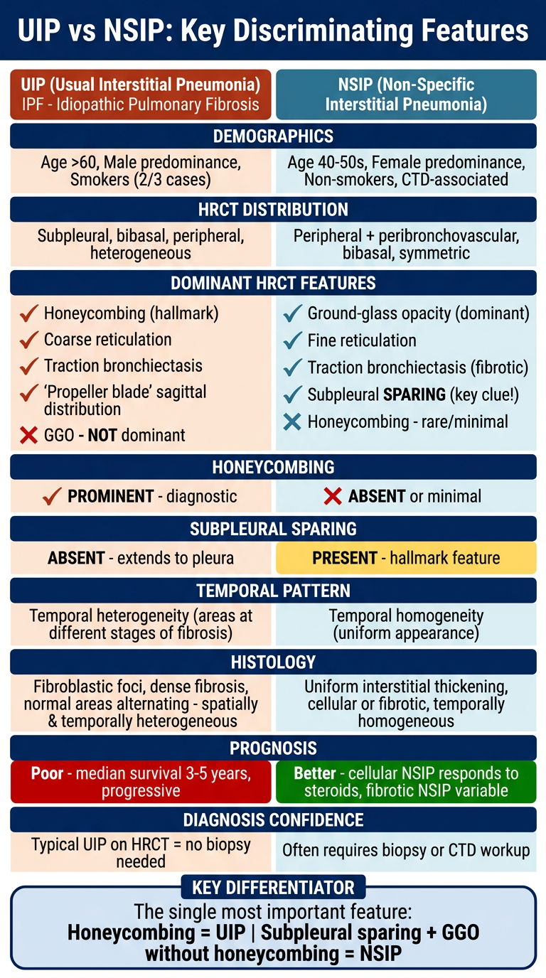
| Feature | UIP | NSIP |
|---|---|---|
| Honeycombing | Prominent, diagnostic | Rare/absent |
| Subpleural sparing | Absent | Present (hallmark) |
| Dominant opacity | Reticulation | Ground-glass |
| Temporal pattern | Heterogeneous | Homogeneous |
| Prognosis | Poor (3-5 yr survival) | Better (especially cellular) |
| Demographics | Older male smokers | Younger females, CTD |