
Basic terminology definition in gynaecology
Basic Terminology in Gynaecology
1. Menstrual Cycle Terminology
| Term | Definition |
|---|---|
| Menarche | The first menstrual period; onset of menstruation at puberty (typically ages 11-14) |
| Menstruation | Cyclic shedding of the functional layer of the endometrium due to declining progesterone at the end of the luteal phase |
| Eumenorrhoea | Normal, regular menstrual cycles occurring every 21-35 days, lasting 2-7 days, with normal flow |
| Menopause | Cessation of menstruation for 12 consecutive months due to ovarian failure; average age ~51 years |
| Perimenopause | The transitional phase leading up to menopause, characterised by irregular cycles |
| Oligomenorrhoea | Infrequent menstrual bleeding with cycles occurring more than 35 days apart |
| Polymenorrhoea | Frequent menstrual bleeding with cycles shorter than 21 days |
| Menorrhagia | Excessive or prolonged menstrual bleeding (>80 mL blood loss per cycle or bleeding lasting >7 days) |
| Metrorrhagia | Irregular uterine bleeding occurring between expected menstrual periods |
| Menometrorrhagia | Excessive, prolonged, and irregular uterine bleeding |
| Intermenstrual bleeding | Any bleeding occurring between regular menstrual periods |
| Postcoital bleeding | Vaginal bleeding occurring after sexual intercourse |
| Postmenopausal bleeding | Any vaginal bleeding occurring more than 12 months after the last menstrual period; always warrants investigation |
| Dysmenorrhoea | Painful menstruation; may be primary (no underlying pathology, due to prostaglandin-mediated uterine cramping) or secondary (due to an underlying condition such as endometriosis or fibroids) |
| Amenorrhoea | Absence of menstruation; classified as primary (no menstruation by age 16) or secondary (cessation of previously established menstruation for >3 months) |
2. Abnormal Uterine Bleeding (AUB) - PALM-COEIN Classification
- PALM (structural): Polyp, Adenomyosis, Leiomyoma, Malignancy/hyperplasia
- COEIN (non-structural): Coagulopathy, Ovulatory dysfunction, Endometrial, Iatrogenic, Not yet classified
3. Ovarian/Hormonal Terminology
| Term | Definition |
|---|---|
| Ovulation | Release of a mature oocyte from the dominant follicle, triggered by the LH surge |
| Anovulation | Failure of ovulation to occur; common in the first 12-18 months after menarche and before menopause |
| Dominant follicle | The one follicle destined to ovulate each cycle; has more FSH receptors and produces higher estrogen concentrations than other follicles |
| Corpus luteum | The post-ovulation remnant of the dominant follicle; secretes progesterone, estrogen, and inhibin A; regresses after 12-16 days if fertilisation does not occur |
| Luteal phase | The post-ovulatory phase of the cycle (approx. days 15-28); characterised by progesterone dominance |
| Follicular phase | The pre-ovulatory phase; characterised by follicular growth and rising estrogen |
4. Obstetric History Notation
| Term | Definition |
|---|---|
| Gravida (G) | Total number of pregnancies (including current), regardless of outcome |
| Para (P) | Number of deliveries after 20 weeks gestation (some systems use >28 weeks), regardless of whether the infant was born alive or stillborn |
| Nulligravida | A woman who has never been pregnant |
| Nulliparous | A woman who has never delivered a viable infant |
| Primigravida | A woman pregnant for the first time |
| Multigravida | A woman who has been pregnant more than once |
| TPAL system | T = Term deliveries (≥37 weeks); P = Preterm deliveries (20-37 weeks); A = Abortions/miscarriages; L = Living children |
5. Vaginal Discharge Terminology
| Term | Definition |
|---|---|
| Leukorrhoea | Normal or physiological white/milky vaginal discharge; physiological leukorrhoea is non-offensive and non-irritating |
| Pathological discharge | Discharge associated with colour change (green, yellow, grey), odour, or irritation - suggests infection |
| Vaginitis | Inflammation of the vaginal mucosa, usually due to infection (bacterial vaginosis, candidiasis, trichomonas) or atrophy |
| Bacterial vaginosis (BV) | Overgrowth of anaerobic bacteria (e.g., Gardnerella vaginalis), replacing normal Lactobacillus flora; produces a fishy-smelling, grey discharge |
6. Anatomical and Structural Terms
| Term | Definition |
|---|---|
| Cervix | The lower, narrow portion of the uterus that opens into the vagina |
| Endocervix | The inner canal of the cervix, lined by columnar epithelium |
| Ectocervix | The outer portion of the cervix visible on speculum examination, lined by squamous epithelium |
| Transformation zone (T-zone) | The area between squamous and columnar epithelium on the cervix; the most common site for cervical dysplasia |
| Cul-de-sac (Pouch of Douglas) | The rectouterine pouch - the most dependent part of the peritoneal cavity in females; fluid or blood can collect here |
| Adnexa | Structures adjacent to the uterus: the ovaries, fallopian tubes, and associated ligaments |
| Fornix | The upper part of the vagina surrounding the cervix; divided into anterior, posterior, and lateral fornices |
| Myometrium | The muscular middle layer of the uterine wall |
| Endometrium | The inner mucous membrane lining of the uterus; shed during menstruation |
| Perimetrium | The outer serosal layer of the uterus |
7. Gynaecological Conditions - Key Terms
| Term | Definition |
|---|---|
| Endometriosis | Presence of endometrial-like tissue outside the uterus; causes dysmenorrhoea, dyspareunia, and infertility |
| Adenomyosis | Presence of endometrial glands and stroma within the myometrium |
| Leiomyoma (fibroid) | Benign smooth muscle tumour of the uterus; the most common benign pelvic tumour in women |
| PCOS (Polycystic Ovary Syndrome) | Endocrine disorder characterised by anovulatory cycles, hyperandrogenism, and polycystic ovaries |
| Endometrial hyperplasia | Excessive proliferation of endometrial glands relative to stroma; a risk factor for endometrial carcinoma |
| Cervical intraepithelial neoplasia (CIN) | Dysplastic changes of cervical epithelium graded I-III; a precursor to cervical carcinoma |
| Pelvic inflammatory disease (PID) | Ascending infection of the upper genital tract (uterus, tubes, ovaries) |
8. Procedures
| Term | Definition |
|---|---|
| Colposcopy | Examination of the cervix under magnification using a colposcope, typically after an abnormal Pap smear |
| LEEP (Loop Electrosurgical Excision Procedure) | Removal of abnormal cervical tissue using a thin wire loop carrying electrical current |
| Hysteroscopy | Direct visualisation of the uterine cavity using a thin telescope introduced through the cervix |
| Laparoscopy | Minimally invasive surgical technique using a camera through a small abdominal incision; used for diagnosis and treatment |
| Dilation and Curettage (D&C) | Dilation of the cervix followed by scraping of the uterine lining; used for diagnosis or to manage miscarriage/bleeding |
| Hysterectomy | Surgical removal of the uterus; may be total (with cervix) or subtotal (cervix retained) |
9. Contraception Terminology
| Term | Definition |
|---|---|
| IUD / IUC | Intrauterine device/contraceptive - a small device placed inside the uterus for contraception |
| Levonorgestrel IUD | A progestogen-releasing IUD that reduces menstrual bleeding and treats dysmenorrhoea |
| Combined oral contraceptive (COC) | A pill containing both estrogen and progestogen |
Describe and discuss the etiology, pathology, clinical features, differential diagnosis, investigations, management and long-term indicators of sexually transmitted infection
sexually transmitted infection genital ulcer syphilis chancre herpes
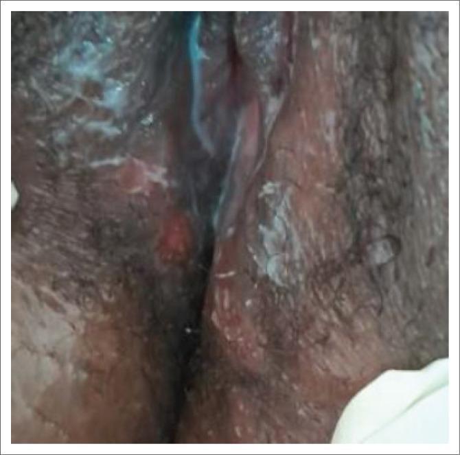
Content Type: Clinical Photograph. Anatomical Region: Female genitalia (vulva). Clinical Findings: The image shows at least two distinct ulcerated lesions on the mucosal surface of the vulva. The primary visible ulcer is erythematous (reddish), roughly circular in shape, and appears slightly depressed below the surrounding skin level. There is no obvious purulent discharge or active bleeding visible. The surrounding skin and mucous membranes show areas of white, macerated tissue or possibly dried topical application. Significance: This is a classic clinical presentation of a genital ulcer, which in a diagnostic context often suggests primary syphilis (chancre), herpes simplex virus, or chancroid. The photograph illustrates the morphology and distribution of lesions relevant for the syndromic management of sexually transmitted infections (STIs). Educational Focus: Identification of genital ulcerative disease (GUD) morphology and its clinical relevance in dermatology and infectious disease specialties.
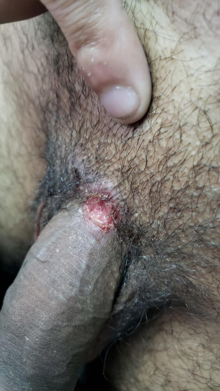
This clinical photograph shows a single, well-defined ulcerated lesion located at the base of the penile shaft, near the junction with the pubic region. The lesion is roughly circular with irregular borders and features a raw, erythematous base with a moist appearance, suggesting potential serous exudate. The surrounding tissue demonstrates mild perilesional inflammation and erythema. Anatomically, the site is characterized by overlying pubic hair and standard genital skin architecture. The visual presentation is highly characteristic of a primary syphilis chancre (Treponema pallidum infection), though the context indicates a coinfection with herpes simplex virus type 1 (HSV-1). This image serves as an educational example of genital ulcer disease (GUD) and primary syphilis manifestations in a clinical setting, highlighting the morphology of a syphilitic chancre which is typically firm, indurated, and painless.
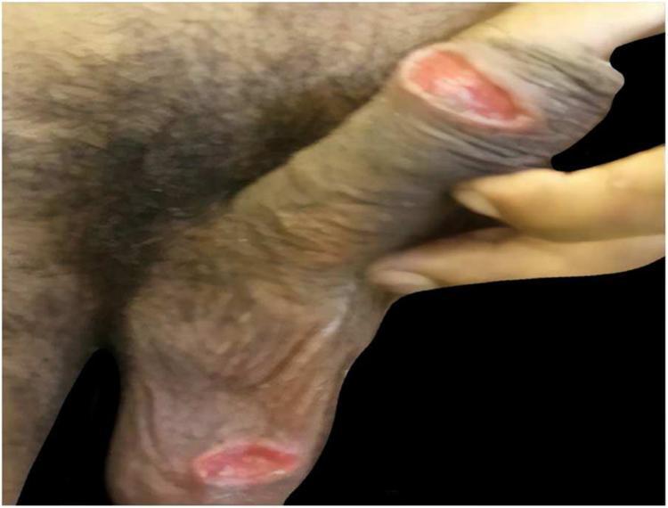
A clinical photograph of the male genital region demonstrating two distinct cutaneous ulcerations associated with American Cutaneous Leishmaniasis (ACL). One ulcer is located on the shaft (body) of the penis, and the second is located on the scrotum. Both lesions are characterized by an oval shape, well-defined margins, and a granular, erythematous (reddish-pink) base. The ulcers appear shallow with slightly raised borders. The surrounding integument shows hyperpigmentation and typical anatomical wrinkling of the scrotal and penile skin. Clinical metadata indicates these lesions are typically painless. This image is used to illustrate the atypical presentation of cutaneous leishmaniasis involving genital mucosa and skin, which can mimic other sexually transmitted infections such as primary syphilis (chancre) or genital herpes. It serves as an educational reference for infectious disease diagnosis and dermatology.
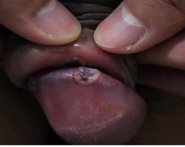
This clinical photograph shows a close-up view of a penile lesion located on the glans penis. The primary finding is a solitary, well-demarcated, circular ulcer approximately 5-10mm in diameter. The lesion exhibits a distinct morphology characterized by a raised, whitish, indurated peripheral border and a central base that is purplish-red or dusky in hue. The surrounding glans tissue appears mildly erythematous and glossy. This visual presentation is characteristic of educational material focused on sexually transmitted infections (STIs), specifically demonstrating an ulcerative condition of the male genitalia. The clinical significance involves the differential diagnosis of genital ulcers, such as primary syphilis (chancre), chancroid, or herpes simplex virus, although the clean-based, sharply defined appearance of the border is particularly illustrative of specific dermatological manifestations of balanitis or infectious ulcers.
condyloma acuminata genital warts HPV
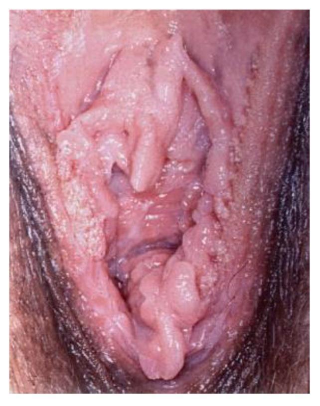
Clinical photograph of the female external genitalia showing extensive human papillomavirus (HPV) infection presenting as condyloma acuminata (genital warts). The lesions are characterized by multiple raised, exophytic, and papillary growths with a verrucous (cauliflower-like) texture. The distribution is widespread across the vulvar area, involving the labia minora, labia majora, and the vaginal introitus. Morphologically, the lesions appear flesh-colored to pale pink, with some areas exhibiting a whitish, hyperkeratotic surface. There is evidence of epithelial thickening and irregular surface contours, deviating from normal smooth mucosal architecture. This image illustrates the typical clinical presentation of low-risk HPV-induced benign neoplasia, which is a key diagnostic feature in gynecology and dermatology for distinguishing between infectious viral warts and other vulvar intraepithelial neoplasia (VIN) or malignant conditions.
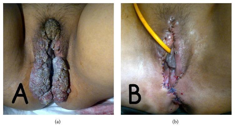
Two-panel clinical photograph demonstrating condyloma acuminata (genital warts) and its surgical management in a patient with systemic lupus erythematosus. Image (a) shows extensive, multifocal, hypertrophic verrucous lesions involving the vulva, perineum, and perianal region. The lesions exhibit a classic 'cauliflower-like' morphology with a rough, filiform texture and varied pigmentation ranging from skin-toned to brownish-grey. Image (b) shows the same anatomical region post-surgical excision. The surgical site features approximated skin edges secured with blue interrupted sutures extending through the labia and perineum. A yellow drainage tube is positioned for fluid management. Evidence of post-operative erythema and inflammation is visible. This comparison illustrates the clinical manifestation of severe HPV-related verrucous disease in an immunocompromised state and the immediate results of surgical debulking and reconstruction.
Sexually Transmitted Infections (STIs): A Comprehensive Discussion
1. Definition and Classification
| Syndrome | Major Organisms |
|---|---|
| Urethritis (gonococcal) | Neisseria gonorrhoeae |
| Urethritis (non-gonococcal) | Chlamydia trachomatis, Trichomonas vaginalis, Mycoplasma genitalium, Ureaplasma urealyticum |
| Genital ulcers | HSV-1/2, Treponema pallidum, Haemophilus ducreyi, Klebsiella granulomatis, C. trachomatis (LGV serovars) |
| Epithelial cell disorders | Human papillomavirus (HPV), Molluscum contagiosum |
| Vaginal discharge / cervicitis | N. gonorrhoeae, C. trachomatis, T. vaginalis, Candida albicans, Bacterial vaginosis |
| Pelvic inflammatory disease | N. gonorrhoeae, C. trachomatis, anaerobes |
| Systemic/blood-borne | HIV, Hepatitis B/C, CMV |
| Ectoparasites | Pthirus pubis (pubic lice), Sarcoptes scabiei (scabies) |
2. Etiology
2a. Bacterial STIs
- Gram-negative diplococcus; has tropism for columnar epithelium (endocervix, urethra, rectum, pharynx)
- Immune evasion via antigenic variation of pili and outer membrane proteins
- Control of dissemination requires complement-mediated immunity
- Gram-negative obligate intracellular bacterium
- Serovars D-K: genital tract infections (urethritis, cervicitis, PID)
- Serovars L1-L3: Lymphogranuloma venereum (LGV)
- Exists in two forms: the infectious elementary body (EB) and the replicating reticulate body (RB)
- A motile spirochete; cannot be cultured in vitro
- Causes syphilis; enters via mucosal breaches or abraded skin
- Disseminates haematogenously early in infection
- Gram-negative bacillus causing chancroid
- Produces a cytotoxin causing local tissue destruction
- Intracellular bacterium causing granuloma inguinale (donovanosis)
2b. Viral STIs
- HSV-2 is the predominant cause of genital herpes; HSV-1 increasingly implicated
- Establishes latency in dorsal root ganglia; reactivation causes recurrent disease
- Shedding occurs both symptomatically and asymptomatically
-
200 genotypes; sexually transmitted types classified as low-risk (6, 11 - genital warts) and high-risk (16, 18 - cervical carcinoma)
- Infects basal layer keratinocytes of stratified squamous epithelium
- Retrovirus targeting CD4+ T-lymphocytes; transmitted via sexual contact (especially anal intercourse), blood, and vertically
- STIs (especially ulcerative) dramatically increase HIV transmission risk by disrupting mucosal barriers
- HBV is highly sexually transmissible; transmitted via blood and bodily fluids
2c. Protozoal STI
- Flagellated protozoan; infects squamous epithelium of the vagina, urethra, and Skene's glands
- Only STI with a flagellated motile form visible on wet mount
3. Pathology
Gonorrhoea
- Infects columnar and transitional epithelium; produces acute purulent inflammation with neutrophil-rich exudate
- Local tissue invasion can lead to salpingitis with tubal scarring; systemic dissemination causes septic arthritis and dermatitis
- In females, often asymptomatic; the cervix may appear normal or show mucopurulent discharge
Chlamydia
- Intracellular infection; causes a lymphocytic, plasma cell infiltration rather than a neutrophilic exudate
- LGV: causes granulomatous inflammation of lymph nodes with stellate abscesses, progressing to fibrosis and lymphoedema
- Chronic pelvic infection can cause peritubal and periovarian adhesions (Fitz-Hugh-Curtis syndrome when involving the liver capsule)
Syphilis (Treponema pallidum)
- Primary: Chancre - epithelial ulceration with plasma cell and lymphocyte infiltrate, proliferative endarteritis of small vessels
- Secondary: Systemic dissemination; skin shows perivascular lymphocytic infiltrates and epidermal changes
- Tertiary: Gummas (granulomatous necrotic lesions) in any organ; obliterative endarteritis affects the vasa vasorum of the aorta causing aortic aneurysm and aortic regurgitation
- Neurosyphilis: Meningovasculitis, or parenchymatous involvement (tabes dorsalis, general paresis)
Herpes Simplex Virus
- Vesicles contain necrotic epithelial cells, fused multinucleated giant cells, and Cowdry type A intranuclear inclusions
- After primary infection, virus travels retrograde along sensory neurons to dorsal root ganglia where it establishes latency
HPV
- Low-risk types: stimulate koilocytic changes (perinuclear vacuolation of squamous cells) and benign papillomatous proliferation (condyloma acuminata)
- High-risk types: E6 and E7 oncoproteins inactivate p53 and Rb tumour suppressor proteins respectively, promoting malignant transformation
Chancroid (H. ducreyi)
- Soft, painful ulcer; histology shows superficial acute inflammation and necrosis overlying a zone of granulation tissue and mononuclear infiltrate
Bacterial Vaginosis
- Polybacterial dysbiosis of the vaginal microbiome with replacement of hydrogen peroxide-producing Lactobacillus by Gardnerella vaginalis and anaerobes including Prevotella species
4. Clinical Features
Gonorrhoea
| Sex | Features |
|---|---|
| Males | Dysuria, profuse purulent urethral discharge (95% symptomatic) |
| Females | Often asymptomatic; mucopurulent cervical discharge, dysuria, intermenstrual bleeding |
| Disseminated | Septic arthritis, tenosynovitis, necrotic skin pustules on distal extremities ("arthritis-dermatitis syndrome"), rare: endocarditis, meningitis |
| Neonates | Ophthalmia neonatorum (conjunctivitis acquired during passage through birth canal) |
Chlamydia
- Clinically indistinguishable from gonorrhoea but typically milder
- Mucopurulent cervicitis, urethritis, proctitis
- Reactive arthritis triad (Reiter's syndrome): urethritis + arthritis + conjunctivitis
- Often entirely asymptomatic (hence major reservoir for silent spread)
Syphilis
| Stage | Features |
|---|---|
| Primary (2-6 weeks post-exposure) | Single, painless, indurated ulcer (chancre) with clean base and well-defined borders on genitalia, anus, or oral mucosa; regional non-tender lymphadenopathy |
| Secondary (6-8 weeks later; 60-90% of untreated) | Fever, generalised lymphadenopathy, headache, sore throat, arthralgias; maculopapular rash classically involving the palms and soles; condylomata lata (moist warty plaques in perineum); alopecia; CNS, eye, liver, and kidney involvement possible |
| Latent | No symptoms; reactive serology only (early vs late latent) |
| Tertiary | Gummas (skin, bone, organs); cardiovascular syphilis (ascending aortitis, aortic regurgitation, aneurysm); neurosyphilis (general paresis, tabes dorsalis, Argyll Robertson pupils) |
| Congenital | Hydrops fetalis, "snuffles" (rhinitis), saddle-nose, interstitial keratitis, Hutchinson's teeth, saber shins |
Genital Herpes (HSV)
- Incubation: <21 days
- Primary episode: clustered vesicles on erythematous base → pustules → shallow painful ulcers → healing by crusting over 2-3 weeks; may have systemic fever, headache, aseptic meningitis
- Only ~20% manifest the classic presentation; 60% have mild/atypical symptoms; 20% are completely asymptomatic
- Viral shedding occurs 20% of days in symptomatic individuals, 10% of days in asymptomatic individuals
- Recurrences shorter (5-7 days) and less severe; prodrome of tingling/burning may precede lesions
HPV
- Condylomata acuminata (genital warts): cauliflower-like, soft, flesh-coloured papillary lesions on external genitalia, perianal region, cervix, vagina
- Often asymptomatic; may cause pruritus, bleeding, or psychological distress
- High-risk HPV: cervical dysplasia (CIN) - usually subclinical until advanced; cervical, vulvar, vaginal, anal, oropharyngeal carcinomas
Trichomonas vaginalis
- Profuse, frothy, yellow-green malodorous vaginal discharge
- Vulval pruritus and burning dysuria
- Pathognomonic "strawberry cervix" (colpitis macularis - punctate haemorrhages of ectocervix) on colposcopy
- Many infections (especially in men) are asymptomatic
Lymphogranuloma Venereum (LGV)
- Small, transient painless ulcer (often missed) followed by inguinal lymphadenopathy ("buboes") that may rupture
- Proctitis and rectal strictures in advanced disease (especially MSM)
Chancroid (H. ducreyi)
- Painful, soft, non-indurated genital ulcer ("soft chancre") - contrast with the painless hard chancre of syphilis
- Inguinal lymphadenopathy (bubo) in 50% of cases; nodes may ulcerate
Granuloma Inguinale (Donovanosis)
- Painless, progressive, beefy-red ulcerative genital lesions
- Pathognomonic Donovan bodies (intracellular organisms in macrophages) on tissue smear
Bacterial Vaginosis (BV)
- Thin, homogeneous, grey-white, fishy-smelling vaginal discharge
- pH >4.5; positive whiff (amine) test; clue cells on wet mount
- Often asymptomatic
5. Differential Diagnosis
For Genital Ulcers
| Feature | Syphilis | Herpes | Chancroid | LGV | Donovanosis |
|---|---|---|---|---|---|
| Pain | Painless | Very painful | Painful | Variable | Painless |
| Ulcer | Indurated, clean base | Shallow, multiple vesicles | Soft, ragged | Small, transient | Beefy-red, progressive |
| Lymphadenopathy | Non-tender, rubbery | Tender | Tender bubo, may suppurate | Tender bubo, grooves sign | Pseudo-bubo (subcutaneous granuloma) |
| Number | Usually single | Multiple | Multiple | Small, often missed | Single or multiple |
- Behcet's disease (recurrent oral and genital ulcers)
- Fixed drug eruption
- Erosive lichen planus
- Squamous cell carcinoma
- Cutaneous leishmaniasis (in endemic regions)
For Vaginal Discharge
| Feature | BV | Candidiasis | Trichomonas | Gonorrhoea/Chlamydia |
|---|---|---|---|---|
| Discharge | Thin, grey, fishy | Thick, white, "cottage cheese" | Frothy, yellow-green | Mucopurulent, yellow |
| pH | >4.5 | <4.5 | >4.5 | Variable |
| Whiff test | Positive | Negative | Negative | Negative |
| Microscopy | Clue cells | Pseudohyphae, budding yeasts | Motile flagellated protozoa | PMNs; intracellular diplococci (GC) |
| Itch | Mild | Severe | Moderate | Mild |
6. Investigations
General Principles
- Nucleic acid amplification tests (NAATs/PCR) have become the gold standard for most STIs due to superior sensitivity. Can be performed on urine, vaginal swabs, or cervical swabs.
- Culture is still important for antibiotic susceptibility testing (especially for gonorrhoea due to resistance issues)
- All patients diagnosed with one STI should be screened for co-infections
Specific Tests
| STI | First-line Investigation | Additional Tests |
|---|---|---|
| Gonorrhoea | NAAT (urine or swab) | Culture + sensitivity (if NAAT+, to guide resistance); Gram stain of urethral discharge (90-95% sensitive in symptomatic males only) |
| Chlamydia | NAAT (urine, vaginal, or cervical swab) | Culture not routinely used |
| Syphilis - Primary | Dark-field microscopy of chancre exudate (motile spirochetes = diagnostic); DFA staining | RPR/VDRL (may be negative in early primary) |
| Syphilis - Secondary/Latent | Non-treponemal: RPR (99% sensitive in 2° syphilis), VDRL | Specific treponemal confirmatory: MHA-TP, TPPA, FTA-ABS |
| Syphilis - Neurosyphilis | CSF VDRL; LP if neurological/cardiovascular symptoms or treatment failure | |
| Herpes (HSV) | NAAT swab of lesion (97% sensitivity) | Viral culture of vesicle fluid; HSV serology (type-specific IgG to distinguish HSV-1 vs HSV-2) |
| HPV/Warts | Clinical diagnosis; colposcopy; cervical cytology (Pap smear); HPV DNA testing | Biopsy of atypical lesions |
| Trichomonas | NAAT (preferred) | Wet mount microscopy (motile protozoa; ~65% sensitive); vaginal pH >4.5 |
| Bacterial Vaginosis | Amsel criteria (3 of 4): thin discharge, pH >4.5, clue cells >20%, positive whiff test | Nugent scoring of Gram stain |
| HIV | 4th-generation HIV Ag/Ab combination test | HIV RNA viral load; CD4 count |
| Hepatitis B | HBsAg; Anti-HBs; Anti-HBc | HBV DNA; LFTs |
7. Management
Gonorrhoea
- Uncomplicated: Ceftriaxone 500 mg IM single dose (1 g if weight ≥150 kg) PLUS doxycycline 100 mg twice daily for 7 days (to cover co-infection with Chlamydia)
- Fluoroquinolones are no longer recommended due to high rates of resistance
- Disseminated gonococcal infection: Ceftriaxone 1 g IV/IM daily for at least 7 days
Chlamydia
- First-line: Doxycycline 100 mg twice daily for 7 days
- Alternative: Azithromycin 1 g orally single dose
Syphilis
- Primary/Secondary: Benzathine penicillin G 2.4 million units IM single dose
- Latent (early, <1 year): Benzathine penicillin G 2.4 million units IM single dose
- Latent (late, >1 year) / Tertiary: Benzathine penicillin G 2.4 million units IM weekly x 3 doses (total 7.2 million units)
- Neurosyphilis: Aqueous penicillin G 18-24 million units IV daily for 10-14 days
- Penicillin-allergic: Doxycycline 100 mg twice daily for 14 days (primary/secondary) - desensitisation preferred in pregnancy
- Test of cure: Monitor RPR titres at 6 and 12 months; treatment failure = failure of RPR to fall fourfold within 6 months
Genital Herpes (HSV)
- Primary episode: Acyclovir 400 mg three times daily x 7-10 days; or Famciclovir 250 mg three times daily x 7-10 days; or Valacyclovir 1 g twice daily x 7-10 days
- Recurrent episode: Acyclovir 800 mg twice daily x 5 days; or Valacyclovir 500 mg twice daily x 3 days
- Chronic suppression (to reduce frequency and asymptomatic shedding): Acyclovir 400 mg twice daily; or Valacyclovir 500 mg-1 g once daily; or Famciclovir 250 mg twice daily
- Patient counselling: discuss asymptomatic viral shedding, partner disclosure, and avoidance of sex during prodrome/outbreaks
HPV / Genital Warts
- Topical (patient-applied): Imiquimod 5% cream (3x/week for up to 16 weeks); Podophyllotoxin 0.5% solution
- Clinician-applied: Trichloroacetic acid (TCA); Podophyllin resin; Cryotherapy; Laser ablation
- Surgical: Excision for large or refractory warts
- Vaccination: HPV vaccine (Gardasil-9) protects against HPV types 6, 11, 16, 18, 31, 33, 45, 52, 58; recommended for all adolescents (ideally before sexual debut) and up to age 26 (catch-up); approved to age 45 in some populations
Trichomonas vaginalis
- Metronidazole 500 mg twice daily for 7 days (preferred in women)
- Tinidazole 2 g orally single dose (alternative)
- Both partners must be treated; high reinfection rate; re-test within 3 months
Bacterial Vaginosis
- Metronidazole 500 mg twice daily for 7 days or 0.75% vaginal gel once daily for 5 days
- Clindamycin 300 mg twice daily for 7 days (alternative)
- Treatment of male partner does not reduce recurrence
Chancroid
- Azithromycin 1 g orally single dose; or Ceftriaxone 250 mg IM single dose; or Ciprofloxacin 500 mg twice daily for 3 days
LGV
- Doxycycline 100 mg twice daily for 21 days
- Alternative: Azithromycin 1 g weekly for 3 weeks
Granuloma Inguinale (Donovanosis)
- Azithromycin 1 g weekly or 500 mg daily for >3 weeks (and until all lesions healed)
- Alternatives: Doxycycline, TMP-SMX, erythromycin
Pelvic Inflammatory Disease (PID)
- Outpatient (mild-moderate): Ceftriaxone 500 mg IM single dose PLUS doxycycline 100 mg twice daily x 14 days PLUS metronidazole 500 mg twice daily x 14 days
- Inpatient (moderate-severe / TOA suspected): Ceftriaxone 1 g IV q24h PLUS doxycycline 100 mg IV/oral q12h PLUS metronidazole 500 mg IV/oral q8h
- Indications for hospitalisation: tubo-ovarian abscess, failure/intolerance of oral therapy, pregnancy, surgical emergency cannot be excluded
HIV
- Combined antiretroviral therapy (cART) is not curative but suppresses viral load to undetectable levels
- Pre-exposure prophylaxis (PrEP): tenofovir disoproxil/emtricitabine for high-risk individuals
- Post-exposure prophylaxis (PEP): within 72 hours of high-risk exposure
General Principles
- Partner notification and treatment (contact tracing) is essential for all STIs
- Condom promotion remains the single most effective prevention measure
- Screening: All sexually active women <25 years should be screened annually for chlamydia and gonorrhoea; syphilis and HIV screening for high-risk individuals
8. Long-Term Complications and Outcomes
Reproductive
- Infertility: Ascending infection with C. trachomatis or N. gonorrhoeae causes salpingitis and peritubal adhesions; each episode of PID increases the risk of tubal-factor infertility - after 3 episodes, up to 40-75% risk
- Ectopic pregnancy: Tubal scarring from salpingitis predisposes to implantation of fertilised ovum in the fallopian tube; risk is increased 6-10-fold after PID
- Chronic pelvic pain: PID can leave adhesions causing persistent pain
- Fitz-Hugh-Curtis syndrome: Perihepatitis from ascending C. trachomatis or N. gonorrhoeae causing right upper quadrant pain and "violin-string" adhesions between liver capsule and anterior abdominal wall
Oncological
- Cervical carcinoma: Persistent high-risk HPV infection (especially types 16 and 18) is the necessary cause of >99% of cervical cancers; also causes vulvar, vaginal, anal, penile, and oropharyngeal cancers
- Hepatocellular carcinoma: Chronic HBV and HCV infection leads to cirrhosis and liver cancer
Neurological
- Neurosyphilis: General paresis (dementia, personality change), tabes dorsalis (lightning pains, sensory ataxia, Argyll Robertson pupils, Charcot joints), meningovasculitis
- HSV meningitis: Recurrent aseptic (Mollaret's) meningitis associated with HSV-2
Cardiovascular
- Cardiovascular syphilis: Aortic aneurysm (ascending aorta), aortic regurgitation, coronary ostial stenosis in tertiary syphilis
Neonatal/Congenital
- Gonorrhoea: Ophthalmia neonatorum (blindness if untreated)
- Chlamydia: Neonatal conjunctivitis and pneumonia
- Syphilis: Congenital syphilis (stillbirth, hydrops, multi-organ disease)
- HSV: Neonatal herpes (generalised disease, encephalitis with high mortality)
- HIV: Vertical transmission to infant
Immunological and Systemic
- HIV facilitation: STIs (especially ulcerative: herpes, syphilis, chancroid) increase HIV transmission risk 2-5-fold by disrupting mucosal barriers; BV also increases HIV acquisition risk
- Reactive arthritis (Reiter's syndrome): Post-chlamydial urethritis in genetically susceptible (HLA-B27+) individuals: arthritis, conjunctivitis, urethritis, mucocutaneous lesions
Social and Psychological
- Stigma and psychological distress, especially with incurable viral infections (HSV, HIV, HPV)
- Recurrent HSV significantly impacts quality of life and relationships
Summary Table: Key STI Comparisons
| STI | Organism | Key Clinical Feature | First-line Treatment | Long-term Risk |
|---|---|---|---|---|
| Gonorrhoea | N. gonorrhoeae | Purulent discharge; arthritis-dermatitis | Ceftriaxone + doxycycline | PID, infertility, ectopic |
| Chlamydia | C. trachomatis | Often silent; cervicitis | Doxycycline 7 days | PID, infertility, Reiter's |
| Syphilis | T. pallidum | Painless chancre → rash → gummas | Benzathine penicillin G | Neuro/cardiovascular disease |
| Genital herpes | HSV-1/2 | Painful vesicles/ulcers; recurrences | Acyclovir/valacyclovir | Recurrent disease; neonatal risk |
| HPV | HPV 6,11,16,18 | Warts; abnormal Pap smear | Topical/ablation + vaccine | Cervical and other cancers |
| Trichomonas | T. vaginalis | Frothy discharge; "strawberry cervix" | Metronidazole 7 days | Preterm delivery; HIV risk |
| Chancroid | H. ducreyi | Painful soft ulcer + bubo | Azithromycin or ceftriaxone | Rare long-term sequelae |
Systemic changes in menopause
menopause estrogen bone loss osteoporosis diagram physiology
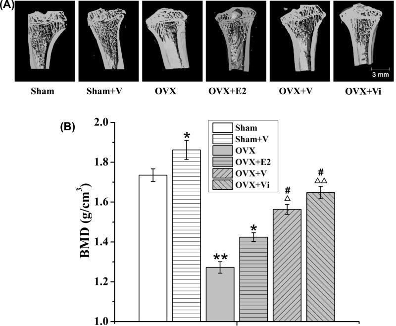
This composite image evaluates the impact of Low-Magnitude Vibration (LMV) and estrogen (E2) on bone mineral density (BMD) in a rat model of postmenopausal osteoporosis. Panel (A) presents micro-CT sectional views of the left proximal tibiae across six experimental groups: Sham, Sham+V (vibration), OVX (ovariectomized), OVX+E2, OVX+V, and OVX+Vi (immediate vibration). Visually, the OVX group shows significant trabecular bone loss and reduced density compared to Sham controls. The OVX+V and OVX+Vi groups demonstrate partial restoration of trabecular architecture, appearing superior to the OVX+E2 group. Panel (B) provides a corresponding bar graph measuring BMD in g/cm³. Statistical significance is noted relative to the Sham group (*, **), the OVX group (Δ, ΔΔ), and the OVX+E2 group (#). The data indicates that while ovariectomy significantly reduces BMD, LMV—particularly when initiated early (Vi)—effectively mitigates bone loss, showing higher efficacy in preserving bone mass and structural integrity than traditional estrogen treatment in this preclinical model.
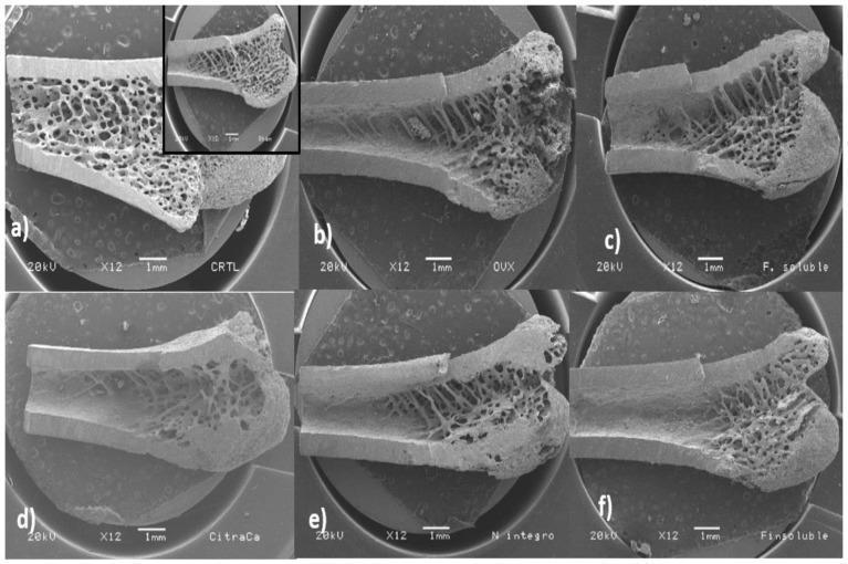
This set of six scanning electron micrographs (SEM) at 12X magnification presents a comparative analysis of rat femur longitudinal cuts, specifically examining trabecular and cortical bone architecture. The images illustrate the pathophysiology of estrogen-deficiency-induced osteoporosis and the potential therapeutic effects of dietary interventions. (a) Control/Sham group shows a robust, dense network of interconnected trabeculae extending significantly into the diaphysis and thick cortical walls. (b) The Ovariectomized (OVX) group exhibits classic osteoporotic changes: markedly reduced trabecular density, loss of connectivity, and thinning of the cortical bone. (c-f) Experimental groups demonstrate varying degrees of bone preservation: (c) soluble fiber and (f) O. ficus-indica powder groups show improved trabecular area and cortical integrity compared to OVX. (d) Calcium citrate and (e) insoluble fiber groups show intermediate recovery, with slightly improved trabecular morphology but thinner cortical structures than the control. The clinical significance focuses on bone mineral density (BMD) maintenance and the role of soluble fiber as a protector against osteoclastic bone resorption.
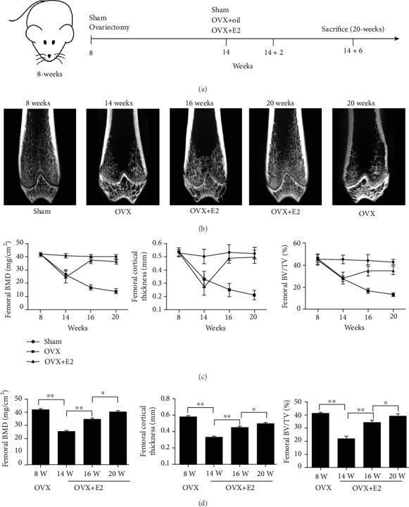
This composite educational image illustrates an experimental study on osteoporosis and estrogen therapy using a rat model. Panel (a) presents a timeline of the experiment from ovariectomy (OVX) at 8 weeks through sacrifice at 20 weeks. Panel (b) displays 3D micro-CT (Diagnostic Imaging) cross-sections of the distal femur, comparing Sham, OVX, and OVX+E2 (17̢-estradiol) groups. The Sham group shows dense trabecular bone, while the OVX group exhibits progressive bone loss and increased porosity. The OVX+E2 group shows preserved structural integrity. Panel (c) contains three longitudinal line graphs quantifying femoral bone mineral density (BMD), femoral cortical thickness (CtTh), and bone volume fraction (BV/TV). The graphs show a sharp decline in all parameters for the OVX group post-8 weeks, while the OVX+E2 group shows significant recovery and stabilization starting at 14 weeks. Panel (d) provides bar chart comparisons of these parameters specifically for the OVX vs. OVX+E2 groups at 14, 16, and 20 weeks, with statistical markers (*P < 0.05, **P < 0.01) highlighting the therapeutic effect of 17̢-E2 on cortical and cancellous bone.
Systemic Changes in Menopause
1. Definition and Hormonal Basis
Hormonal Profile at Menopause
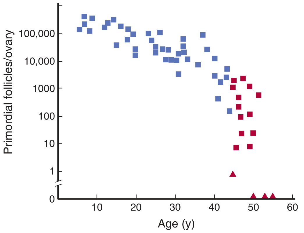
- Oestrogen (17β-oestradiol): falls by up to 90%; residual oestrogen comes from peripheral aromatisation of androstenedione (in adipose tissue, muscle, liver)
- Progesterone: falls to near zero (no corpus luteum formation)
- FSH: rises markedly (loss of negative feedback from inhibin B and oestradiol); FSH ≥25 IU/L is consistent with menopause
- LH: rises moderately; episodic bursts coincide with hot flushes
- Anti-Müllerian hormone (AMH): becomes undetectable (best marker of ovarian reserve)
- Inhibin B: undetectable
Stages of Reproductive Ageing (STRAW Framework)
| Stage | Features |
|---|---|
| Early menopausal transition (age ~47-49) | Minor menstrual irregularity; FSH sporadically elevated; AMH low |
| Late menopausal transition (age ~49-52) | ≥60 consecutive days of amenorrhoea; FSH consistently elevated |
| Early postmenopause (0-5 years after FMP) | Amenorrhoea; FSH consistently ≥25 IU/L; AMH undetectable; most symptoms most severe |
| Late postmenopause | Symptoms may diminish but systemic sequelae progress |
2. Systemic Changes
2a. Vasomotor System - Hot Flushes and Night Sweats
- Loss of ovarian oestrogen leads to dysregulation of the hypothalamic thermoregulatory set point
- Aberrant activation of the tachykinin system (specifically neurokinin B signalling via NK3 receptors in the arcuate nucleus) triggers vasodilation and heat dissipation responses
- Each flush coincides with a large pulsatile LH secretion burst - however, LH itself is not the cause, since flushes persist after hypophysectomy; rather, an estrogen-sensitive event in the hypothalamus simultaneously triggers both LH release and the flush
- Median duration: 7.4 years after the final menstrual period; ~50% of women have symptoms for >10 years; Black women are disproportionately affected with longer duration
- Sudden sensation of warmth spreading from the trunk to the face and neck
- Cutaneous flushing and profuse sweating
- Night sweats causing sleep disruption
- Associated palpitations and anxiety
- Each episode lasts 1-5 minutes; may occur multiple times daily
2b. Genitourinary System - Genitourinary Syndrome of Menopause (GSM)
- Thinning and atrophy of vaginal squamous epithelium
- Reduced vaginal glycogen (less Lactobacillus colonisation) → rise in vaginal pH from ~4.0 to >5.0
- Decreased vaginal secretions and lubrication
- Thinning of urethral and bladder epithelium
- Vaginal dryness, chronic irritation, burning, and itching (vulvovaginal atrophy)
- Dyspareunia (painful intercourse) and post-coital bleeding
- Loss of vaginal rugae; labia minora may become atrophic
- Urinary urgency, frequency, dysuria, and recurrent urinary tract infections
- Urethral caruncle formation
- Sexual dysfunction: reduced libido, arousal, and orgasm
2c. Skeletal System - Osteoporosis and Fracture Risk
- Oestradiol normally suppresses bone resorption by inhibiting osteoclast formation and activity via regulation of the RANK-L / osteoprotegerin axis
- At menopause, falling oestrogen increases RANK-L and decreases osteoprotegerin, tipping the balance toward increased osteoclast activity and accelerated bone resorption
- Oestradiol levels fall by 90% over the menopausal transition, closely paralleling the period of accelerated and progressive bone loss
- Spine: approximately 1.8 to 2.3% per year during early menopause
- Hip: approximately 1.0 to 1.4% per year
- Over 6-10 perimenopausal years: up to 30% loss of trabecular bone and 10% loss of cortical bone
- Results in a 50-100% higher fracture rate compared with premenopausal women
2d. Cardiovascular System
- Lipid profile deteriorates: LDL and triglycerides rise; HDL falls
- Blood pressure rises: loss of oestrogen-mediated vasodilation; increased arterial stiffness
- Central adiposity increases: visceral fat accumulation promotes insulin resistance and the metabolic syndrome
- Inflammatory markers rise: CRP, fibrinogen, and other pro-atherogenic markers increase
- Endothelial dysfunction: reduced nitric oxide production
2e. Central Nervous System - Cognitive and Mood Changes
- Hypogonadism of menopause explains frequent complaints of memory loss, "brain fog," and inability to focus
- Sex differences in hippocampal and temporal lobe structure and function are linked to oestradiol - these change after menopause
- Higher executive functioning in women (pre-menopause) is partly oestrogen-mediated
- Many executive functions spontaneously recover within a few years of the final menstrual period
- Long-term: menopause is associated with increased risk for dementia (including Alzheimer's disease); surgical menopause (bilateral oophorectomy) carries a particularly elevated risk
- The "critical window hypothesis": oestrogen neuroprotection may only be effective if initiated within 5-10 years of the final menstrual period
- Perimenopausal and recently menopausal women have higher rates of anxiety, irritability, and depression compared with premenopausal counterparts
- Especially in women with a history of mood dysfunction (PMS, postnatal depression), or adverse childhood experiences
- The relationship is complex: sleep disruption from hot flushes independently worsens mood
- Not simply "psychological" - abrupt oestrogen withdrawal using GnRH agonists in premenopausal women reproducibly produces mood symptoms
2f. Sleep Architecture
- Increased sleep fragmentation, especially in the second half of the night, with disproportionate disruption of REM sleep
- Nighttime hot flushes and night sweats are a direct cause of nocturnal awakenings
- Independent sleep disorders become more prevalent: obstructive sleep apnoea (worsened by central adiposity), restless legs syndrome, and insomnia
- Chronic poor sleep contributes to mood deterioration, reduced cognitive performance, and metabolic dysfunction
2g. Body Composition and Metabolic Changes
- Weight gain: midlife women gain an average of 2-5 pounds (with wide variation) during the menopausal transition, but this is partly age-related
- Redistribution of fat: shift from gynoid (gluteal-femoral) to android (central/visceral) distribution, regardless of weight change
- Visceral adiposity promotes:
- Insulin resistance and type 2 diabetes
- Metabolic syndrome
- Chronic low-grade inflammation (elevated TNF-α, IL-6, CRP)
- Hypogonadism disrupts adipocyte lipolysis and adipogenesis via adipose oestrogen receptors
- The gut microbiome is also affected by hypogonadism, potentially contributing to weight gain
2h. Skin and Hair
- Skin: collagen content declines after menopause (oestrogen maintains dermal collagen synthesis); skin becomes thinner, drier, and less elastic; wound healing may be impaired
- Hair: pubic and axillary hair thins; paradoxically, androgen excess relative to oestrogen may cause increased facial hair (hirsutism) in some women
- Scalp hair may thin or develop a female-pattern androgenic alopecia pattern
- Skin sebaceous gland activity decreases, contributing to dryness and pruritus
2i. Urological and Pelvic Floor
- Loss of oestrogen weakens pelvic floor support structures
- Stress urinary incontinence (increased with coughing, sneezing, physical activity)
- Urge incontinence and overactive bladder symptoms
- Prolapse of pelvic organs (uterovaginal, cystocele, rectocele) becomes more common with ageing and oestrogen deficiency
- Recurrent urinary tract infections due to urethral mucosal atrophy and loss of protective vaginal flora
2j. Haematological and Coagulation Changes
- Postmenopausal state is associated with a mild pro-thrombotic tendency: increased fibrinogen, factor VII, factor VIII, and reduced fibrinolysis
- This contributes to the elevated cardiovascular risk of the postmenopausal period
3. Diagnosis
- FSH ≥25 IU/L (confirmatory, but fluctuates - less useful than clinical criteria alone)
- AMH - undetectable (best proxy for ovarian reserve; does not fluctuate with cycle)
- Oestradiol - very low (<20 pg/mL)
- Thyroid function tests (to exclude hypothyroidism mimicking menopausal symptoms)
- DEXA scan to assess baseline bone mineral density
- Fasting lipids, glucose, and blood pressure screening
4. Management Overview
Vasomotor Symptoms
| Approach | Examples |
|---|---|
| Hormone therapy (most effective) | Oestradiol ± progestogen (oral, patch, gel, spray) |
| SSRI/SNRIs | Paroxetine mesylate (FDA-approved), escitalopram, venlafaxine |
| NK3 receptor antagonists | Fezolinetant (FDA-approved; specifically targets the tachykinin pathway driving hot flushes) |
| Gabapentin/pregabalin | Moderate evidence |
| Clonidine | Limited evidence |
| CBT | ~50% efficacy; negligible harm |
Genitourinary Syndrome
- Local (vaginal) oestrogen: tablets, cream, ring, pessary - minimal systemic absorption; safe long-term
- Vaginal moisturisers and lubricants (over the counter)
- Ospemifene (oral SERM with oestrogenic effects on urogenital epithelium)
- DHEA (prasterone) vaginal insert
Bone Protection
- Adequate calcium (1200 mg/day) and vitamin D (800-1000 IU/day)
- Weight-bearing exercise
- Bisphosphonates (first-line): alendronate, risedronate, zoledronate
- Denosumab (RANK-L inhibitor): second-line
- Hormone therapy: preserves bone density if started within 10 years of FMP, but not given primarily for bone protection due to other risks
Cardiovascular Risk Reduction
- Screening and treatment of hypertension and hyperlipidaemia
- Lifestyle modification (exercise, weight loss, smoking cessation)
- Hormone therapy is not recommended for cardiovascular risk reduction (Women's Health Initiative data)
Risks of Combined Hormone Therapy (WHI trial data)
- Increased coronary heart disease (HR 1.29)
- Increased breast cancer (HR 1.26; significant after 5+ years of use)
- Increased stroke (HR 1.41)
- Increased venous thromboembolism
5. Summary: Organ Systems Affected by Menopause
| System | Change | Clinical Consequence |
|---|---|---|
| Vasomotor | Loss of hypothalamic thermoregulation | Hot flushes, night sweats |
| Genitourinary | Vaginal/urethral atrophy | Dyspareunia, UTIs, incontinence |
| Skeletal | Increased osteoclast activity | Osteoporosis, fractures |
| Cardiovascular | Dyslipidaemia, arterial stiffness | IHD, stroke, hypertension |
| CNS | Loss of neuroprotective oestrogen signalling | Memory loss, mood changes, dementia risk |
| Sleep | Fragmented sleep architecture | Insomnia, fatigue |
| Metabolic | Central adiposity, insulin resistance | Metabolic syndrome, T2DM |
| Skin/hair | Reduced collagen, androgen dominance | Thinning skin, hair changes |
| Pelvic floor | Weakened support structures | Prolapse, stress incontinence |
| Haematological | Pro-thrombotic tendency | VTE risk |