
Nasopharyngeal swab for RTPCR for respiratory panel
nasopharyngeal swab RTPCR respiratory panel procedure pathogens detected 2025
nasopharyngeal swab technique procedure
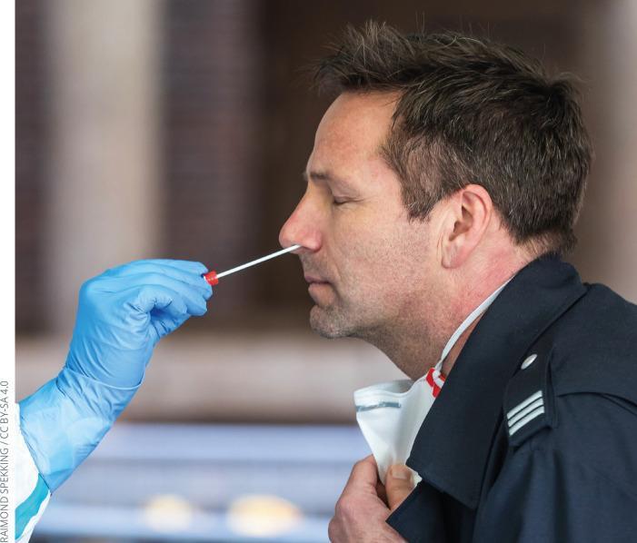
A clinical photograph demonstrating the collection of a nasopharyngeal specimen for infectious disease testing, such as COVID-19 (SARS-CoV-2). The image depicts a healthcare provider, wearing a blue nitrile glove, inserting a specialized nasopharyngeal swab into the right nostril of an adult male patient. The swab features a long, flexible white shaft and a red handle, characteristic of devices designed to reach the posterior nasopharynx by following the nasal floor. The patient is shown in profile with eyes closed and a lowered face mask, facilitating clear access to the nasal cavity. This visual illustrates the standard technique for obtaining respiratory samples for nucleic acid amplification tests (NAAT) or rapid antigen tests. The procedure is a critical component of diagnostic algorithms for respiratory viruses, requiring precise horizontal insertion to ensure adequate cellular collection from the nasopharyngeal mucosa while minimizing patient discomfort.
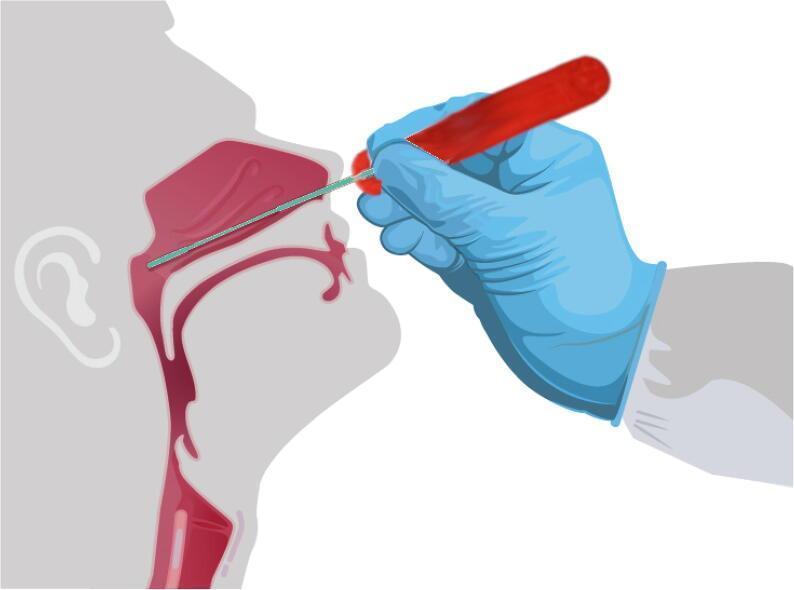
This medical illustration demonstrates the correct clinical procedure for nasopharyngeal swab insertion. The image features a sagittal cross-section of a human head, highlighting the nasal cavity, hard palate, and nasopharynx. A clinician's hand, wearing a blue medical glove, is shown manipulating a 3D-printed red press-fit handle attached to a semi-flexible nasopharyngeal swab. The swab is inserted through the naris, following a trajectory parallel to the nasal floor and palate rather than upwards toward the bridge of the nose. The tip of the swab is positioned deep within the nasopharynx, at the back of the nasal passage, to reach the site for respiratory specimen collection. The illustration serves as a pedagogical guide for healthcare professionals to visualize the anatomical pathway and depth required to reach the nasopharyngeal mucosa for diagnostic testing, such as for viral pathogens.
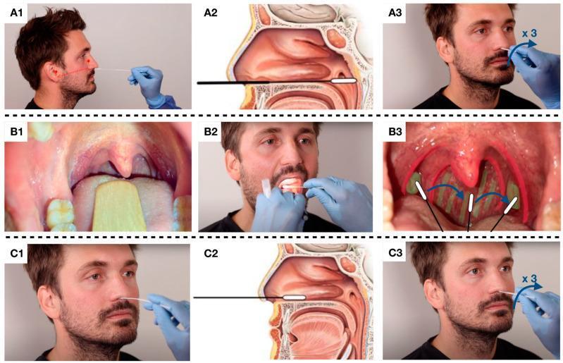
A multi-panel clinical instructional graphic demonstrating three types of upper respiratory tract specimen collection: nasopharyngeal swab (NPS), oropharyngeal swab (OPS), and nasal swab. Row A (NPS) shows a side-view clinical photo with a trajectory line toward the earlobe, followed by an anatomical cross-section illustrating a flexible swab reaching the posterior wall of the nasopharynx (8–11 cm depth), and a rotation step. Row B (OPS) displays intraoral views using a tongue depressor to visualize the oropharynx, with diagrammatic overlays highlighting the swabbing of the palatine tonsils and the posterior pharyngeal wall. Row C (nasal swab) illustrates a shallow insertion (1–3 cm) into the anterior nasal cavity, with an anatomical diagram showing contact with the septum and inferior nasal concha, followed by a triple-rotation technique. This infographic serves as a clinical guide for proper diagnostic sampling techniques in infectious disease screening, such as for COVID-19.
https://www.mayocliniclabs.com/test-catalog/overview/609409
Nasopharyngeal Swab - RT-PCR Respiratory Panel
What Is It?
Specimen Collection - Technique
- Position patient with head tilted slightly back (~70°)
- Insert a flexible Dacron or rayon flocked swab horizontally along the nasal floor (NOT upward toward the bridge of the nose) - trajectory aimed toward the ear
- Advance 8-11 cm until resistance is met at the posterior nasopharyngeal wall
- Rotate the swab gently (2-3 rotations) and hold for 5-10 seconds to absorb secretions
- Withdraw slowly while rotating
- Place swab into viral transport medium (VTM)
Important: Calcium alginate swabs are inhibitory to PCR - use Dacron or rayon flocked swabs only. - Red Book 2021, AAP
Pathogens Detected (BioFire FilmArray RP2.1 - 22 targets)
| Category | Pathogen |
|---|---|
| Influenza | Influenza A (H1, H3, H1-2009), Influenza B |
| Coronaviruses | SARS-CoV-2, Coronavirus HKU1, NL63, OC43, 229E |
| RSV | RSV A, RSV B |
| Parainfluenza | PIV 1, 2, 3, 4 |
| Other viruses | Adenovirus, Human Metapneumovirus (hMPV), Rhinovirus/Enterovirus, Human Bocavirus |
| Bacteria | Bordetella pertussis, Bordetella parapertussis, Chlamydia pneumoniae, Mycoplasma pneumoniae |
Technology & How It Works (FilmArray System)
- Nucleic acid purification - from the NP swab lysate
- First-stage PCR - broad amplification
- Second-stage PCR (nested multiplex) - target-specific amplification
- Melt curve analysis - pathogen identification
Clinical Indications
- Acute respiratory illness requiring pathogen identification (influenza-like illness, bronchiolitis, pneumonia)
- Immunocompromised patients with respiratory symptoms
- Hospitalized patients needing cohorting decisions
- Outbreak investigation
- Atypical pneumonia (covers Mycoplasma, Chlamydia pneumoniae)
- Suspected whooping cough (Bordetella pertussis) - panel preferred over culture given higher sensitivity - Red Book 2021
Important Limitations & Interpretation Notes
| Point | Detail |
|---|---|
| False negatives | Poor collection technique, late sampling (>1 week), sequence variability in target region |
| False positives | Asymptomatic carriage (e.g., rhinovirus), viral shedding post-recovery |
| Coinfection | Positive result does NOT rule out co-infection with other pathogens |
| Untargeted pathogens | A negative panel does not exclude infection - pathogens not on the panel will be missed |
| Not for test-of-cure | Should not be repeated within 7 days (residual nucleic acid persists after recovery) |
| Specimen type | Validated for NP swabs; BAL/bronchial washings require a different assay |
Specimen Handling & Storage
| Condition | Timeframe |
|---|---|
| Refrigerated transport | Within 72 hours |
| Frozen (if delayed) | Up to 30 days |
| Avoid | Calcium alginate swabs, dry swabs, prolonged room temperature |
Multiplex RT-PCR vs. Conventional Testing
| Feature | Multiplex RT-PCR Panel | Conventional (culture/RAT) |
|---|---|---|
| Turnaround time | ~45-60 min | Hours to days (culture) / 15 min (RAT) |
| Number of targets | 15-22+ simultaneously | Single or few pathogens |
| Sensitivity | High (superior to RAT) | Variable |
| Cost | Higher | Lower |
| Clinical utility | High (guides isolation, antivirals, antibiotic stewardship) | Moderate |