
Paget disease of nipple
Reading File
Now let me search the medical library and PubMed in parallel.
Finding Sources
Searching PubMed
"Paget disease" nipple breast
Finding Sources
Good results. Now let me read the key sections from multiple textbooks in parallel.
Reading File
Reading File
Reading File
Reading File
Reading File
Excellent - I have rich, comprehensive data from multiple authoritative textbooks. Let me also fetch the clinical image from Bailey & Love.
Loading Image
Loading Image
I now have comprehensive information from multiple authoritative surgical and dermatology textbooks, along with a clinical image. Here is the full answer:
Paget Disease of the Nipple
Definition and Historical Background
First described by Sir James Paget in 1874, who reported 15 women with chronic nipple ulcerations who developed breast cancer within 2 years. Paget disease of the nipple accounts for 1-3% of all new breast cancers in females.
- Bailey and Love's Short Practice of Surgery, 28th Ed.
- Mulholland and Greenfield's Surgery, 7th Ed., p. 3975
Pathogenesis
Paget disease is essentially a unique form of DCIS (ductal carcinoma in situ) arising in the nipple epidermis. There are two main theories:
- Epidermotropic theory (favored): Malignant ductal cells migrate from underlying DCIS via the lactiferous ducts and invade upward into the epidermis of the nipple.
- In-situ transformation theory: Keratinocytes of the nipple epidermis undergo malignant transformation independently.
The carcinoma cells spread into the lactiferous sinuses, invade across the junction of epidermal and ductal epithelial cells, and enter the epidermal layer. Paget cells do not invade through the dermal basement membrane, so they remain categorized as carcinoma in situ until invasion occurs.
- Sabiston Textbook of Surgery, p. 3022-3031
Clinical Features
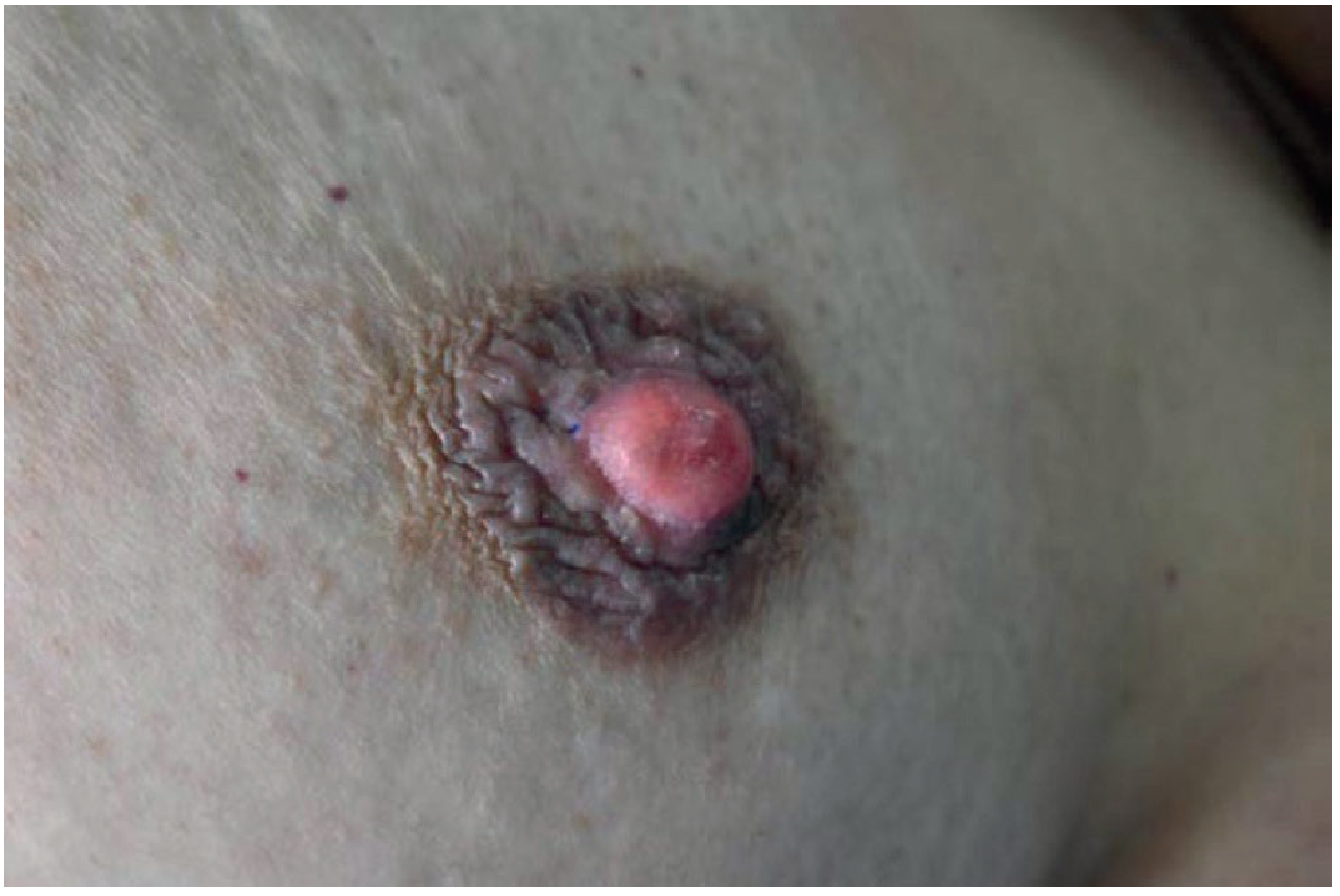
The hallmark is a scaly, raw, vesicular, or ulcerated lesion that begins on the nipple and spreads secondarily to the areola (unlike eczema, which typically begins on the areola).
Key features:
- Chronic erythematous, scaling, eczematous eruption of the nipple
- Progressive erosion/destruction of the nipple
- Pruritus, burning, or pain (may precede visible disease)
- Bloody nipple discharge
- May progress to crusting, ulceration, and spread onto areola/breast skin
- Usually unilateral
Important: Paget disease starts on the nipple, whereas most benign skin conditions (eczema, contact dermatitis) begin on the areola.
Associated Malignancy
This is clinically very important:
| Finding | Association |
|---|---|
| Overall association with underlying breast carcinoma | >95% of cases (Sabiston) / 85-88% (Mulholland) |
| Palpable mass present | >50% of patients |
| Palpable mass + mammographic abnormality | >90% have invasive carcinoma |
| No palpable mass, normal mammogram | More likely DCIS than invasive |
- Sabiston Textbook of Surgery, p. 3029
Pathology (Histology)
The pathognomonic finding is the Paget cell - a large, pale (vacuolated) cell with round or oval nuclei and large nucleoli, found between normal keratinocytes of the nipple epidermis (in the rete pegs of the epithelium).
Immunohistochemistry:
-
CK7 positive (low-molecular-weight cytokeratins) - characteristic
-
CEA (carcinoembryonic antigen) positive - distinguishes from melanoma
-
S-100 negative (positive in melanoma) - key differential
-
HER2 overexpression in 70-90% of cases
-
Hormone receptor (ER/PR) positive in approximately 50% of cases
-
Schwartz's Principles of Surgery, 11th Ed., p. 352
-
Mulholland and Greenfield's Surgery, 7th Ed., p. 3400
Differential Diagnosis
| Condition | Distinguishing Feature |
|---|---|
| Eczema / Atopic dermatitis | Starts on areola, bilateral, responds to steroids |
| Contact dermatitis | Bilateral, history of irritant exposure |
| Post-radiation dermatitis | History of prior RT |
| Superficial spreading melanoma | S-100 positive, CEA negative on IHC |
| Erosive adenomatosis | Benign proliferation of lactiferous ducts |
- Sabiston Textbook of Surgery, Fitzpatrick's Dermatology
Investigations / Workup
- Wedge or punch biopsy of nipple - confirms diagnosis by demonstrating Paget cells
- Bilateral breast examination
- Mammography - abnormal in ~50%; abnormalities can occur distant from the nipple-areolar complex
- MRI - if mammography and breast imaging are normal (highly sensitive but not specific; detected lesions must be biopsied to confirm)
- Triple assessment to exclude/characterize underlying malignancy
Treatment
| Scenario | Treatment |
|---|---|
| Paget disease without underlying mass/malignancy | Central core excision (nipple + areola + cone of major ducts down to pectoralis major) + adjuvant radiotherapy |
| Paget disease with underlying malignancy | Mastectomy with axillary staging |
| Breast-conserving option | Wide local excision (nipple-areolar complex + underlying cancer to clear margins) + whole breast RT ± Grisotti flap reconstruction |
-
For patients treated with breast-conserving surgery, nipple-areolar reconstruction can be performed 4-6 months after radiotherapy, or via 3D tattooing.
-
Axillary staging and adjuvant systemic therapy are determined by the nature of the underlying breast cancer.
-
Bailey and Love's, 28th Ed.; Sabiston, p. 3031; Mulholland, p. 3404
Extramammary Paget Disease (for comparison)
Paget disease can also occur outside the breast (most commonly anogenital skin, axillae, penis). It is classified into 3 types:
- Primary intraepidermal adenocarcinoma (most common)
- Associated with visceral malignancy (GI or genitourinary) - 25%
- Associated with adjacent sweat gland carcinoma (least common)
- Fitzpatrick's Dermatology, Vol. 1&2
Recent Literature
Recent reviews confirm the above framework. A 2026 systematic review of dermatologic mimickers (PMID: 40814164) highlights that Paget disease can be confused with several benign skin conditions, reinforcing the need for biopsy in any persistent unilateral nipple eruption. A 2024 review describes it as "an often-overlooked malignancy" (PMID: 42273835), emphasizing that delay in diagnosis is common due to its eczema-like appearance.
Key exam points to remember:
- Starts on the nipple (not areola), spreads outward
- Paget cells = large pale vacuolated cells in nipple epidermis (pathognomonic)
-
95% have underlying breast carcinoma
- CEA positive, S-100 negative (distinguishes from melanoma)
- HER2 overexpressed in 70-90%
- Always biopsy any persistent unilateral nipple skin change
This is a shared conversation. Sign in to Orris to start your own chat.