
Write a five mark short note on Steps of superficial parotidectomy Make it compatible, compact but high yielding. So that... I can easily remember and, uh, write it in a... Systematic way being a 2nd year resident. make it in a way its interesting and easy to learn visually and Create a high density, high yielding image based(generate image) Flashcard I can use Before exam to revise and remember all the important points regarding this short-note. but it should be in a way so its easy to understand and remember also use flowcharts or mindmaps in the response wherever possible dont use code
superficial parotidectomy steps surgical technique facial nerve identification
A high-yield medical exam flashcard for "Superficial Parotidectomy - Steps" designed for a surgery resident. Clean white background with color-coded sections. Layout includes: TOP HEADER (bold, dark blue): "SUPERFICIAL PAROTIDECTOMY - High Yield Steps" LEFT SIDE - A vertical numbered flowchart with arrows connecting each step in colored boxes: 1. POSITION & INCISION (orange box) - Supine, head turned. Modified Blair incision (lazy-S) — preauricular crease → around ear lobule → down SCM 2. SKIN FLAP ELEVATION (yellow box) - Subplatysmal plane in neck, superficial to parotid fascia anteriorly. Identify Greater Auricular Nerve (GAN) — preserve posterior branch if possible 3. EXPOSE LANDMARKS (green box) - Free parotid from: Tragal cartilage | SCM | Posterior belly of digastric | Mastoid tip 4. IDENTIFY FACIAL NERVE TRUNK (red box, most prominent) - Tragal pointer → 1-1.5 cm deep & inferior = Main trunk. Other landmarks: Tympanomastoid suture | PBD muscle | Stylomastoid artery. USE NERVE MONITOR! 5. ANTEGRADE DISSECTION (blue box) - Dissect gland off nerve from trunk → branches. Identify pes anserinus (bifurcation into temporofacial & cervicofacial divisions) 6. REMOVE SUPERFICIAL LOBE (purple box) - All parotid tissue LATERAL to facial nerve removed en bloc 7. CLOSURE (grey box) - Drain placement | Layered closure | Watch for Frey's syndrome RIGHT SIDE - Small inset anatomy diagram showing parotid region with labeled: Tragal pointer, Facial nerve trunk, Pes anserinus, SCM, Digastric (posterior belly) BOTTOM BANNER (yellow, bold): "KEY PEARL: Never sacrifice facial nerve! Landmarks = Tragal pointer + Tympanomastoid suture + Posterior belly of digastric" Color-coded mnemonic at bottom: "PSELF-RC" in bold red Clean, readable fonts, organized like a study flashcard an ENT/surgery resident would use before exams.
Superficial Parotidectomy - Steps (5 Mark Short Note)
Definition: Superficial (lateral) parotidectomy = removal of all parotid tissue lateral to the facial nerve, preserving the nerve and deep lobe. Most commonly done for pleomorphic adenoma or benign tumors of the superficial lobe.
FLOWCHART OF STEPS
POSITION & GA
      ↓
INCISION (Modified Blair / Lazy-S)
      ↓
SKIN FLAP ELEVATION (subplatysmal + supra-parotid fascia plane)
      ↓
EXPOSURE OF LANDMARKS (Tragal cartilage, SCM, Mastoid tip, PBD)
      ↓
IDENTIFY GREATER AURICULAR NERVE → preserve posterior branch
      ↓
★ IDENTIFY FACIAL NERVE MAIN TRUNK (KEY STEP)
      ↓
ANTEGRADE DISSECTION → TRUNK → PES ANSERINUS → ALL BRANCHES
      ↓
REMOVAL OF SUPERFICIAL LOBE EN BLOC (all tissue lateral to FN)
      ↓
HEMOSTASIS + DRAIN + CLOSURE
      ↓
MONITOR FOR COMPLICATIONS
STEP-BY-STEP BREAKDOWN
Step 1 - Anesthesia, Position & Preparation
- General anesthesia - use short-acting or NO muscle relaxant (needed for facial nerve monitoring)
- Position: Supine, head turned to opposite side, slight head-up (reverse Trendelenburg - reduces bleeding)
- Infiltrate with 1:100,000 adrenaline - reduces haemorrhage, slightly eases nerve identification
Step 2 - Incision
- Modified Blair incision ("Lazy S" / "Question mark" incision)
- Preauricular crease → curves around ear lobule → over mastoid tip → down along SCM → forward in neck crease
- Alternative: Facelift/Rhytidectomy incision - better cosmesis, used in benign tumors (scar hidden in hairline)
  Preauricular crease
       ↓ (curves under lobule)
  Mastoid tip
       ↓ (down over SCM)
  Neck crease (forward)
Step 3 - Skin Flap Elevation
- Plane: Superficial to parotid fascia anteriorly; subplatysmal plane in cervical region
- SMAS layer is included in the flap
- Identify Greater Auricular Nerve (GAN) over SCM
- Sacrifice anterior branch (supplies parotid skin - numbness inevitable)
- Preserve posterior branch if possible (supplies ear lobule - reduces post-op numbness)
Step 4 - Mobilize Parotid & Expose Landmarks
| Structure | What you do |
|---|---|
| Tragal cartilage | Dissect parotid off it superiorly - exposes tragal pointer |
| SCM | Free parotid off its anterior surface |
| Mastoid tip | Dissect off parotid tissue |
| Post. belly of digastric (PBD) | Exposed proximally near mastoid attachment |
Step 5 - ★ IDENTIFY FACIAL NERVE MAIN TRUNK (MOST CRITICAL STEP)
TRAGAL POINTER
  → Facial nerve lies 1 - 1.5 cm DEEP & INFERIOR to its tip
  (NB: Tragal pointer is slightly mobile - use with caution)

TYMPANOMASTOID SUTURE LINE
  → Follow medially → leads directly to main trunk
  (More constant than tragal pointer!)

POSTERIOR BELLY OF DIGASTRIC
  → Superior border near mastoid → nerve lies just above/anterior
  → Nerve ~1 cm from PBD attachment to mastoid

STYLOMASTOID ARTERY
  → Accompanies facial nerve at stylomastoid foramen
  → Useful additional landmark
- Use intraoperative facial nerve monitor (stimulator)
- Antegrade (anterograde) dissection is standard = trunk → branches
- Retrograde approach (branches → trunk) used in revision surgery
Step 6 - Antegrade Dissection of Facial Nerve & Superficial Lobe Removal
Main Trunk (stylomastoid foramen)
         ↓
   PES ANSERINUS (bifurcation)
    /              \
Temporofacial     Cervicofacial
  division          division
    /                    \
Temporal              Marginal mandibular
Zygomatic             Cervical
Buccal
- Parotid tissue is elevated off each nerve branch using blunt dissection (small hemostat, McCabe dissector)
- Use bipolar cautery only - NEVER monopolar near facial nerve
- Gland is removed en bloc lateral to all branches
Step 7 - Closure & Post-op
- Hemostasis - meticulous
- Drain placement (suction drain)
- Layered closure (deep, SMAS, skin)
- No pressure dressing that compresses drain
COMPLICATIONS MIND MAP
                    SUPERFICIAL PAROTIDECTOMY
                         COMPLICATIONS
                              |
        ┌──────────────────────────────────────┐
        |                    |                  |
   NERVE RELATED         SKIN/WOUND         FREY'S SYNDROME
        |                    |            (Auriculotemporal nerve
  ┌─────────┐          ┌─────────┐         injury → anomalous
  |         |          |         |          reinnervation)
Transient  Permanent  GAN       Scar    Gustatory sweating
 palsy      palsy    numbness  |        Tx: Botulinum toxin
 (common)  (rare)   (lobule)  Salivary fistula
                              Wound infection
                              Haematoma
HIGH-YIELD EXAM PEARLS 🔑
| Point | Fact |
|---|---|
| Incision type | Modified Blair (Lazy S) / Facelift incision |
| Best landmark for FN | Tympanomastoid suture (most constant) |
| Tragal pointer landmark | FN lies 1-1.5 cm deep and inferior |
| Anesthesia tip | NO long-acting relaxants - need nerve monitor |
| Most feared complication | Permanent facial nerve palsy |
| Late complication | Frey's syndrome (auriculotemporal nerve) |
| Treatment of Frey's | Botulinum toxin injection |
| GAN posterior branch | Preserve - reduces ear lobule numbness |
| Dissection type | Antegrade (trunk → branches) standard |
| Cautery rule | Bipolar ONLY near facial nerve |
MNEMONIC - "P-I-E-F-D-R-C"
Position → Incision → Elevate flap → Facial nerve identify → Dissect antegrade → Remove lobe → Close
📸 INTRAOPERATIVE REFERENCE
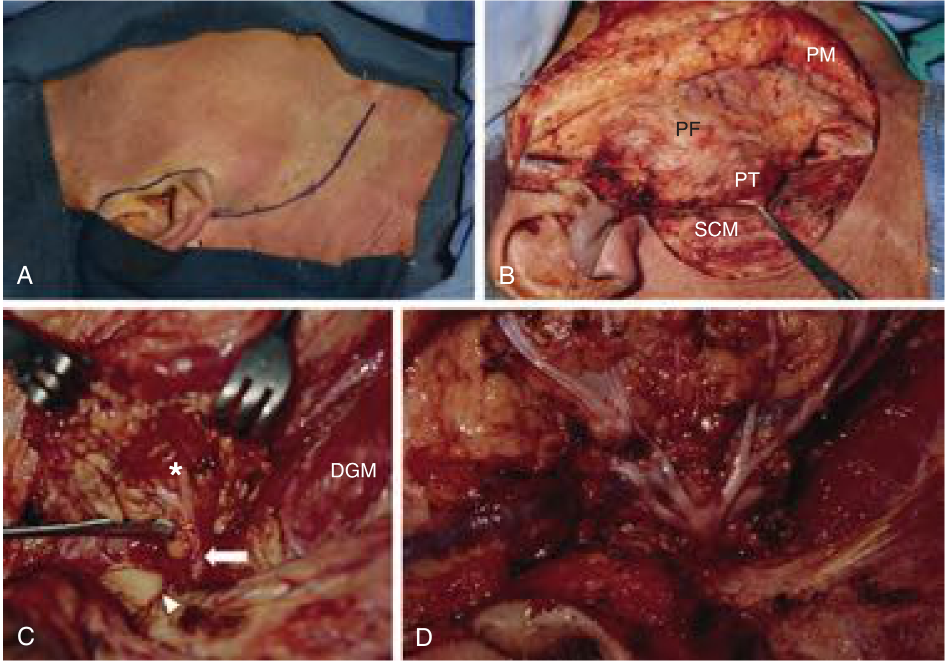
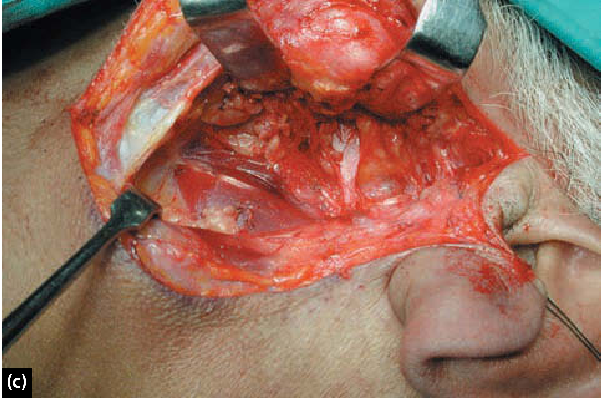
FLASHCARD (Pre-Exam Revision)
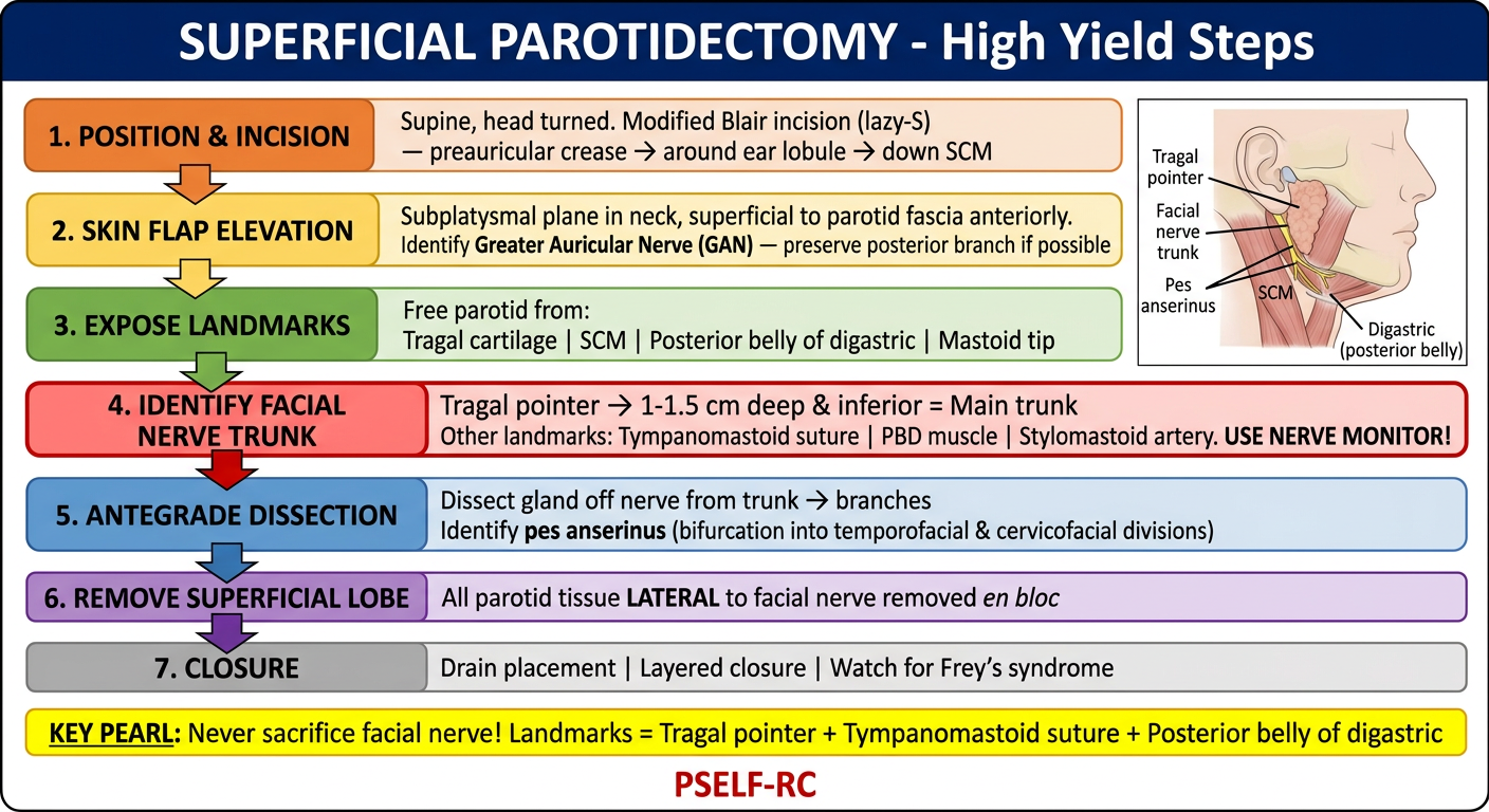
QUICK-RECALL TABLE (Write in exam in 2 min)
STEP        ACTION                           KEY POINT
1. Position  Supine, head turned, head-up     No long relaxants
2. Incision  Modified Blair (Lazy S)          Facelift option = better cosmesis
3. Flap      Subplatysmal, supra-fascia       GAN: cut anterior, preserve posterior
4. Landmarks Tragal cart, SCM, Mastoid, PBD   Wide exposure = easier nerve ID
5. FN trunk  Tympanomastoid suture/           1-1.5 cm deep/inf to tragal pointer
             Tragal pointer/PBD attachment    Bipolar only. Use nerve monitor.
6. Dissect   Antegrade: trunk→pes anserinus   Blunt dissection superficial to nerve
             →temporofacial+cervicofacial
7. Remove    En bloc, all tissue lateral FN   Capsule intact (esp. pleomorphic)
8. Close     Drain + layered closure          Watch: Frey's, haematoma, palsy