
Causes of migraine 2nd year mbbs pharmacology
migraine pathophysiology trigeminovascular diagram
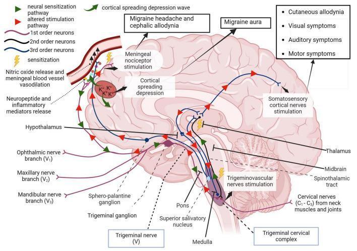
A pathophysiology diagram illustrating the trigeminovascular pathway and its role in migraine development. The diagram depicts a sagittal view of the human brain, highlighting key anatomical structures including the trigeminal ganglion, trigeminal cervical complex (medulla/pons), thalamus, hypothalamus, and somatosensory cortex. It maps the neural hierarchy from 1st-order neurons (trigeminal branches V1, V2, V3 and cervical nerves C1-C3) through the 2nd-order spinothalamic tract to 3rd-order thalamic projections. Visual indicators identify areas of sensitization and altered stimulation pathways. Key educational concepts include the cortical spreading depression wave, meningeal nociceptor stimulation, and neuropeptide release leading to vasodilation. The diagram correlates specific neural activity with clinical symptoms, specifically migraine aura, cephalic and cutaneous allodynia, and visual, auditory, and motor impairments. It further demonstrates the descending pathway via the superior salivatory nucleus involving nitric oxide release in meningeal blood vessels, providing a comprehensive visual of central and peripheral sensitization in migraine pathology.
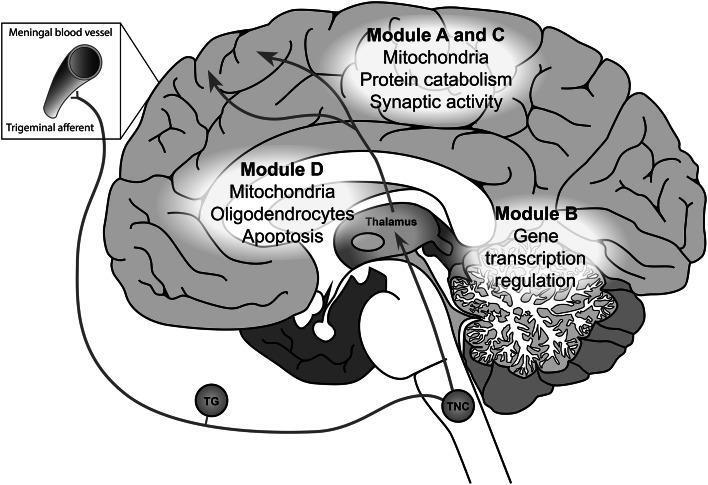
A medical schematic illustrating the pathophysiology of migraine, specifically the trigeminovascular pathway and associated gene co-expression modules in the brain. The diagram depicts a sagittal view of the human brain with key anatomical structures labeled, including the cortex, cerebellum, thalamus, and trigeminal nucleus caudalis (TNC). The trigeminovascular pathway is traced from meningeal blood vessels via trigeminal afferents through the trigeminal ganglion (TG) and TNC, ascending to the thalamus and ultimately the cerebral cortex. Overlaid on the anatomy are localized gene expression modules (A-D) highlighting specific cellular functions and components: Modules A and C in the cortex are associated with mitochondria, protein catabolism, and synaptic activity; Module B in the cerebellum is linked to gene transcription regulation; and Module D in the subcortical white matter and thalamic region is associated with mitochondria, oligodendrocytes, and apoptosis. The illustration serves as an educational tool for understanding the integration of genetic expression patterns with neuroanatomical pain pathways in migraine headache disorders.
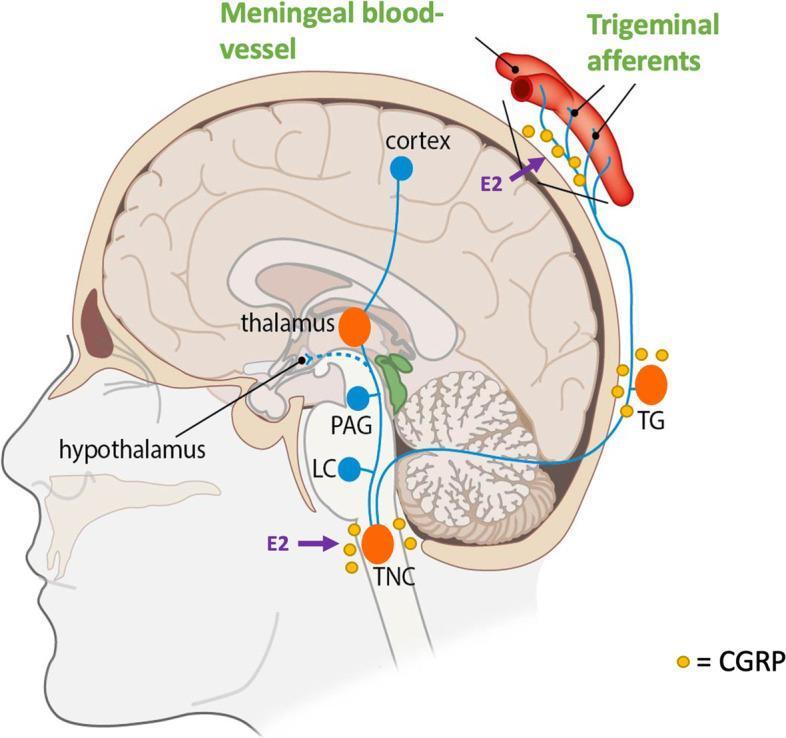
This pathophysiology diagram illustrates the human trigeminovascular system and its activation during a migraine attack. Presented in a sagittal cross-section of the head, the diagram highlights key anatomical structures including the meningeal blood vessels, cerebral cortex, thalamus, hypothalamus, and brainstem components such as the periaqueductal gray (PAG), locus coeruleus (LC), and trigeminal nucleus caudalis (TNC). The trigeminal ganglion (TG) is shown as a peripheral relay point. The diagram emphasizes the role of Calcitonin Gene-Related Peptide (CGRP), represented by yellow icons, localized at the meningeal vessels, TG, and TNC, indicating its role in neurotransmission and vasodilation. Purple arrows denote the influence of estradiol (E2) on CGRP release at the level of the TNC and the trigeminal afferents. Blue lines trace the ascending and descending pathways between these centers, demonstrating the functional connectivity involved in headache phase signaling. This visual is designed for medical education regarding migraine pathophysiology and the neuro-hormonal modulation of the trigeminal system.
Causes of Migraine - 2nd Year MBBS Pharmacology
Overview
1. Neurological (Brain-Based) Mechanisms
A. Cortical Spreading Depression (CSD)
- CSD is the electrophysiological basis of migraine aura
- A slow wave of neuronal depolarization starts in the occipital lobe and spreads anteriorly at 2-4 mm/min
- The CSD wave causes disruption of ionic gradients (Na+/K+ ATPase pump failure), leading to mass neuronal depolarization followed by prolonged hyperpolarization (suppression)
- This correlates with the classic aura: initial "positive" symptoms (flashing lights/scintillations) followed by "negative" symptoms (visual scotoma/blind spot)
- CSD triggers meningeal nociceptors and may activate the trigeminovascular system, initiating the headache phase
B. The Trigeminovascular System (Key Pharmacological Mechanism)
- The trigeminal nerve innervates intracranial blood vessels (meningeal/dural vessels) via small unmyelinated C-fibers and thinly myelinated Aδ-fibers
- Activation of these trigeminal afferents causes release of:
- CGRP (Calcitonin Gene-Related Peptide) - potent vasodilator; key mediator of migraine pain
- Substance P
- Neurokinin A
- These neuropeptides cause neurogenic inflammation: vasodilation, plasma protein extravasation, and mast cell degranulation around meningeal vessels
- Pain signals travel: Trigeminal ganglion → Trigeminal nucleus caudalis (brainstem) → Thalamus → Somatosensory cortex
- This is why triptans (5-HT1B/1D agonists) and CGRP antagonists (gepants) are effective - they block this pathway
C. Central Sensitization
- Recurrent trigeminovascular activation leads to peripheral and then central sensitization
- Sensitized neurons have lower activation thresholds, increased spontaneous activity, and expanded receptive fields
- ~75% of migraineurs show central sensitization during attacks
- Clinically manifests as cutaneous allodynia (skin becomes painful to light touch) in ~2/3 of patients
- This is why delaying triptan use (once allodynia develops) reduces effectiveness
D. Brainstem and Hypothalamic "Migraine Generator"
- Functional MRI shows activation of dorsal rostral pons and hypothalamus before cortical changes and before symptoms begin
- The hypothalamus likely explains premonitory (prodromal) symptoms: mood changes, food cravings, yawning, thirst, drowsiness
- These regions modulate pain perception through descending pain modulatory pathways (periaqueductal gray, locus coeruleus)
2. Neurotransmitter / Biochemical Causes
| Neurotransmitter | Role in Migraine |
|---|---|
| Serotonin (5-HT) | Decreased levels during attack; implicated in vascular spasm and pain modulation. Basis for triptan therapy (5-HT1B/1D agonists) |
| CGRP | Released from trigeminal afferents; causes vasodilation and pain. Elevated in blood during attacks. Target of new anti-CGRP antibodies (erenumab, fremanezumab) |
| Dopamine | Hypersensitivity to dopamine explains yawning, nausea, vomiting in prodrome |
| Nitric oxide (NO) | A potent vasodilator; nitroglycerin (NO donor) can reliably trigger migraine in susceptible individuals |
| Glutamate | Excitatory; may contribute to cortical hyperexcitability and CSD initiation |
| β-endorphins | Reduced levels may lower pain threshold |
3. Vascular Mechanisms (Historical but Still Relevant)
- The old "vascular theory" (Wolff's hypothesis): aura caused by vasoconstriction → headache caused by rebound vasodilation of cranial vessels
- This is now considered incomplete - pure vasodilation is not the primary cause
- However, vascular changes DO occur: dilation of meningeal arteries (especially middle meningeal), extracerebral temporal artery, and middle cerebral artery during headache phase
- Blood flow reduction (oligemia) spreading from occipital cortex at ~2-3 mm/min confirms CSD as the substrate
- Complication: cerebral infarction can occur in a small subset of migraineurs
4. Genetic Causes
- Migraine has strong genetic predisposition: first-degree relatives of patients with migraine with aura are ~4x more likely to be affected
- Familial Hemiplegic Migraine (FHM) - the best-studied genetic model:
- FHM1: Mutation in CACNA1A (chromosome 19p13) - voltage-gated P/Q-type calcium channel α subunit
- FHM2: Mutation in ATP1A2 (chromosome 1q23) - Na+/K+ ATPase pump α2 subunit
- FHM3: Mutation in SCN1A (chromosome 2q24) - neuronal voltage-gated sodium channel α subunit
- Common migraine is polygenic - GWAS studies identified 44 variants at 38 loci, enriched in genes for vascular biology, synaptic regulation, and ion channels
- ~50% of susceptibility is genetic; the other ~50% is environmental
5. Trigger Factors
| Category | Examples |
|---|---|
| Hormonal | Estrogen fluctuation (menstrual migraine), oral contraceptives, menopause |
| Dietary | Tyramine (aged cheese, red wine), nitrates (processed meats), MSG, alcohol, caffeine withdrawal, skipping meals |
| Sleep disturbance | Oversleeping or sleep deprivation ("weekend migraine") |
| Stress | Emotional stress - and paradoxically, "let-down" after stress |
| Sensory stimuli | Bright lights, loud noise, strong odors (perfumes, gasoline) |
| Medications | Proton-pump inhibitors, reserpine, nifedipine, vasodilators (nitrates), estrogens |
| Environmental | Weather changes, high altitude, heat |
| Behavioral | Irregular meals, dehydration, excess caffeine |
Diagram: Trigeminovascular Pathway in Migraine
Summary for Exam (Pharmacological Relevance)
CSD → Triggers trigeminal nerve activation
         ↓
Trigeminal afferents release CGRP + Substance P
         ↓
Neurogenic inflammation of meningeal vessels
         ↓
Sensitization of trigeminal nucleus caudalis
         ↓
Pain signals → Thalamus → Cortex = MIGRAINE HEADACHE
- Triptans (sumatriptan) - 5-HT1B/1D agonists → inhibit CGRP release + cause vasoconstriction
- Ergotamine/DHE - also act on 5-HT1B/1D receptors
- CGRP monoclonal antibodies (erenumab) - block CGRP receptor for prevention
- NSAIDs - reduce neurogenic inflammation
- Beta-blockers, valproate, topiramate - reduce cortical hyperexcitability (prophylaxis)