
Examination of pupil
Reading File
Searching sources...
Finding Sources
Finding Sources
Reading File
Finding Sources
Reading File
Reading File
Reading File
Reading File
Loading Image
Loading Image
Loading Image
Finding Sources
Finding Sources
Now I have comprehensive material. Let me compile the full answer.
Examination of the Pupil
1. Neuroanatomical Basis
Pupil size is governed by a tonic balance between sympathetic (dilates) and parasympathetic (constricts) innervation of the iris.
Parasympathetic (constrictor) pathway:
- Light stimulates retinal rods and cones → optic nerve → pregeniculate optic tracts → pretectal nucleus (midbrain-diencephalic junction) → bilateral Edinger-Westphal nuclei → travel with CN III → ciliary ganglion → ciliary nerves → pupillary sphincter muscle.
Sympathetic (dilator) pathway (3-neuron chain):
- 1st-order neuron: Ventral lateral hypothalamus → descends ipsilaterally through brain stem → synapses at C8-T2 (intermediolateral column)
- 2nd-order (preganglionic) neuron: Exits with ventral roots of C8, T1, T2 → ascends the neck → superior cervical ganglion
- 3rd-order (postganglionic) neuron: Follows the internal carotid artery → ophthalmic branch of CN V → pupillodilator muscle
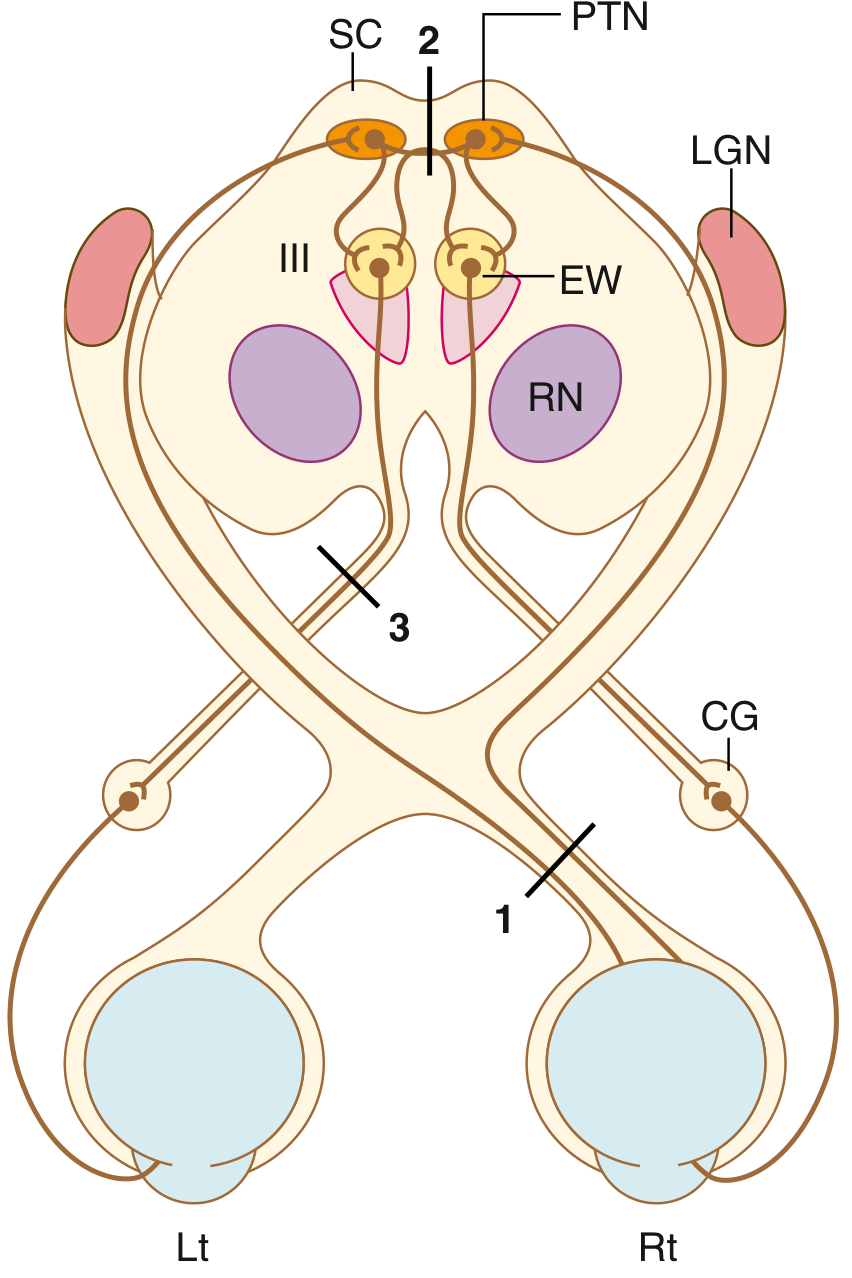
(Goldman-Cecil Medicine)
2. Method of Examination
Step 1 - Assess Size and Shape
- Examine in a dimly lit room (pupils naturally dilated).
- Shine a dim light from below so both pupils are seen simultaneously in indirect illumination.
- Record size in millimeters (normal: 2-6 mm) and note whether pupils are equal, round, and regular.
- Anisocoria (unequal pupils): measure in both darkness and bright light to distinguish cause.
Step 2 - Direct and Consensual Light Reflex
- Ask the patient to fixate on a distant target (to minimize accommodative miosis).
- Shine a bright light directly into one eye - note direct constriction in that eye.
- Note consensual constriction in the opposite eye simultaneously.
- Repeat for the other eye.
Step 3 - Swinging Flashlight Test (for RAPD)
The relative afferent pupillary defect (RAPD), formerly called the Marcus Gunn pupil, detects unilateral or asymmetric optic neuropathy.
Technique (Box 16.1):
- Patient fixates on an immobile distant target.
- Use the brightest light available for maximum constriction.
- Grade each pupil's direct light response on a 1-4 scale.
- Swing the light between eyes at ~1 second intervals (swinging flashlight test).
- Observe each pupil for initial constriction or dilation.
- Limit to 3-4 swings to avoid retinal bleaching.
Interpretation: Normally, direct and consensual reactions are equal. With an optic neuropathy, the affected pupil dilates (rather than constricts) when the light swings to it, because the direct reaction is weaker than the consensual. Both pupils are actually "resetting" at the larger size dictated by the damaged nerve.
Note: RAPD is absent when both optic nerves are equally damaged. Severe inner retinal pathology can also produce RAPD.
Step 4 - Near (Accommodation) Reflex
- Ask the patient to look at a distant object, then quickly at their thumb held 15-30 cm away.
- Normal: pupil constricts (as part of the accommodation-convergence-miosis triad).
- A near reaction can be elicited even in a blind patient via proprioceptive influences.
Recording Format
| Size (mm) | Direct Light | Consensual Light | Near Reaction | |
|---|---|---|---|---|
| Right eye | 4.0 | 4+ | 2+ | 4+ |
| Left eye | 4.0 | 2+ | 4+ | 4+ |
(Bradley & Daroff's Neurology in Clinical Practice, Table 16.1)
3. Pupillary Responses with Lesions
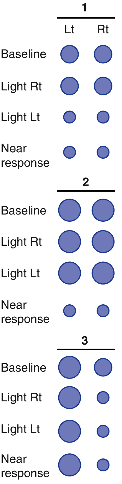
Lesion 1 - Optic nerve (afferent defect): Direct light reaction lost in affected eye; consensual lost when that eye is illuminated; both intact when opposite eye illuminated. RAPD present.
Lesion 2 - Pretectal region: Afferent light reflex interrupted bilaterally → both pupils dilated and light-fixed; near response preserved (light-near dissociation). Causes: pineal tumors (dysgerminomas), localized midbrain infarcts.
Lesion 3 - Oculomotor nerve (CN III): Pupil dilated 6-7 mm, unresponsive to light or near. Associated with ptosis, external ophthalmoplegia, and diplopia. Key causes: posterior communicating artery aneurysm, uncal herniation, midbrain compression.
4. Light-Near Dissociation
The pupil fails to react to light but does constrict to near (accommodation). Causes:
- Adie tonic pupil
- Dorsal midbrain syndrome (Parinaud syndrome)
- Severe bilateral optic neuropathies
- Argyll Robertson pupils
5. Common Pupillary Abnormalities
Physiologic Anisocoria
- Long-standing unequal pupils with normal reflex reactions in both eyes.
- Disparity remains constant in both light and dark.
- Benign; no localizing significance.
Adie Tonic Pupil
- Medium-to-large pupil (3-6 mm) that constricts little/not at all to light, very slowly to accommodation.
- Constricts with dilute pilocarpine 0.125% (denervation hypersensitivity).
- Usually unilateral, more common in women aged 25-45.
- Likely post-viral denervation of ciliary ganglion.
- Benign; may have associated areflexia (Adie syndrome).
Horner Syndrome
Classic triad of sympathetic paresis:
- Miosis (small pupil)
- Ptosis (partial, from Müller muscle paresis)
- Anhidrosis (of forehead/face, variable with postganglionic lesions)
- Also: enophthalmos, lower eyelid elevation ("upside-down ptosis")
Anisocoria is greater in the dark (affected pupil cannot dilate). Lesion may be anywhere along the 3-neuron pathway.
Argyll Robertson Pupils
- Small (1-2 mm), unequal, irregular pupils.
- Fixed to light (no direct or consensual reaction).
- Constrict briskly to near (light-near dissociation).
- Classical cause: tertiary neurosyphilis.
Pharmacologic Mydriasis
- Dilated, unresponsive pupils from mydriatic drugs (atropine, scopolamine patches).
- Fails to constrict with 1% pilocarpine - distinguishes from CN III palsy (which does constrict with pilocarpine).
Third Nerve Compression
- Dilated pupil (6-7 mm), unresponsive to light or near.
- Always associated with other CN III signs.
- Causes: posterior communicating artery aneurysm, brain herniation, neoplasia.
Adie tonic pupil - right eye (affected pupil larger):
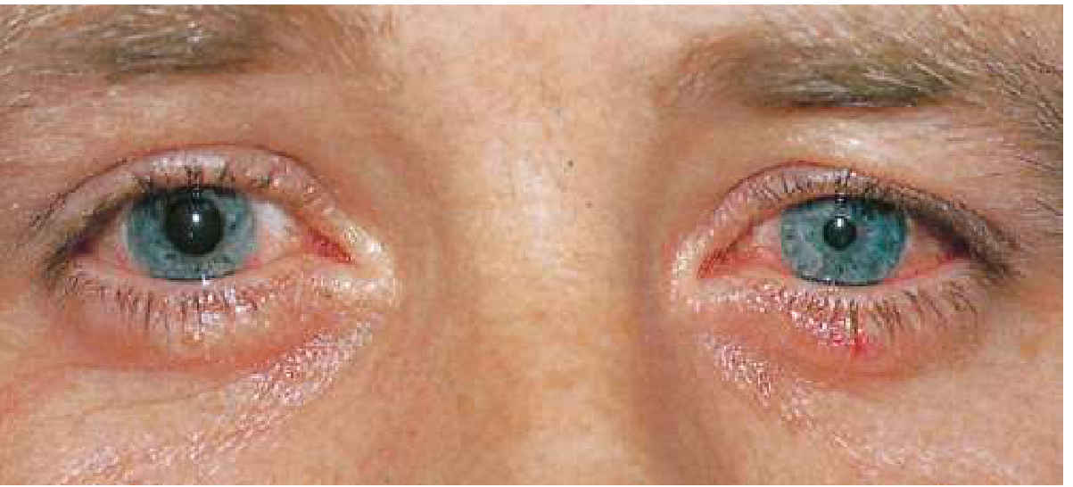
(Goldman-Cecil Medicine, Fig. 392-3 - Adie tonic pupil in the right eye. The affected pupil appears larger and responds sluggishly to light.)
Sources:
- Goldman-Cecil Medicine - Examination of the Pupil, Pupillary Pathways
- Bradley & Daroff's Neurology in Clinical Practice - Examination of the Pupils, Box 16.1
- S Das: A Manual on Clinical Surgery, 13th Edition - Oculomotor Nerve Examination
This is a shared conversation. Sign in to Orris to start your own chat.