
Pain recereptors and pain pathway m
pain pathway spinothalamic tract diagram nociception
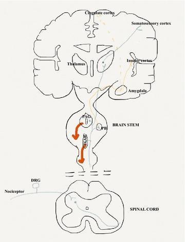
This medical anatomical diagram illustrates the neuroanatomical pathways of pain (nociception), including ascending sensory and descending modulatory systems. The diagram spans the spinal cord, brainstem, and cerebral cortex. The 'sensory-discriminative' pathway (blue line) originates from primary afferent nociceptors and the dorsal root ganglia (DRG), ascends the spinal cord to the thalamus, and terminates in the somatosensory cortex to convey stimulus location and nature. The 'affective-emotional' pathway (orange line) projects through the parabrachial nucleus (PB) in the brainstem and the amygdala, reaching the cingulate and insular cortices. Additionally, a descending inhibitory control pathway (red line/arrows) is shown originating in the periaqueductal grey (PAG) and rostroventral medulla (RVM) of the brainstem, projecting back to the spinal dorsal horn to modulate nociceptive output. This illustration serves as an educational tool for understanding the multi-dimensional processing of pain within the central nervous system.
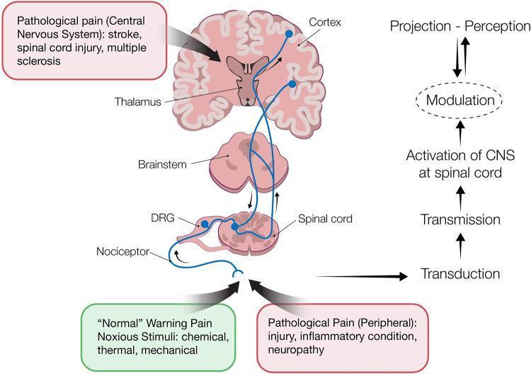
This pathophysiology diagram illustrates the ascending and descending pathways of pain processing in the human nervous system. The visual is divided into an anatomical illustration on the left and a functional flowchart on the right. Anatomical structures shown include peripheral nociceptors, the dorsal root ganglion (DRG), the spinal cord, brainstem, thalamus, and cerebral cortex. The diagram traces the sequence of nociception through four key stages: 1) Transduction (conversion of noxious stimuli like chemical, thermal, or mechanical signals into electrical impulses), 2) Transmission (conduction of action potentials via the DRG to the spinal dorsal horn), 3) Perception (projection of signals through the brainstem and thalamus to the cortex), and 4) Modulation (the enhancement or inhibition of pain signals via descending pathways, represented by a bidirectional arrow). Clinical context is provided through text boxes distinguishing between 'Normal' warning pain and 'Pathological Pain,' citing causes such as inflammation, neuropathy, stroke, spinal cord injury, and multiple sclerosis. This diagram is designed for medical students to understand the neuroanatomy of the pain pathway and the functional distinction between different pain types.
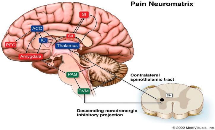
This pathophysiology diagram illustrates the 'Pain Neuromatrix,' detailing the complex network of cortical and subcortical structures involved in pain perception. The schematic features a sagittal view of the human brain and a cross-section of the spinal cord. An ascending pathway is shown via the contralateral spinothalamic tract, originating from the dorsal horn (DH) of the spinal cord and projecting to the Thalamus. Subcortical nodes including the Periaqueductal Gray (PAG) and Rostral Ventral Medulla (RVM) are depicted in the brainstem, with a labeled descending noradrenergic inhibitory projection returning to the DH. Cortical regions highlighted include the Primary Somatosensory Cortex (SI), Secondary Somatosensory Cortex (SII), Anterior Cingulate Cortex (ACC), Prefrontal Cortex (PFC), and Insular Cortex (IC), alongside the Amygdala. Interconnecting arrows demonstrate the bidirectional flow between the Thalamus, limbic structures, and sensory-cognitive cortical areas, emphasizing that pain is a distributed neural process rather than a linear pathway. This resource is suitable for medical education on neuroanatomy and chronic pain mechanisms.
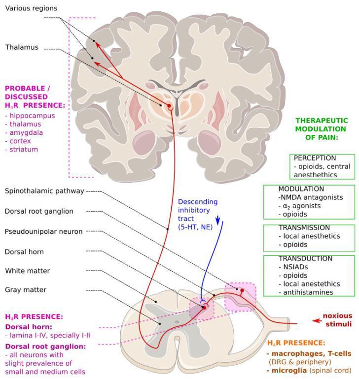
This medical illustration depicts a simplified schematic of the ascending and descending neuroanatomical pain pathways. The ascending pathway (red) illustrates nociceptive transmission from peripheral noxious stimuli through pseudounipolar neurons in the dorsal root ganglion to the dorsal horn. The signal travels via the spinothalamic tract to the thalamus and cortical regions for perception. The descending inhibitory tract (blue) originates in the brain and projects to the spinal cord, modulating pain transmission via serotonin (5-HT) and norepinephrine (NE). A green side panel categorizes therapeutic modulation of pain into four physiological processes: transduction (NSAIDs, local anesthetics), transmission (local anesthetics, opioids), modulation (NMDA antagonists, alpha-2 agonists), and perception (opioids, central anesthetics). The diagram also highlights the localization of Histamine H4 Receptors (H4R) in the brain (thalamus, hippocampus, cortex), spinal cord (dorsal horn lamina I-IV), and immune cells (macrophages, microglia), indicating their potential role as targets for analgesic pharmacology.
Pain Receptors (Nociceptors) and the Pain Pathway
1. Pain Receptors - Nociceptors
Types of Nociceptive Fibers
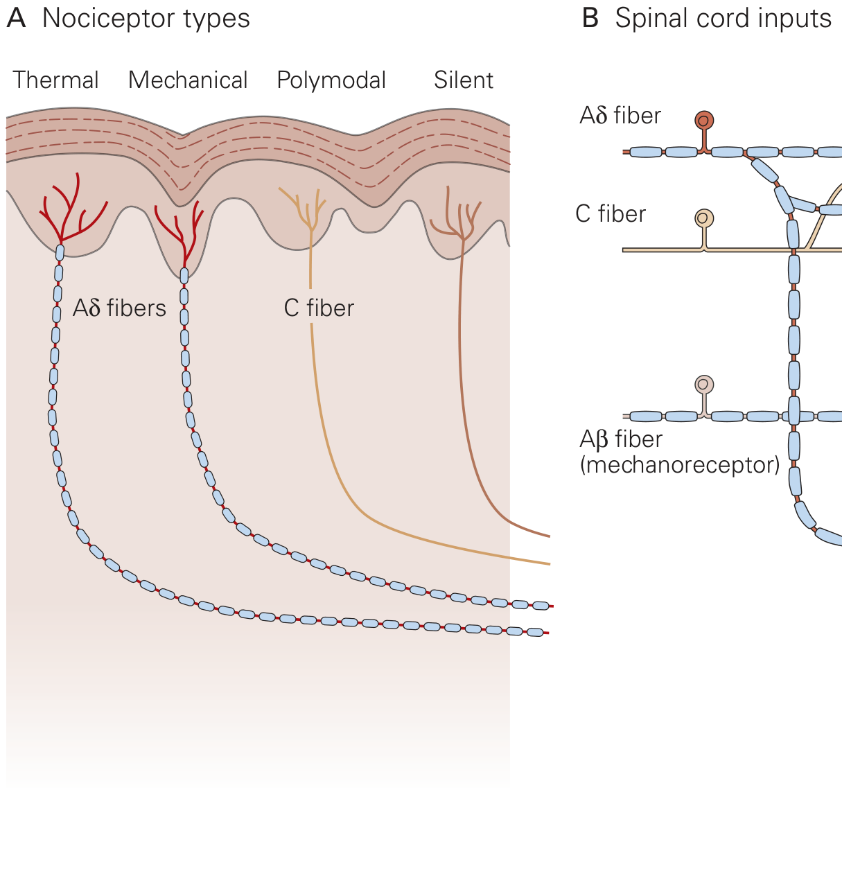
| Fiber | Myelination | Diameter | Conduction velocity | Pain quality |
|---|---|---|---|---|
| A-beta (Aβ) | Heavily myelinated | Large | Fast (~30-70 m/s) | Touch/pressure - NOT pain under normal conditions |
| A-delta (Aδ) | Thinly myelinated | Small | Moderate (~5-30 m/s) | Sharp, well-localized, fast/first pain; thermal and mechanical |
| C fibers | Unmyelinated | Smallest | Slow (~0.5-2 m/s) | Burning, dull, poorly localized, slow/second pain; polymodal |
"Most Aδ and C fiber afferents respond maximally to intense (painful) stimuli and produce the subjective experience of pain when they are activated; this defines them as primary afferent nociceptors. The ability to detect painful stimuli is completely abolished when conduction in Aδ and C fiber axons is blocked." - Harrison's Principles of Internal Medicine 22E
Classes of Nociceptors
- Thermal nociceptors - respond to extreme heat (>45°C) or cold; largely Aδ fibers
- Mechanical nociceptors - respond to intense mechanical distortion (pinching, cutting); Aδ fibers
- Polymodal nociceptors - respond to heat, cold, mechanical, AND chemical stimuli; predominantly C fibers
- Silent (sleeping) nociceptors - normally unresponsive, but become sensitized during inflammation and chemical insults
Key Receptor Molecules
- TRPV1 (vanilloid receptor) - activated by heat (>43°C), acidic pH, endogenous mediators, and capsaicin. Mediates thermal hyperalgesia.
- Voltage-gated Na+ channels - set threshold for action potential generation (Na-v 1.7, 1.8, 1.9 are nociceptor-specific)
- Voltage-gated Ca2+ channels - control neurotransmitter release at central terminals
- Bradykinin (B2) receptors, prostaglandin receptors, P2X3 receptors - chemical mediators that sensitize nociceptors
2. Peripheral Sensitization
- Hyperalgesia - increased pain to a normally painful stimulus (e.g., sunburned skin)
- Allodynia - pain produced by a normally non-painful stimulus
3. The Pain Pathway - Overview
Step 1 - Transduction
Step 2 - Transmission (First-order neuron)
Step 3 - Dorsal Horn Processing
| Lamina | Key neurons | Input received |
|---|---|---|
| Lamina I (marginal layer) | Nociceptive-specific neurons, Wide Dynamic Range (WDR) neurons | Aδ and C fibers |
| Lamina II (substantia gelatinosa) | Excitatory and inhibitory interneurons | C fibers; key site for pain modulation |
| Lamina III-IV | Mix of interneurons and projection neurons | Aβ fibers (light touch) |
| Lamina V | Wide Dynamic Range (WDR) neurons | Aβ, Aδ, and C fibers; also visceral input |
"Activation of nociceptors causes the release of glutamate from presynaptic terminals in the spinal cord dorsal horn; this release acts on the ionotropic glutamate receptor AMPA postsynaptically to cause rapid depolarization of dorsal horn neurons." - Firestein & Kelley's Textbook of Rheumatology
4. Ascending Pain Pathways (Second-order neurons)
| Tract | Origin | Destination | Function |
|---|---|---|---|
| Spinothalamic tract (STT) | Laminae I, V-VII | VPL/VPM thalamus → somatosensory cortex | Discriminative pain: localization, intensity, quality |
| Spinoreticular tract | Laminae VII, VIII | Reticular formation + medial thalamus | Diffuse, poorly localized pain; arousal |
| Spinoparabrachial tract | Laminae I, V | Parabrachial nucleus (pons) → amygdala | Affective/emotional component of pain |
| Spinohypothalamic tract | Laminae I, V, VII, VIII | Hypothalamus | Autonomic and neuroendocrine responses to pain |
"The spinothalamic tract is the most prominent ascending nociceptive pathway in the spinal cord... Lesioning this tract (anterolateral cordotomy) can result in a marked reduction in pain sensation on the contralateral side." - Kandel, Principles of Neural Science
5. Thalamus and Cortical Processing (Third-order neurons)
- Lateral thalamic nuclei (VPL/VPM): relay to primary somatosensory cortex (S1, S2) - gives pain its precise location, intensity, and quality (sensory-discriminative dimension)
- Medial thalamic nuclei: relay to anterior cingulate cortex (ACC) and insular cortex - mediates the unpleasantness and emotional suffering (affective dimension)
- Prefrontal cortex: cognitive-evaluative dimension - how the patient interprets and responds to pain based on culture, past experience, and context
6. Gate Control Theory (Melzack and Wall, 1965)
- Large-diameter Aβ fibers (non-nociceptive touch) activate inhibitory interneurons in the substantia gelatinosa (lamina II), which suppress the output of projection neurons - this closes the gate
- Small-diameter C fibers (nociceptive) inhibit those same interneurons - this opens the gate, increasing pain transmission
"Activation of nonnociceptive sensory neurons, by engaging inhibitory interneurons in the dorsal horn, closes a gate for afferent transmission of nociceptive signals." - Kandel, Principles of Neural Science
- Rubbing an injured area provides temporary relief (activates Aβ)
- TENS (transcutaneous electrical nerve stimulation) - activates large Aβ fibers to "close the gate"
- Spinal cord stimulation for chronic pain management
7. Descending Pain Modulation
- Periaqueductal Gray (PAG) - the "master control" center; activated by stress, opioids, or stimulation
- PAG projects to the Rostral Ventromedial Medulla (RVM)
- RVM projects back down to the dorsal horn via serotonergic and noradrenergic pathways
- These pathways inhibit second-order pain neurons, reducing pain signal transmission
Summary Diagram
Quick Reference Summary
Noxious stimulus
      ↓
Nociceptor (free nerve ending) — TRPV1, Na+ channels
      ↓
Aδ fibers (fast, sharp) / C fibers (slow, burning)
      ↓
Dorsal Root Ganglion (cell body)
      ↓
Dorsal Horn (laminae I, II, V) — Glutamate + Substance P
      ↓
Decussation in spinal cord
      ↓
Anterolateral system:
  • Spinothalamic tract → VPL thalamus → Somatosensory cortex (localization)
  • Spinoparabrachial → Amygdala (emotion)
  • Spinoreticular → Reticular formation (arousal)
  • Spinohypothalamic → Hypothalamus (autonomic)
      ↓
Conscious pain perception (multidimensional)
      ↑
Descending modulation: PAG → RVM → Dorsal Horn (inhibition)