
let's answer
ls "/tmp/workspace/attachments/5eba3352-9574-4987-9143-a6a66e87f053/"
python3 -c " from docx import Document doc = Document('/tmp/workspace/attachments/5eba3352-9574-4987-9143-a6a66e87f053/Y3_Neuro_non-infectious_student1 (1).docx') for p in doc.paragraphs: style = p.style.name if p.style else 'None' if p.text.strip(): print(f'[{style}] {p.text}') "
python3 -c " from docx import Document doc = Document('/tmp/workspace/attachments/5eba3352-9574-4987-9143-a6a66e87f053/Y3_Neuro_non-infectious_student1 (1).docx') for i, table in enumerate(doc.tables): print(f'=== TABLE {i+1} ===') for row in table.rows: cells = [cell.text.strip() for cell in row.cells] print(' | '.join(cells)) print() "
UGHE Neurology Case - Complete Answers
Question 1: Top 3 Diagnoses and Why
| # | Diagnosis | Reasoning |
|---|---|---|
| 1 | Acute ischemic stroke (left MCA territory) - most likely | Sudden-onset expressive aphasia with right arm/leg weakness (3/5) and left facial droop. She is right-handed, so the LEFT hemisphere is language-dominant. The left MCA supplies Broca's area (inferior frontal gyrus). Hypertension (12 years) plus an irregular heart rhythm (likely AF) are major risk factors. |
| 2 | Intracerebral hemorrhage (ICH) | Also causes sudden focal neurological deficits. Long-standing hypertension is the classic risk factor for hypertensive bleeds (typically basal ganglia, thalamus). Cannot be excluded without non-contrast CT. |
| 3 | Embolic stroke from atrial fibrillation | The heart is described as "irregular" - highly suspicious for atrial fibrillation. AF allows thrombus formation in the left atrial appendage; emboli travel preferentially into the MCA territory because it receives the largest proportion of carotid flow. This may overlap with diagnosis #1 (the mechanism behind the ischemic stroke). |
Key point: CT head without contrast is the first-line imaging - it rules out hemorrhage immediately. If negative, the diagnosis shifts to ischemic stroke and time-to-treatment (tPA window) becomes urgent.
Question 2: Compare and Contrast Stroke Syndromes
Table A: Mechanism & Clinical Features
| Feature | Intracerebral Hemorrhage | Ischemic - Thrombotic | Ischemic - Embolic |
|---|---|---|---|
| Basic mechanism | Rupture of a cerebral blood vessel; blood accumulates within brain tissue | Thrombus forms over an atherosclerotic plaque within a cerebral artery, causing progressive occlusion | Embolus forms elsewhere (heart, aorta, carotid) and lodges in a distal cerebral artery |
| Typical onset | Sudden; often worsens over minutes-hours as hematoma expands | May be gradual or "stuttering" - symptoms can fluctuate or build over hours | Abrupt, often maximal at onset (the embolus arrives all at once) |
| Headache | Common and often severe ("worst headache") | Usually absent or mild | Usually absent |
| Vomiting | More common (raised ICP) | Less common | Less common |
| Level of consciousness | More likely impaired, especially with large hemorrhage or intraventricular extension | Usually preserved unless massive infarction | Usually preserved initially |
| Neurological deficits | Focal + mass effect; hemiparesis, aphasia, visual deficits; often severe | Focal deficits matching the vascular territory | Focal cortical deficits (aphasia, visual field cut, neglect) matching the artery |
| Major risk factors | Chronic hypertension, cerebral amyloid angiopathy, anticoagulation, AVM | Atherosclerosis, hypertension, diabetes, hyperlipidemia, smoking, older age | Atrial fibrillation, recent MI, valvular disease (mitral stenosis), ventricular thrombus |
| Typical locations | Basal ganglia, thalamus, pons, cerebellum (hypertensive); lobar (amyloid angiopathy) | Large cerebral arteries (MCA, ACA); can cause lacunar infarcts in small penetrating vessels | MCA most common (receives ~80% of ICA flow); any artery possible |
| CT imaging | Hyperdense (bright white) blood immediately visible | Normal initially; may show subtle early ischemic signs (loss of gray-white differentiation, dense MCA sign) | Normal initially; infarction appears as hypodensity after 6-24 hours |
| Classic example | Hypertensive patient with sudden headache, vomiting, hemiparesis, declining consciousness | Patient with known carotid stenosis develops gradually worsening unilateral limb weakness | Patient with known AF suddenly develops aphasia and hemiplegia - as in this case |
Table B: Presentation / Risk Factors / Associated Conditions (for the blank student table)
| Presentation | Risk factors | Associated conditions | |
|---|---|---|---|
| Intracerebral hemorrhage | Sudden severe headache, nausea/vomiting, focal deficits, decreased consciousness; may worsen rapidly | Hypertension (most common), anticoagulant use, amyloid angiopathy, AVM, cocaine/stimulants | Hypertension, coagulopathy, cerebral amyloid angiopathy (elderly), AVMs |
| Ischemic (thrombotic) | Focal deficits in a vascular territory; may be preceded by TIA; onset can be stuttering; often on waking | Atherosclerosis, hypertension, diabetes mellitus, dyslipidemia, smoking, older age | Carotid artery disease, coronary artery disease, peripheral vascular disease, metabolic syndrome |
| Ischemic (embolic) | Abrupt onset, often maximal at onset; cortical signs prominent (aphasia, neglect, hemianopia); may have sudden improvement (lysis) | Atrial fibrillation, valvular disease, recent MI, dilated cardiomyopathy, endocarditis | Atrial fibrillation, structural heart disease, deep vein thrombosis (paradoxical embolism via PFO) |
Question 3: Bell's Palsy vs. Stroke on Physical Exam
The Neuroanatomical Basis
-
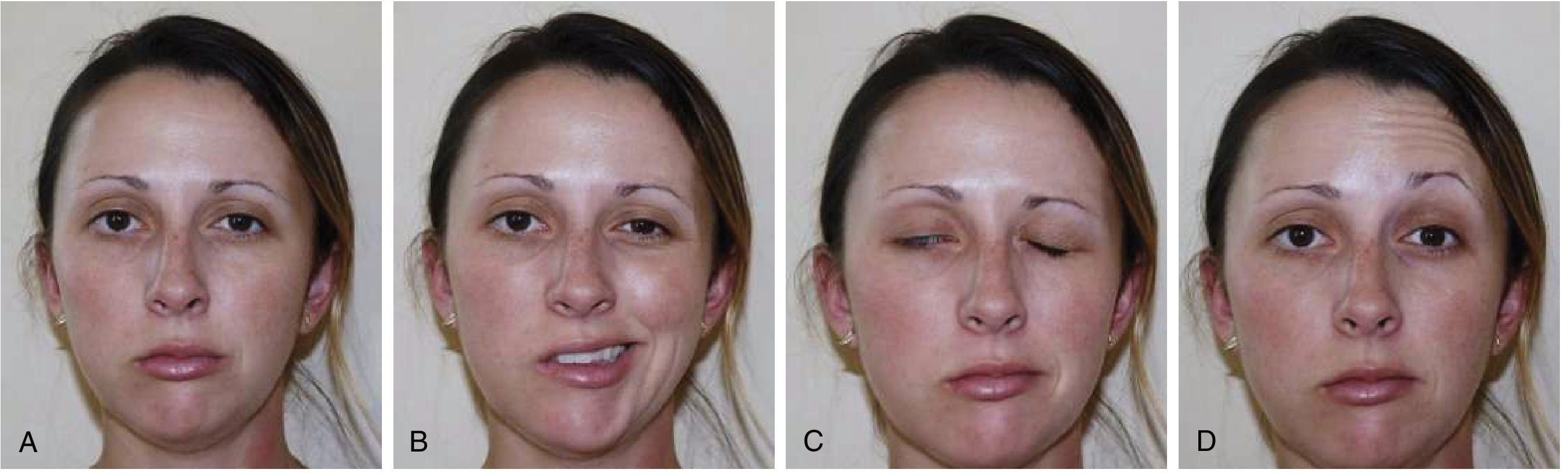
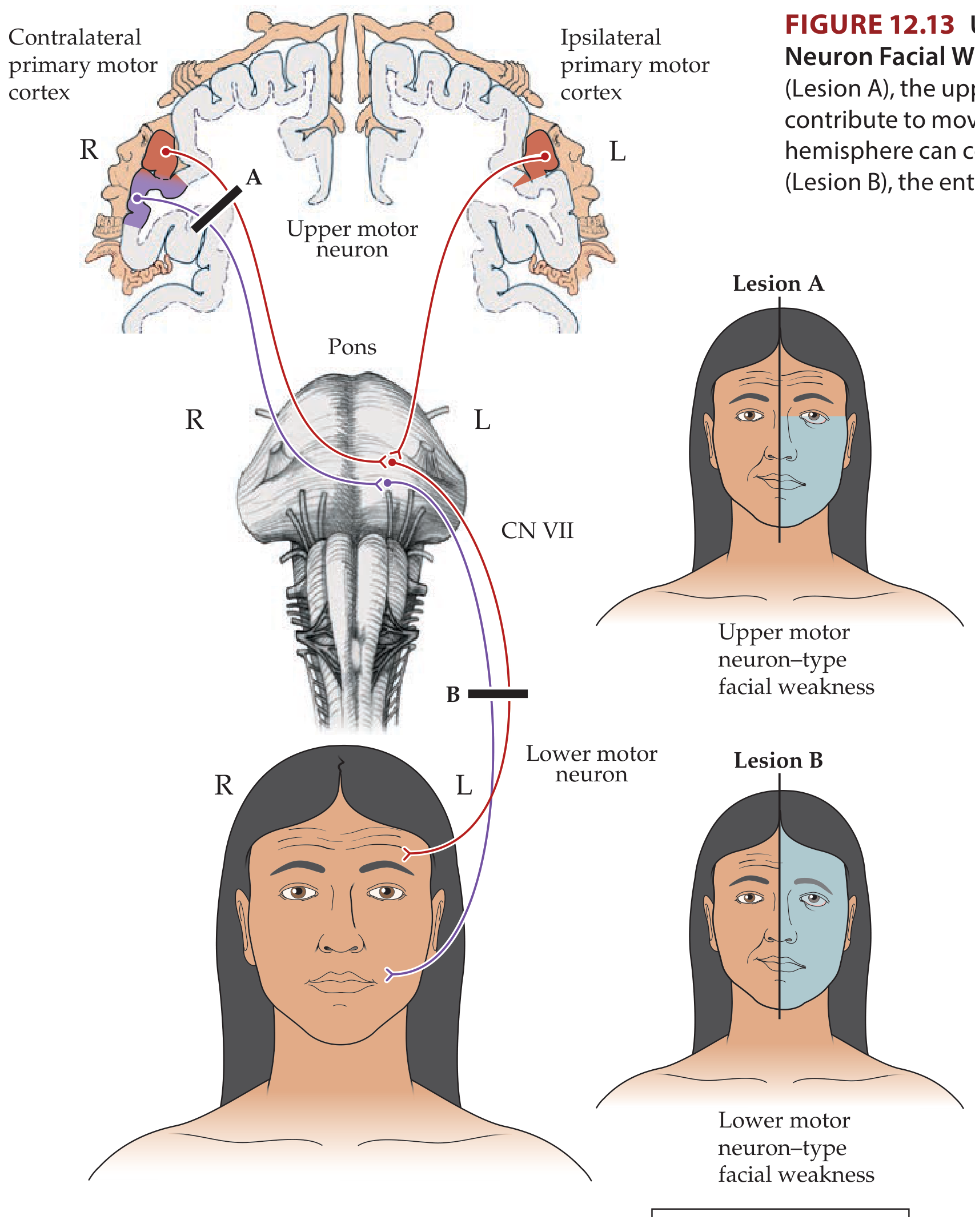
Stroke (Upper Motor Neuron lesion): Damages the cortex or internal capsule on one side. The forehead is spared because the intact opposite hemisphere can compensate. Weakness is confined to the lower face contralateral to the lesion. The patient can still wrinkle their forehead and raise their eyebrow on the weak side.
-
Bell's Palsy (Lower Motor Neuron lesion): Damages CN VII (the facial nerve) itself, after it exits the facial nucleus. ALL branches are affected. Weakness involves the entire ipsilateral face - forehead AND lower face. The patient cannot raise the eyebrow or wrinkle the forehead on the affected side. Eye closure is also impaired (risk of corneal damage).
Quick Exam Comparison Table
| Finding on Exam | Stroke (UMN) | Bell's Palsy (LMN) |
|---|---|---|
| Forehead wrinkle (raise eyebrow) | Preserved on affected side | Absent - cannot wrinkle forehead |
| Eye closure | Mild weakness; can usually close eye | Incomplete eye closure (lagophthalmos) |
| Lower face droop | Present (contralateral to lesion) | Present (ipsilateral to nerve lesion) |
| Tongue deviation | May be present (CN XII involvement) | Not affected |
| Other neurological signs | Often present: arm/leg weakness, aphasia, sensory loss | Absent (pure facial nerve lesion) |
| Hyperacusis | Absent | May be present (stapedius weakness) |
| Taste loss (anterior 2/3 tongue) | Absent | May be present (chorda tympani) |
| Ear pain (retroauricular) | Absent | Common early symptom |
Question 4: Artery Occlusion → Symptoms
4a. Behavior changes, left lower leg weakness → Anterior Cerebral Artery (ACA)
- The ACA supplies the medial surface of the frontal and parietal lobes (the "parasagittal" cortex) - this includes the motor and sensory homunculus for the leg.
- Left lower leg weakness → lesion in the right hemisphere (contralateral control).
- The frontal lobe is also the seat of executive function and personality; ACA strokes (especially bilateral or involving the anterior communicating artery) cause abulia, disinhibition, and personality/behavior changes.
- Arm and face are usually spared or minimally affected because they are represented more laterally (supplied by MCA).
4b. Vision loss → Posterior Cerebral Artery (PCA)
- The PCA supplies the occipital lobe (primary visual cortex, V1) and the inferior temporal lobe.
- Occlusion causes contralateral homonymous hemianopia (loss of the same half of the visual field in both eyes) because the visual cortex processes the contralateral visual field.
- It is the classic "stroke you can miss" - patients may not notice or just report "blurry vision." Always check visual fields formally.
- Source: Harrison's Principles, 22nd Ed.; Adams and Victor's Principles of Neurology: "occlusion of the posterior cerebral artery causes loss of vision in both eyes in the half of the retina on the opposite side."
4c. Headache, nystagmus, slurred speech → Basilar Artery (posterior circulation / vertebrobasilar)
- The basilar artery supplies the brainstem (pons, midbrain) and cerebellum via the AICA and PICA branches.
- Nystagmus - brainstem/cerebellar vestibular pathways.
- Dysarthria (slurred speech) - cranial nerve nuclei in the pons and cerebellar connections.
- Headache - common in posterior circulation strokes; severe headache + posterior signs should raise suspicion for basilar artery stroke or vertebral artery dissection.
- Additional posterior circulation signs: vertigo, diplopia, dysphagia, ataxia, crossed deficits (ipsilateral face + contralateral body), "locked-in syndrome" with basilar occlusion.
4d. Right facial droop, right arm weakness → Left Middle Cerebral Artery (MCA)
- The MCA supplies the lateral cortex including: motor strip (face, arm, hand > leg), sensory strip, and in the dominant hemisphere - language areas (Broca's area anteriorly, Wernicke's area posteriorly).
- Motor control is contralateral - so RIGHT face and arm weakness = LEFT hemisphere lesion.
- The face and arm are affected more than the leg because they are represented laterally on the cortex (MCA territory), while the leg is medial (ACA territory).
- This is exactly the presentation of the patient in this case: left MCA stroke causing right-sided face + arm weakness and aphasia.
Summary Table - Question 4
| Symptoms | Artery | Side of Occlusion |
|---|---|---|
| Behavior changes + left lower leg weakness | Anterior Cerebral Artery (ACA) | Right ACA |
| Vision loss | Posterior Cerebral Artery (PCA) | Contralateral to the visual field deficit |
| Headache + nystagmus + slurred speech | Basilar Artery | N/A (midline/bilateral brainstem) |
| Right facial droop + right arm weakness | Middle Cerebral Artery (MCA) | Left MCA |
labs and tests if symptoms resolved
Labs and Tests When Symptoms Have Resolved: TIA Workup
Why the Workup Still Matters
- Confirm it was vascular (rule out mimics)
- Find the cause (cardiac? large vessel? small vessel? hematologic?)
- Stratify risk and decide on admission vs. outpatient
- Start secondary prevention immediately
The Full Workup
IMAGING (most urgent)
| Study | What it tells you | Priority |
|---|---|---|
| Non-contrast CT head | Rules out hemorrhage, mass lesion, tumor (TIA mimics); usually normal in TIA | First, immediate |
| MRI brain with DWI (diffusion-weighted imaging) | Detects acute ischemic infarction even when CT is normal. About 33% of "TIAs" show infarction on DWI - changing the diagnosis to stroke. Also detects old infarcts and leukoaraiosis. | Urgent, same-day if possible |
| CT Angiography (CTA) or MR Angiography (MRA) of head and neck | Evaluates for carotid stenosis, intracranial stenosis, arterial dissection - the structural cause of ischemia | Urgent |
| Carotid duplex ultrasound | Non-invasive assessment of carotid bifurcation stenosis; relevant because carotid endarterectomy is most beneficial within 2 weeks of TIA | Urgent (if CTA/MRA not available) |
Key point from Tintinalli's: "positive findings on DWI have predictive value for subsequent early stroke risk in TIA patients, as does cervical vascular imaging by MRA."
CARDIAC WORKUP (critical in this patient - she has an irregular heart rhythm)
| Study | What it tells you |
|---|---|
| ECG (12-lead) | Most important first cardiac test. Rules in/out atrial fibrillation, recent MI, other arrhythmias. This patient has an "irregular" heart - AF must be confirmed or excluded. |
| Telemetry / continuous cardiac monitoring | AF is paroxysmal and can be missed on a single ECG. Inpatient monitoring catches intermittent AF. |
| Prolonged cardiac monitoring (Holter 24-48h or 30-day event monitor) | Detects occult AF in up to 25% of cryptogenic strokes when short monitoring is negative. Especially relevant here given the irregular rhythm. |
| Transthoracic echocardiogram (TTE) | Evaluates left ventricular function (thrombus), valvular disease (mitral stenosis), wall motion abnormalities (post-MI source), patent foramen ovale (PFO). |
| Transesophageal echocardiogram (TEE) | Better visualization of the left atrial appendage (where AF thrombi form), aortic arch plaques, and PFO. Indicated when TTE is non-diagnostic and cardioembolic source is suspected. |
LABORATORY TESTS
| Lab | Rationale |
|---|---|
| Complete Blood Count (CBC) | Thrombocytosis (clotting risk), polycythemia (hyperviscosity), anemia (reduced O2 delivery), thrombocytopenia (bleeding risk before antiplatelet therapy) |
| Basic Metabolic Panel (BMP) | Electrolytes and glucose - hypoglycemia is the most important TIA/stroke mimic and must be excluded immediately. Renal function guides contrast use for CTA. |
| Glucose / HbA1c | Diabetes is a major stroke risk factor; HbA1c establishes long-term glycemic control |
| Lipid panel (fasting) | LDL level guides statin therapy (high-intensity statin is part of secondary prevention after TIA/stroke) |
| PT/INR and aPTT | Coagulation status; baseline before initiating anticoagulation or antiplatelet therapy |
| Thyroid-stimulating hormone (TSH) | Hyperthyroidism causes AF; hypothyroidism contributes to atherosclerosis |
| Cardiac biomarkers (troponin) | Rule out concurrent ACS (stroke and MI can co-occur); also, large strokes can cause troponin elevation via catecholamine surge |
- Hypercoagulability panel: Factor V Leiden, Prothrombin G20210A, protein C/S, antithrombin III, antiphospholipid antibodies (lupus anticoagulant, anticardiolipin)
- ESR/CRP (vasculitis)
- Blood cultures (if endocarditis suspected)
- Urine toxicology (cocaine in young patients)
- Sickle cell screen (if appropriate population)
ABCD2 Score - Risk Stratification Tool
| Factor | Points |
|---|---|
| Age ≥ 60 years | 1 |
| Blood pressure ≥ 140/90 mmHg at presentation | 1 |
| Clinical features: unilateral weakness | 2 |
| Clinical features: speech disturbance without weakness | 1 |
| Duration ≥ 60 minutes | 2 |
| Duration 10-59 minutes | 1 |
| Diabetes mellitus | 1 |
| Total | 0-7 |
Important caveat: The American College of Emergency Physicians (2016 guidelines) stated that ABCD2 alone should NOT be used to decide safe discharge - it is inadequate as a standalone risk stratification tool. Most high-risk TIA patients should be admitted.
Disposition Decision for This Patient
- ABCD2 score ≥ 4 (high risk)
- Irregular heart rhythm (likely AF) = high-risk embolic source
- Hypertension (chronic, inadequately controlled: BP 155/89)
- Symptoms lasted ~1 hour with both motor AND speech involvement
- Age 72
retry
Labs and Tests When Symptoms Have Resolved: TIA Workup
What is happening now?
- ~10% stroke risk within 90 days after TIA
- 50% of those strokes occur within 48 hours of TIA onset
- 1 in 3 untreated TIA patients will have a stroke within 5 years
Imaging
| Study | Purpose | Timing |
|---|---|---|
| Non-contrast CT head | First-line: rules out hemorrhage, tumor, abscess (TIA mimics). Usually normal in TIA. | Immediate |
| MRI brain with DWI (diffusion-weighted imaging) | The key test. Detects acute ischemic infarction even when CT is normal - ~33% of "TIAs" show infarction on DWI, reclassifying them as strokes. Also shows old infarcts and white matter disease. Patients with DWI lesions have significantly higher early stroke risk. | Same-day, urgent |
| MRA or CTA of head and neck | Evaluates for carotid stenosis, intracranial stenosis, arterial dissection - identifies the structural source of ischemia and guides whether carotid endarterectomy is needed. Carotid ultrasound is an alternative if CTA/MRA unavailable. | Urgent |
From Bradley and Daroff's Neurology: "Patients with TIA and DW-MRI lesions are at greater early risk for experiencing a subsequent stroke than patients without a lesion."
Cardiac Workup
| Study | Purpose |
|---|---|
| 12-lead ECG | First cardiac test. Confirms or rules out atrial fibrillation, identifies recent MI, LVH from hypertension. |
| Continuous telemetry monitoring | AF is paroxysmal - a single ECG misses it. Inpatient monitoring detects intermittent AF. |
| Transthoracic echocardiogram (TTE) | Evaluates LV function, wall motion abnormalities, valvular disease (mitral stenosis), intracardiac thrombus, and patent foramen ovale (PFO). |
| Transesophageal echocardiogram (TEE) | Better visualization of the left atrial appendage (where AF thrombi form), aortic arch plaques, and PFO with bubble study. Used when TTE is non-diagnostic. |
| Prolonged cardiac monitoring (24-48h Holter or 30-day event monitor) | Detects occult paroxysmal AF in up to 25% of cryptogenic strokes when initial monitoring is negative. |
Blood Tests
| Test | Rationale |
|---|---|
| Glucose (STAT) | Hypoglycemia is the #1 TIA/stroke mimic - must be excluded immediately at bedside |
| CBC with differential and platelets | Thrombocytosis or polycythemia (hyperviscosity/clotting risk); thrombocytopenia (bleeding risk before antiplatelet therapy); anemia (reduced O2 delivery) |
| BMP (electrolytes, BUN, creatinine) | Electrolyte abnormalities can cause neurological symptoms; renal function guides use of contrast for CTA and dosing of medications |
| PT / INR and aPTT | Baseline coagulation before starting antithrombotic therapy |
| Fasting lipid panel | LDL level guides intensity of statin therapy - high-intensity statin is part of secondary prevention |
| HbA1c | Establishes baseline glycemic control; diabetes is a major stroke risk factor |
| TSH | Hyperthyroidism causes AF; hypothyroidism accelerates atherosclerosis |
| Cardiac troponin | Rules out concurrent ACS; large strokes can also elevate troponin via catecholamine surge |
| Test | Rationale |
|---|---|
| Antiphospholipid antibodies (lupus anticoagulant, anticardiolipin Ab) | Hypercoagulable state, especially in younger patients |
| Factor V Leiden, Prothrombin G20210A, Protein C/S, Antithrombin III | Inherited thrombophilia screen |
| ESR / CRP, ANA | Vasculitis |
| Blood cultures / echocardiography | If infective endocarditis suspected |
| Urine toxicology screen | Cocaine/stimulants cause vasospasm and stroke |
| RPR / VDRL | Neurosyphilis |
ABCD2 Score - How Does This Patient Score?
| Factor | This Patient | Points |
|---|---|---|
| Age > 60 | 72 years | 1 |
| BP > 140/90 | 155/89 | 1 |
| Clinical features: unilateral weakness | Yes (R arm/leg 3/5) | 2 |
| Duration ≥ 60 min | ~1 hour | 2 |
| Diabetes | Not mentioned | 0 |
| Total | 6 / 7 |
Important caveat: The American College of Emergency Physicians (2016) states ABCD2 alone should not be used to discharge patients. It is a helpful tool but cannot independently confirm safety for outpatient management.
Disposition: Admit or Discharge?
- ABCD2 score of 6 (high risk)
- Likely atrial fibrillation (irregular heart rhythm = high-risk embolic source)
- Symptoms lasted ~1 hour with both speech AND motor deficits
- Age 72, uncontrolled hypertension
- Needs urgent DWI-MRI, cardiac monitoring, and echocardiogram that cannot wait for outpatient
Immediate Treatment While Being Worked Up
- Dual antiplatelet therapy (aspirin + clopidogrel): start immediately in non-cardioembolic TIA. Pooled data from the POINT and CHANCE trials show dual therapy reduces major ischemic events vs. aspirin alone, with the greatest benefit in the first 21 days.
- If AF confirmed → switch to oral anticoagulation (DOAC preferred over warfarin) rather than antiplatelets - this is the most impactful secondary prevention step for cardioembolic TIA.
- High-intensity statin (e.g. atorvastatin 40-80 mg) regardless of baseline LDL.
- Blood pressure management: do not aggressively lower acutely; optimize long-term control.
- Carotid endarterectomy if ipsilateral carotid stenosis ≥70% found on imaging - most beneficial within 2 weeks of TIA.