
Lateral wall of nose very short answer mbbs
Reading File
The lateral wall of the nose has a complex structure formed by multiple bones and features three scroll-like projections called conchae (turbinates). Let me pull the textbook content for a precise short answer.
Searching sources...
Finding Sources
Finding Sources
Finding Sources
Reading File
Reading File
Loading Image
Loading Image
Reading File
Here is a complete short answer on the Lateral Wall of the Nose, suitable for MBBS exams:
Lateral Wall of the Nose
(Gray's Anatomy for Students, p. 1217-1219)
Introduction
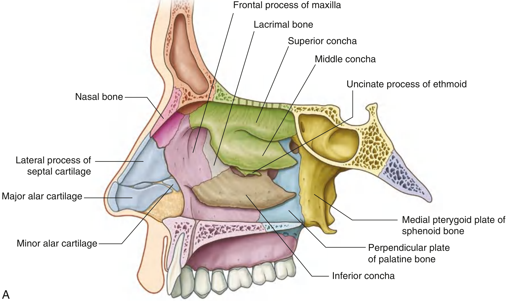
The lateral wall of each nasal cavity is the most complex wall. It is formed by bone, cartilage, and soft tissues, and bears three scroll-like projections called conchae (turbinates) that divide the cavity into air channels (meati).
Bony Skeleton
The bony framework is contributed by six bones/structures:
| Bone | Contribution |
|---|---|
| Ethmoid bone | Ethmoidal labyrinth, superior concha, middle concha, uncinate process |
| Palatine bone | Perpendicular plate |
| Sphenoid bone | Medial pterygoid plate |
| Lacrimal bone | Medial surface |
| Maxilla | Medial surface |
| Inferior concha | A separate bone (one of the facial bones) |
In the external nose, the lateral wall is additionally supported by cartilage: the lateral process of the septal cartilage and the major alar cartilages.
Conchae (Turbinates)
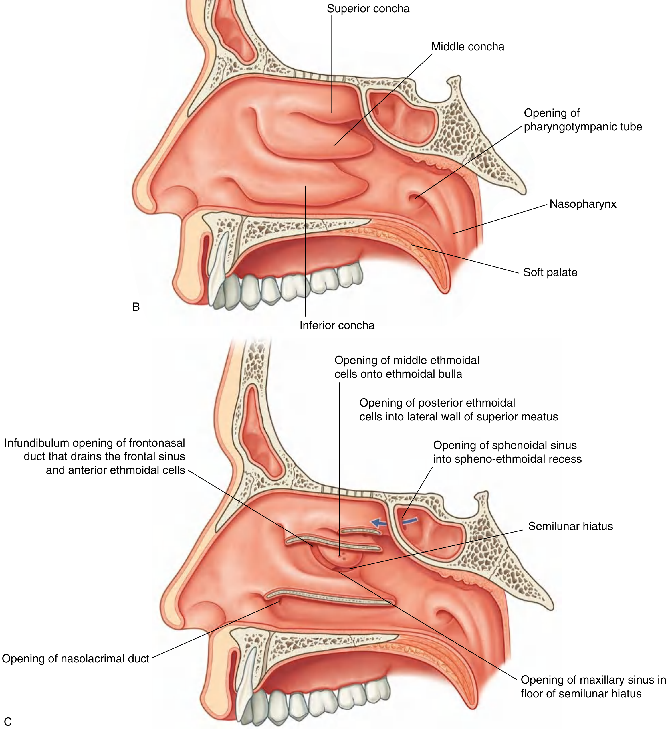
Three shelf-like projections extend medially into the nasal cavity:
- Superior concha - part of ethmoid
- Middle concha - part of ethmoid
- Inferior concha - independent bone
These divide the cavity into four air channels:
- Inferior meatus (below inferior concha)
- Middle meatus (below middle concha)
- Superior meatus (below superior concha)
- Spheno-ethmoidal recess (above superior concha)
The anterior end of each concha curves inferiorly to form a lip that overlies the end of the related meatus.
Important Landmarks in the Middle Meatus
- Ethmoidal bulla: dome-shaped elevation formed by underlying middle ethmoidal air cells, just below the attachment of the middle concha
- Semilunar hiatus: curved gutter inferior to the ethmoidal bulla, between the bulla above and the uncinate process below
- Ethmoidal infundibulum: funnel-shaped channel at the anterior end of the semilunar hiatus, leading upward as the frontonasal duct into the frontal sinus
Openings onto the Lateral Wall
| Structure Opening | Location |
|---|---|
| Nasolacrimal duct | Inferior meatus (under anterior lip of inferior concha) |
| Frontal sinus | Anterior end of semilunar hiatus (via frontonasal duct/infundibulum) |
| Anterior ethmoidal cells | Frontonasal duct or ethmoidal infundibulum |
| Middle ethmoidal cells | Onto or just above ethmoidal bulla |
| Maxillary sinus | Floor of semilunar hiatus (near roof of maxillary sinus) |
| Posterior ethmoidal cells | Lateral wall of superior meatus |
| Sphenoidal sinus | Spheno-ethmoidal recess (on posterior roof - NOT lateral wall) |
Key exam point: The sphenoidal sinus is the only paranasal sinus that does NOT open onto the lateral wall - it opens into the spheno-ethmoidal recess on the sloping posterior roof.
Diagrams
Quick Memory Aid for Bones (ELPSMI)
Ethmoid - Lacrimal - Palatine (perpendicular plate) - Sphenoid (medial pterygoid plate) - Maxilla - Inferior concha
Source: Gray's Anatomy for Students, p. 1217-1219
Epistaxis short answer mbbs book
Epistaxis (nosebleed) is bleeding from the nasal mucosa, most commonly from Little's area (Kiesselbach's plexus) on the anteroinferior nasal septum. Let me get the full textbook content.
Searching sources...
Finding Sources
Reading File
Finding Sources
Reading File
Finding Sources
Reading File
Reading File
Reading File
Finding Sources
Reading File
Loading Image
Reading File
I now have comprehensive textbook content from both Bailey & Love's Short Practice of Surgery and Cummings Otolaryngology. Here is the complete short answer:
Epistaxis (Nosebleed) - Short Answer
(Bailey & Love's Short Practice of Surgery, 28th ed. | Cummings Otolaryngology Head & Neck Surgery)
Definition
Epistaxis is bleeding from the nasal mucosa. It is the most common otorhinolaryngologic emergency, affecting >60% of the population at some point, with 6% requiring medical attention.
Anatomy - Sites of Bleeding
1. Anterior Epistaxis (>90% of cases)
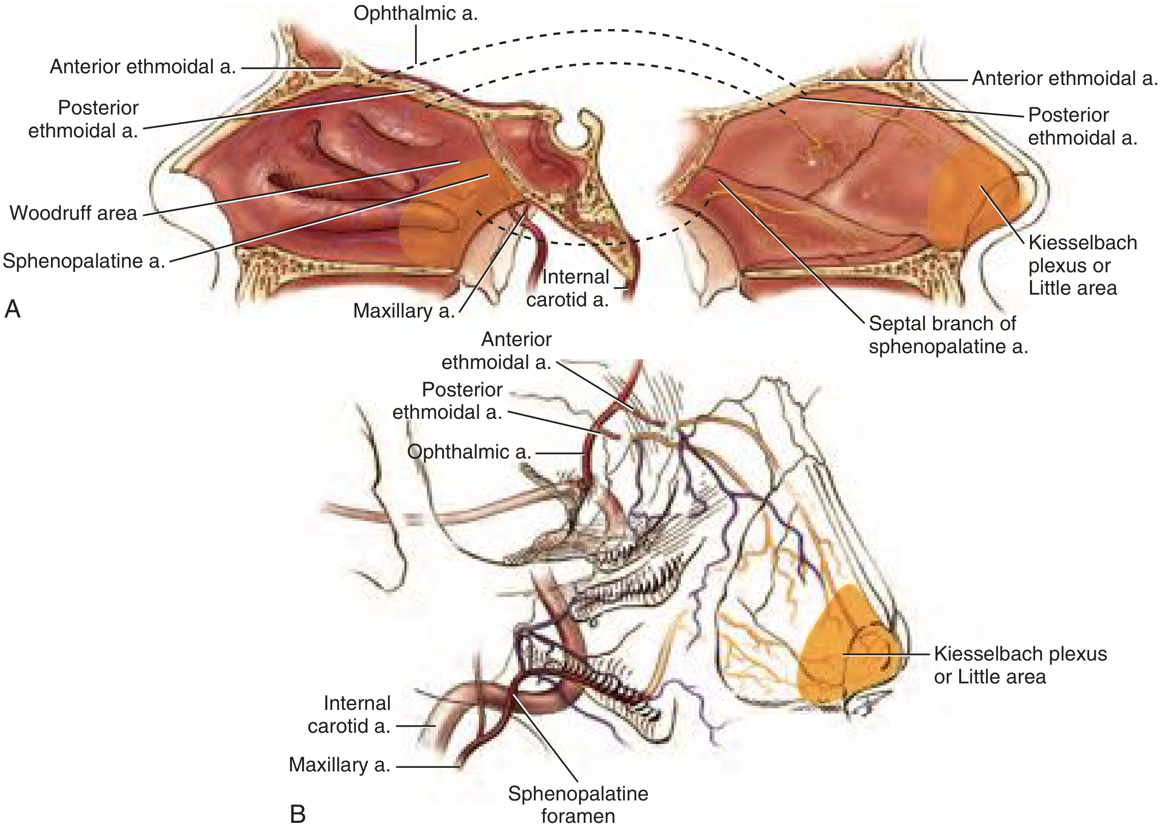
- Bleeds from Kiesselbach's plexus (Little's area) on the anteroinferior nasal septum
- This is a vascular confluence of four arteries:
- Anterior ethmoidal artery (from internal carotid via ophthalmic)
- Sphenopalatine artery (from external carotid via maxillary)
- Greater palatine artery
- Superior labial artery
2. Posterior Epistaxis
- Bleeds from Woodruff's plexus - posterior to the inferior turbinate root
- Fed by branches of the sphenopalatine artery and ascending pharyngeal artery
- Seen in elderly patients; more severe, harder to control
Causes
Local Causes
- Nose picking (digital trauma) - most common
- Nasal trauma / fracture
- Foreign bodies
- Infection / rhinitis
- Granulomatous disorders (Wegener's, sarcoidosis)
- Tumours - Juvenile angiofibroma (adolescent boys; can cause massive bleeding)
- Hereditary haemorrhagic telangiectasia (HHT/Osler's disease)
- Septal perforation / septal deviation
Systemic Causes
- Hypertension (posterior epistaxis in elderly)
- Anticoagulants - warfarin, rivaroxaban, heparin
- Antiplatelet drugs - aspirin, clopidogrel, NSAIDs
- Bleeding disorders - haemophilia, von Willebrand disease, thrombocytopenia
- Leukaemia / lymphoma
- Liver cirrhosis, chronic kidney disease
- Cold, dry climate (mucosal drying)
Key point: Anterior epistaxis = children and young adults (nose picking). Posterior epistaxis = elderly (hypertension, anticoagulants).
Clinical Features
- Bleeding from one or both nostrils
- Blood may flow posteriorly down the throat (posterior epistaxis)
- Signs of hypovolaemia in severe cases: tachycardia, orthostatic hypotension, pallor
- First sign of acute blood loss = tachycardia
Management
Step 1 - First Aid (Conservative)
- Patient sits upright, leaning forward ("chin-to-chest" position) - prevents aspiration/swallowing of blood
- Pinch the soft cartilaginous part of the nose for 10-15 minutes
- Ice pack over forehead/face
- Topical vasoconstrictor: oxymetazoline (α1-agonist) or traditional cocaine
Step 2 - Cautery
- Silver nitrate cautery for anterior bleeding (Kiesselbach's plexus) under local anaesthesia
- Electrocautery under rigid nasendoscopy for identified posterior bleeding points
- Important: Never cauterise both sides of septum simultaneously (risk of perforation)
Step 3 - Nasal Packing
| Type | Indication | Method |
|---|---|---|
| Anterior pack | Moderate posterior bleeding | Vaseline-impregnated ribbon gauze / non-absorbable sponge / inflatable epistaxis balloon catheter |
| Posterior pack | Refractory severe bleeding | Gauze pack in nasopharynx under GA |
- Absorbable haemostatic materials (e.g., Floseal) can also be used
Step 4 - Surgical / Interventional
| Procedure | Indication |
|---|---|
| Endoscopic sphenopalatine artery ligation/clipping | Significant epistaxis not responding to cautery/packing (success rate ~97%) |
| Internal maxillary artery ligation (in pterygopalatine fossa) | Life-threatening, uncontrolled bleeding |
| Anterior & posterior ethmoidal artery ligation | If ethmoidal source identified |
| External carotid artery ligation | Above origin of lingual artery, as last resort |
| Embolisation (interventional radiology) | Alternative to surgical ligation in specific cases |
Treatment of Underlying/Predisposing Causes
| Condition | Management |
|---|---|
| Warfarin | Hold drug; give Vitamin K + FFP |
| Thrombocytopenia | Platelet transfusion (target >50,000/μL) |
| Von Willebrand disease | Desmopressin (types 1 & 2A); Factor VIII |
| Haemophilia | Factor replacement |
| Renal failure/uremia | Desmopressin; haemodialysis |
| Liver cirrhosis | FFP; monitor PT/INR |
Summary Box (Bailey & Love's Key Points)
- Most common causes: nose picking, hypertension, anticoagulant therapy
- Young people bleed from anterior septum - Kiesselbach's plexus
- Elderly bleed from posterior part of nose
- Ideally treated with direct cautery under endoscopic guidance
- Silver nitrate for anterior; packing for moderate; bilateral packing for severe
- Persistent bleeding requires endoscopic sphenopalatine artery ligation
Sources: Bailey & Love's Short Practice of Surgery 28th Ed., pp. 788-789 | Cummings Otolaryngology Head & Neck Surgery, Ch. 47
This is a shared conversation. Sign in to Orris to start your own chat.