
Anatomical basis of lateral squint
Reading File
Lateral squint (divergent strabismus / exotropia) results from an imbalance in the extraocular muscles, particularly involving the lateral and medial recti. Let me search the library for the full anatomical basis.
Searching sources...
Finding Sources
Finding Sources
Finding Sources
Finding Sources
Reading File
Finding Sources
Reading File
Finding Sources
Reading File
Finding Sources
Finding Sources
Finding Sources
Reading File
Reading File
Reading File
Loading Image
I now have sufficient information from the library. Let me compile a comprehensive answer on the anatomical basis of lateral squint.
Anatomical Basis of Lateral Squint (Exotropia / Divergent Squint)
Lateral squint refers to a condition where the eye deviates outward (temporally) - also called exotropia or divergent strabismus. Understanding its anatomical basis requires knowing the muscles, their innervation, and the mechanisms governing ocular alignment.
1. Relevant Anatomy of the Extraocular Muscles
The key muscles for horizontal gaze are the two horizontal recti:
| Muscle | Origin | Insertion | Action | Nerve Supply |
|---|---|---|---|---|
| Medial rectus | Annulus of Zinn (orbital apex) | 5.5 mm behind nasal limbus | Adduction (pulls eye inward) | CN III (oculomotor) |
| Lateral rectus | Annulus of Zinn (orbital apex) | 6.9 mm behind temporal limbus | Abduction (pulls eye outward) | CN VI (abducens) |
When the eye is in the primary position, both horizontal recti act purely on the vertical (Z) axis with no subsidiary actions - they are the only muscles with a single, clean horizontal pull.
- Kanski's Clinical Ophthalmology 10th ed., p. 723
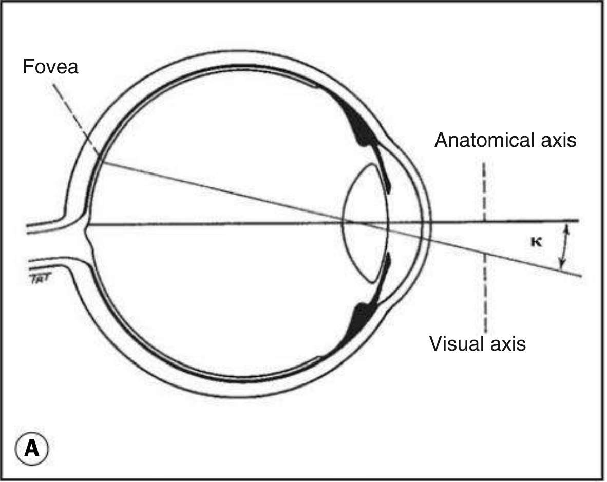
The visual axis forms an angle (kappa) of ~23° with the orbital/anatomical axis. In exotropia, a large positive angle kappa can mimic or contribute to apparent lateral deviation.
2. The Orbital Geometry
The lateral and medial orbital walls form a 45° angle with each other. The orbital axis therefore forms 22.5° (~23°) with both walls. In the primary position of gaze, the visual axis is 23° lateral to the orbital axis.
This geometry means the extraocular muscles pull along the orbital axis, not the visual axis - so their net action on the eye depends on the gaze position at the time of contraction. For the horizontal recti, this effect is minimal because their pull is nearly collinear with horizontal gaze.
3. Normal Ocular Alignment - the Balance That Is Disrupted
Normal alignment (orthophoria) requires a tonic balance between the medial and lateral recti, maintained by:
- Tonic convergence - inherent innervational tone to both medial recti
- Fusional (disparity) convergence - activated by retinal disparity to maintain binocular single vision (BSV)
- Accommodative convergence - linked to accommodation; controlled by the AC/A ratio
- Proximal convergence - triggered by awareness of nearness
Lateral squint arises when the outward pull of the lateral rectus (or loss of inward pull by the medial rectus) is not adequately counterbalanced by these convergence mechanisms.
4. Anatomical Mechanisms of Lateral Squint
A. Weakness / Paralysis of the Medial Rectus (CN III Palsy)
The medial rectus is supplied by the oculomotor nerve (CN III). Third nerve palsy causes loss of adduction, allowing the lateral rectus (CN VI, intact) to pull the eye unopposed laterally - producing a large-angle exotropia. Additional signs of CN III palsy include ptosis, dilated pupil, and loss of elevation/depression.
B. Overaction of the Lateral Rectus
The lateral rectus is supplied by the abducens nerve (CN VI). Overactive or relatively hypertonous lateral rectus tilts the balance toward divergence. In divergence excess exotropia, the angle at distance is greater than at near, reflecting the relative insufficiency of fusional convergence at distance (where accommodative convergence is relaxed).
C. Weakness of Convergence Mechanisms
- Convergence insufficiency - failure of fusional/accommodative convergence at near; eye drifts laterally when convergence demand is high. This is the most common cause of intermittent exotropia in young adults.
- Reduced accommodation - with age or with cycloplegia, accommodative convergence drops, reducing the tonic inward pull mediated by the medial recti.
D. Innervational / Central Causes
The medial recti are yoked with the contralateral lateral recti via Hering's law - equal and simultaneous innervation to paired muscles. The pontine paramedian reticular formation (PPRF) coordinates horizontal saccades through the abducens nucleus. The medial longitudinal fasciculus (MLF) connects CN VI nucleus to the contralateral CN III nucleus for conjugate gaze.
Internuclear ophthalmoplegia (INO) - a lesion of the MLF impairs adduction of the ipsilateral eye while the contralateral eye abducts normally, producing a "wall-eyed" divergent appearance (WEMINO / WEBINO syndromes represent bilateral INO with exotropia). The one-and-a-half syndrome (combined PPRF/MLF lesion) can produce paralytic pontine exotropia - the only horizontal movement remaining is abduction of the contralateral eye.
- Localization in Clinical Neurology 8e, p. 417
E. Sensory / Structural Causes
Any condition that reduces vision in one eye (cataract, corneal opacity, retinal disease) deprives that eye of the sensory stimulus for fusional convergence. Without fusion drive, the resting position is determined by tonic muscle balance, which in most adults is slightly divergent - producing sensory exotropia.
F. Pulley System Disruption
The rectus muscle pulleys act as the functional origins of the muscles and coordinate horizontal-vertical interactions. Pulley displacement can cause abnormal muscle vectors, resulting in A-pattern or V-pattern deviations with associated horizontal misalignment.
5. Types of Exotropia and Their Anatomical Correlate
| Type | Key Mechanism |
|---|---|
| Constant (infantile) exotropia | Large innervational imbalance; frequently associated with neurological anomalies |
| Intermittent exotropia | Fusional convergence normally adequate; breaks down under fatigue, bright light, or inattention |
| Distance excess exotropia | Accommodative convergence absent at distance; AC/A ratio effect |
| Convergence insufficiency | Fusional convergence fails at near despite normal accommodation |
| Sensory exotropia | Unilateral visual loss eliminates fusion drive |
| Consecutive exotropia | Overcorrected esotropia (surgical or spontaneous) |
| Third nerve palsy exotropia | Medial rectus denervation; large, constant, with associated ptosis and pupil signs |
| Paralytic pontine exotropia | MLF + PPRF lesion; contralateral abduction only remains |
6. Practical Summary
The fundamental anatomical basis of lateral squint is an imbalance between the abducting force of the lateral rectus (CN VI) and the adducting force of the medial rectus (CN III), compounded by failure of the central convergence mechanisms that normally maintain ocular alignment. This imbalance can arise at the level of:
- The muscle itself (weakness, restriction, overaction)
- The cranial nerve supply (CN III > CN VI in terms of adduction)
- The brainstem pathways (MLF, PPRF, abducens nucleus)
- The sensory/fusional drive (accommodative, fusional, tonic convergence)
Surgical correction targets this balance directly: recession of the lateral rectus (weakens abduction) and/or resection of the medial rectus (strengthens adduction) are the standard procedures.
- Kanski's Clinical Ophthalmology 10th ed., p. 744 (exotropia treatment)
This is a shared conversation. Sign in to Orris to start your own chat.