
Persistant Reddish to pinkish discolouration of left foot with no itching no pain Pt INR and aptt CBC Rft with in normal limit
Reading File
Now let me search the medical library for this clinical presentation - persistent reddish/pinkish discoloration of foot with no itching, no pain, normal coagulation and labs.
Finding Sources
Finding Sources
Finding Sources
Searching the Web
persistent reddish pink discoloration foot no pain no itching normal INR CBC RFT differential diagnosis
Good. Now let me pull the key textbook sections on erythromelalgia, acrocyanosis, and related vascular conditions of the foot in parallel.
Reading File
Reading File
Reading File
Finding Sources
Now let me fetch the erythromelalgia image from the textbook and also search for livedo reticularis and other causes of painless reddish discoloration of the foot.
Loading Image
Finding Sources
Finding Sources
Now let me pull more details on the management/diagnosis of erythromelalgia and also look for livedo reticularis and pernio sections.
Reading File
Finding Sources
Finding Sources
I now have sufficient information to provide a comprehensive clinical analysis. Let me also read the pernio/erythema ab igne section briefly.
Reading File
I now have a comprehensive picture. Here is the full clinical analysis:
Persistent Reddish-Pink Discoloration of the Left Foot - Clinical Analysis
Key clinical features in this case:
- Unilateral (left foot only)
- Persistent reddish to pinkish discoloration
- No itching, no pain
- Normal INR, aPTT, CBC, RFT
The absence of pain, itching, and the normal labs are the most important discriminating features.
Differential Diagnosis
1. Erythromelalgia (Most Visually Matching - but PAIN is typically present)
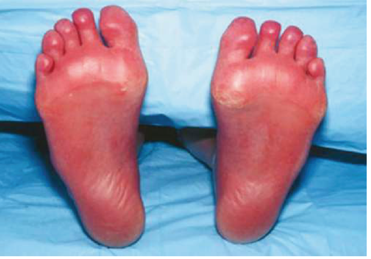
Erythromelalgia classically presents with the triad: erythema + increased warmth + burning pain of the extremities, with the lower limbs (soles/entire foot) most commonly affected. It is episodic and aggravated by heat, dependency, alcohol, or exercise.
Against this diagnosis: The classic feature is intense burning pain. Your patient has no pain, which makes classical erythromelalgia unlikely. However, very mild or atypical forms can present with just erythema and warmth.
- Goldman-Cecil Medicine, p. 779
2. Erythema ab Igne - Top Consideration (painless, persistent)
Strongly consider this if there is a history of heat exposure to that foot/leg. This condition results from repeated/chronic exposure to a heat source (heating pad, hot water bottle, electric blanket, sitting near a heater, laptop on the lap/leg).
Features:
- Persistent reticular or diffuse erythematous/brownish-pink skin discoloration
- Generally asymptomatic - no itching, no pain (patient may report slight burning occasionally)
- Involves the area of heat exposure
- Diagnosis is purely clinical - no labs needed
- Lab tests (INR, CBC, RFT) are normal and expected to be normal
Key question to ask: Is the patient regularly applying a hot water bottle, heating pad, or sitting close to a heat source over that foot?
- Goldman-Cecil Medicine, p. 779
3. Livedo Reticularis (Benign/Physiologic)
Presents as a mottled, reticulate (net-like) pinkish-reddish discoloration of the skin due to dermal arteriolar spasm. Can be physiologic (cold exposure) or secondary to underlying conditions.
Features of benign livedo reticularis:
- Painless, non-itchy
- Improves on warming
- Symmetrical, net-like pattern
- Normal labs (coagulation, CBC, RFT all normal in benign form)
Against this: Livedo reticularis is typically bilateral and has a classic net-like/mottled pattern. Your patient has unilateral involvement, which raises suspicion for a secondary or structural cause.
Secondary causes to consider if the livedo pattern is present: antiphospholipid antibody syndrome, vasculitis, polyarteritis nodosa - but your normal coagulation studies (INR, aPTT) and CBC make most serious causes less likely. Consider ANA, antiphospholipid antibodies if livedo pattern persists.
4. Acrocyanosis (typically bluish, not red/pink)
Acrocyanosis presents with persistent, painless, symmetrical bluish discoloration of hands and feet. It is a benign vasospastic disorder in young women. The color is typically blue to cyanotic, not red or pink. This is less likely given the red-pink color described.
5. Chronic Venous Insufficiency / Stasis Dermatitis
- Can cause reddish-pink discoloration around ankle/foot
- Typically associated with dependent oedema, varicosities, and itching (hemosiderin staining gives a brownish-red color)
- The absence of itching makes active stasis dermatitis less likely, but early CVI can be asymptomatic
6. Early Cellulitis (Infectious)
- Usually warm, tender, with systemic signs (fever, raised WBC)
- Your patient's CBC is normal - makes cellulitis unlikely
- However, very early or resolving cellulitis can be painless
7. Complex Regional Pain Syndrome (CRPS) Type 1
- Can present with redness and skin color changes of a limb
- Typically associated with some degree of pain and allodynia
- The complete absence of pain makes this less likely
Most Likely Diagnosis
Given no pain, no itching, unilateral, normal all labs:
| Priority | Diagnosis | Key Discriminator |
|---|---|---|
| 1st | Erythema ab igne | History of local heat exposure to that foot |
| 2nd | Benign livedo reticularis | Net-like mottled pattern, bilateral usually |
| 3rd | Early CVI / Stasis | Look for subtle swelling, varicosities |
| 4th | Atypical erythromelalgia | Check skin temperature - is foot warm? |
Recommended Next Steps
History to elicit:
- Any use of heating pad, hot water bottle, or heat source on that foot/leg?
- Does the discoloration worsen with heat or improve with cold?
- Any pattern change (worse when feet dependent vs elevated)?
- Any systemic symptoms (joint pains, Raynaud's episodes)?
Examination:
- Skin temperature of the affected vs normal foot (warm = erythromelalgia/CVI; normal/cool = livedo)
- Pattern of discoloration: net-like/reticular = livedo; diffuse/homogenous = erythema ab igne or CVI
- Look for varicosities, pitting oedema
If diagnosis remains unclear, add:
- ANA, antiphospholipid antibody panel (especially if livedo pattern)
- Skin biopsy if erythema ab igne suspected with atypical features
- Doppler venous ultrasound if CVI suspected
- Platelet count (already in CBC - rule out thrombocytosis for secondary erythromelalgia)
Note: The normal INR, aPTT effectively rules out coagulopathy-related vascular occlusion. The normal CBC essentially rules out myeloproliferative disorders as a cause of secondary erythromelalgia. The normal RFT excludes renal-related skin changes (calciphylaxis, etc.).
Sources: Goldman-Cecil Medicine International Edition (pp. 778-780); Dermatology 2-Volume Set 5e (pp. 2224, 1868); Fitzpatrick's Dermatology (pp. 487-514)
This is a shared conversation. Sign in to Orris to start your own chat.