
Ecmo shirt notes
Reading File
ECMO (Extracorporeal Membrane Oxygenation) is a form of prolonged cardiopulmonary bypass used for patients with severe, refractory cardiac and/or respiratory failure. "Short notes" on ECMO is a common medical exam/clinical topic.
Searching sources...
Finding Sources
Searching the Web
ECMO extracorporeal membrane oxygenation short notes clinical indications types management
Reading File
Reading File
Reading File
Reading File
I now have comprehensive textbook content. Let me fetch the ECMO circuit diagram image for the answer.
Loading Image
Here are concise short notes on ECMO:
ECMO (Extracorporeal Membrane Oxygenation) - Short Notes
Definition
ECMO (also called Extracorporeal Life Support - ECLS) is a form of prolonged cardiopulmonary bypass that provides temporary support for patients with severe, refractory cardiac and/or respiratory failure. It allows time for intrinsic recovery of the heart and lungs, or serves as a bridge to transplantation or a durable mechanical device.
Basic Circuit
The ECMO circuit consists of 4 key components:
- Inflow cannula - drains venous blood from the patient into the circuit
- Centrifugal pump - maintains blood flow through the circuit
- Membrane oxygenator - adds O₂ and removes CO₂
- Outflow cannula - returns oxygenated blood to the patient
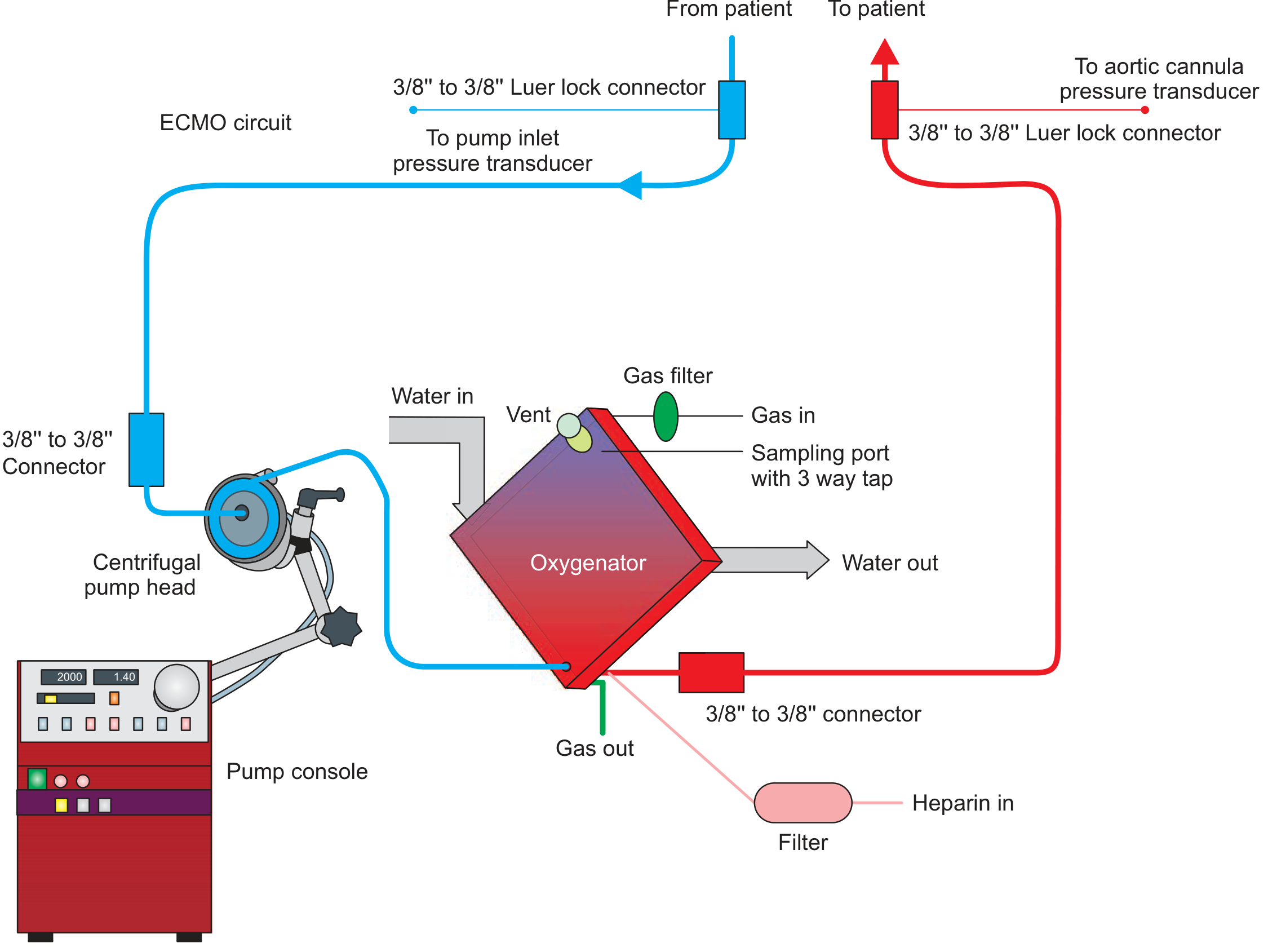
Blood is drained (blue) from patient → pump → oxygenator → returned (red) to patient. Heparin is infused continuously to prevent clotting.
Types of ECMO
| Feature | VV ECMO (Veno-Venous) | VA ECMO (Veno-Arterial) |
|---|---|---|
| Support provided | Lungs only | Heart + Lungs |
| Indications | Severe respiratory failure (ARDS, pneumonia) | Cardiogenic shock, cardiac arrest, biventricular failure |
| Cannulation | Femoral vein (inflow) + Right IJV (outflow) | Femoral/jugular vein (inflow) + Femoral/subclavian artery (outflow) |
| Cardiac output | Dependent on native cardiac output | Augments cardiac output |
| Recirculation | Can occur | Less of an issue |
Hybrid Configurations
- V-AV (Veno-Arteriovenous): Combined respiratory and partial cardiac support; used when standard VV fails to maintain oxygenation in a patient with mild cardiac dysfunction
Indications
VV ECMO (Respiratory)
- Severe ARDS (PaO₂/FiO₂ < 80 despite optimal ventilation)
- Murray Lung Injury Score > 3.0 or pH < 7.20
- Refractory hypoxemia or hypercapnia
- Bridge to lung transplantation
- COVID-19-related severe ARDS
VA ECMO (Cardiac)
- Cardiogenic shock refractory to medical therapy
- Cardiac arrest (ECPR - extracorporeal CPR)
- Acute myocarditis or myocardial stunning
- Refractory arrhythmias
- Bridge to VAD (Ventricular Assist Device) or cardiac transplant
- Post-cardiotomy shock (failure to wean from bypass)
Cannulation
Central Cannulation
- Direct cannulation of large central vessels (usually via sternotomy)
- Reserved for patients already on cardiopulmonary bypass for cardiac surgery
- Allows higher flows but carries higher bleeding and infection rates
Peripheral Cannulation
- Preferred and most common approach
- Can be performed by non-surgeons (even at bedside)
- Uses peripheral vessels (femoral, jugular)
- Preferred VV cannulation sites: Right common femoral vein (inflow) + Right IJV (outflow)
ECMO vs. Cardiopulmonary Bypass
| ECMO | CPB | |
|---|---|---|
| Duration | Days to weeks (3-10 days typical) | Hours |
| Cannulation | Often cervical (local anaesthesia) | Transthoracic (general anaesthesia) |
| Purpose | Allow intrinsic recovery | Support during cardiac surgery |
| Setting | ICU | Operating theatre |
Management on ECMO
Ventilator settings (Lung-Rest Strategy)
- Reduce ventilator support to allow injured lungs to rest
- Low tidal volumes, low PEEP, low FiO₂
Anticoagulation
- Continuous heparin infusion (circuit thrombosis prevention)
- Target ACT 180-220 seconds or anti-Xa levels per institutional protocol
- Risk of bleeding vs. thrombosis must be balanced
Cardiovascular
- Maintain systemic perfusion with inotropes/vasopressors as needed
- Echocardiography to monitor LV function and volume status
- VA ECMO can cause LV afterload increase, pulmonary oedema - may need LV unloading (IABP, Impella)
Neurological
- Avoid paralytic agents; perform regular neurological exams
- Sedation vacations to assess CNS
- Serial head ultrasounds in neonates (screen for IVH)
- Aggressive treatment of seizures (phenobarbital)
Renal
- ~60% of ECMO patients require CRRT (Continuous Renal Replacement Therapy)
- Need for CRRT is associated with significantly increased mortality (17% vs 53% 3-month survival)
Contraindications
Absolute:
- Irreversible organ failure (when ECMO cannot serve as a bridge)
- Aortic dissection (VA ECMO)
- Severe aortic regurgitation (VA ECMO)
Relative:
- Prolonged pre-ECMO CPR without adequate perfusion
- Uncontrolled bleeding or coagulopathy
- Advanced age with poor baseline functional status
Complications
| System | Complication |
|---|---|
| Hemorrhagic | Intracranial hemorrhage (most serious), cannula site bleeding, GI bleed |
| Thrombotic | Circuit thrombosis, limb ischemia, stroke |
| Cardiac | LV distension, pulmonary oedema (VA ECMO) |
| Renal | AKI requiring CRRT |
| Infectious | Line infection, sepsis |
| Circuit | Air embolism, oxygenator failure, pump failure |
Key Trials
- CESAR Trial (Peek et al., 2009): 180 adults with severe ARF. Referral to ECMO centre improved 6-month survival without severe disability (63% vs 47%) compared to conventional management.
- EOLIA Trial: RCT of VV ECMO vs conventional management in severe ARDS. Did not meet primary endpoint (60-day mortality), but high crossover rate (28%) confounded results. Bayesian reanalysis favoured ECMO.
Weaning from ECMO
- VV ECMO: Gradually reduce sweep gas flow; trial of "ECMO holiday" (clamping sweep gas while maintaining blood flow)
- VA ECMO: Gradual reduction in pump flow while monitoring haemodynamics and echo
- Decannulate when patient demonstrates ability to sustain adequate gas exchange and/or cardiac output independently
Sources: Fishman's Pulmonary Diseases and Disorders, 2-Vol Set (Ch. 149); Fischer's Mastery of Surgery, 8th ed. (p. 346)
This is a shared conversation. Sign in to Orris to start your own chat.