
Difference between the spinal ganglia amd sympathetic ganglia
Reading File
Spinal (dorsal root) ganglia contain pseudounipolar sensory neurons, while sympathetic ganglia contain multipolar autonomic motor neurons. The key differences lie in neuron type, location, function, fiber composition, and histology.
Searching sources...
Finding Sources
Finding Sources
Reading File
Finding Sources
Reading File
Reading File
Reading File
Finding Sources
Now let me fetch the histology plate image that shows both ganglia side by side, and also read the dorsal root ganglion section more closely.
Reading File
Loading Image
I now have excellent textbook material. Let me compile the comprehensive answer.
Spinal (Dorsal Root) Ganglia vs. Sympathetic Ganglia
Here is a detailed comparison across all major parameters:
1. Definition and Classification
| Feature | Spinal (Dorsal Root) Ganglia | Sympathetic Ganglia |
|---|---|---|
| Type | Sensory ganglia | Autonomic (motor) ganglia |
| Division | Somatic sensory / visceral sensory afferent | Sympathetic division of the ANS |
| Role | Relay sensory signals into the CNS | Relay motor signals from CNS to visceral effectors |
2. Location
Spinal ganglia lie on the dorsal root of each spinal nerve, just outside the spinal cord. They are closely adjacent to the CNS but remain peripheral.
Sympathetic ganglia are found in two main groups:
- Paravertebral (sympathetic chain) ganglia - a paired chain running on either side of the vertebral column (from the cervical to the sacral region)
- Prevertebral ganglia - located on the anterior surface of the aorta, adjacent to major unpaired branches (celiac, superior mesenteric, inferior mesenteric, aorticorenal ganglia)
3. Neuron Type
This is the single most important histological distinction:
| Feature | Spinal Ganglia | Sympathetic Ganglia |
|---|---|---|
| Neuron shape | Pseudounipolar | Multipolar |
| Processes | Single process that divides into a peripheral and a central segment | Multiple dendrites + one axon |
| Function | Afferent (sensory) | Efferent (motor/postganglionic) |
| Synapse in ganglion? | No synapses in the ganglion | Yes - synapse between pre- and postganglionic neurons occurs here |
As stated in the textbook:
"Dorsal root ganglia differ from autonomic ganglia in a number of ways. Whereas the latter contain multipolar neurons and have synaptic connections, dorsal root ganglia contain pseudounipolar sensory neurons and have no synaptic connections in the ganglion."
- Histology: A Text and Atlas
4. Neuron Function
-
Spinal ganglia neurons: Each pseudounipolar neuron has one process that bifurcates - the peripheral segment collects signals from a receptor (skin, muscle, joint, viscera) and the central segment carries them into the gray matter of the spinal cord. These are purely afferent (sensory) neurons.
-
Sympathetic ganglia neurons: These are postganglionic neurons. They receive synaptic input from preganglionic neurons (whose cell bodies are in the intermediolateral horn of the spinal cord, T1-L3) and send long axons to target organs (smooth muscle, cardiac muscle, glands).
5. Histological Appearance
Sympathetic ganglion (silver + H&E stain):
- Cell bodies (CB) are large, scattered, with multiple processes (P) visible - confirming multipolar morphology
- Large pale nucleus with a prominent nucleolus - reflects high metabolic activity
- Lipofuscin (L) pigment accumulations are common
- Nerve fiber bundles (NF) run in random patterns between cell bodies
- Connective tissue less conspicuous on silver stain
Dorsal root ganglion (H&E stain):
- Cell bodies are large and rounded, with a large pale nucleus and prominent nucleolus
- Satellite cells (Sat C) form a complete cellular capsule around each cell body - they are continuous with the Schwann cells of the axon
- Nerve fibers run in organized parallel bundles alongside cell bodies
- No synaptic contacts visible between neurons
- Clusters of satellite cells appear as epithelioid clusters in tangential sections
Below is the histology plate showing sympathetic ganglion (top panels) and dorsal root ganglion (bottom panels) side by side:
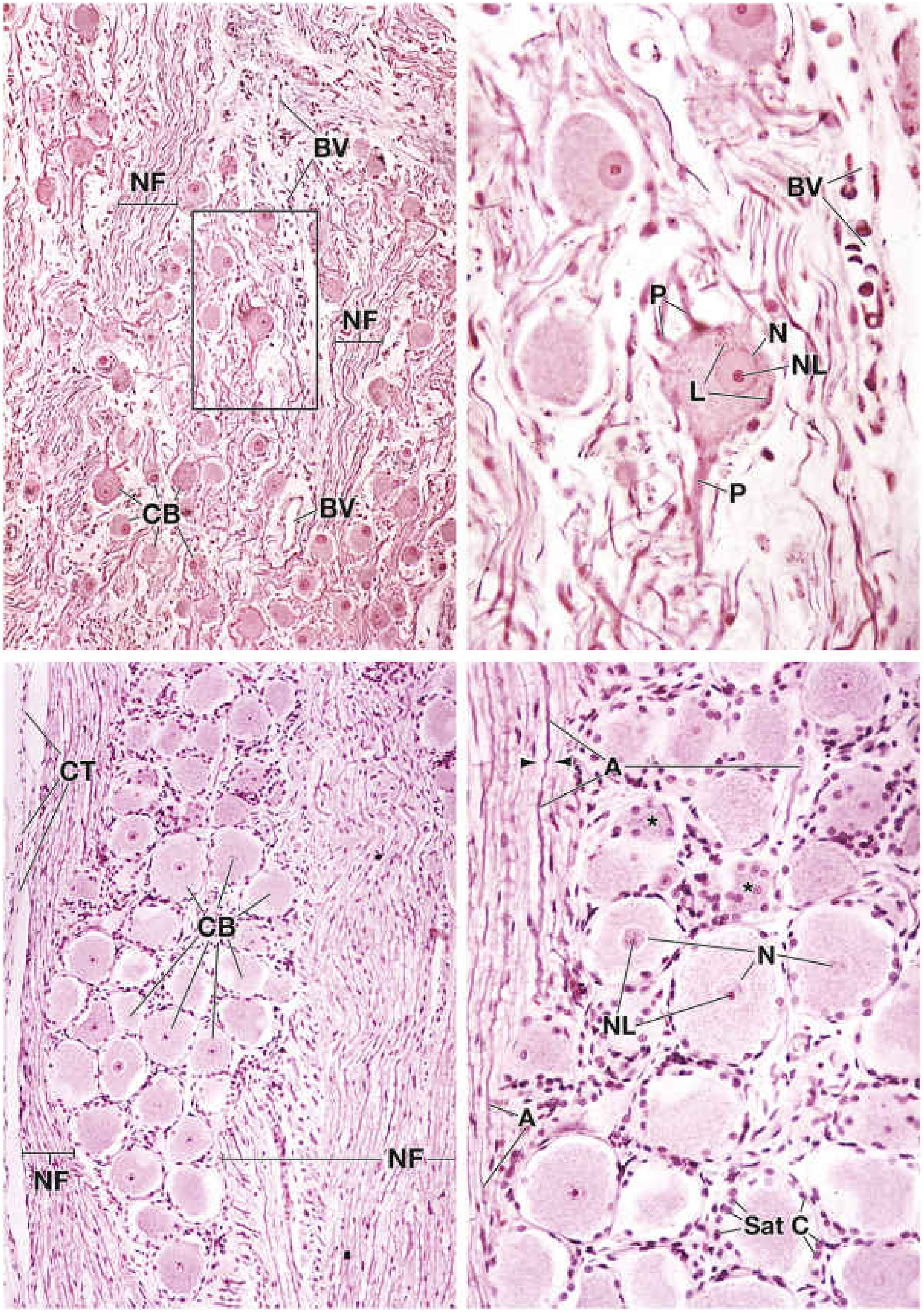
Top-left: Sympathetic ganglion ×160 - multipolar neurons with randomly arranged nerve fibers. Top-right: Higher magnification showing processes (P), nucleus (N), nucleolus (NL), and lipofuscin (L). Bottom-left: Dorsal root ganglion ×160 - rounded pseudounipolar neurons in organized rows. Bottom-right: Higher magnification showing satellite cells (Sat C) encapsulating each neuron, and axons (A) with neurilemma.
6. Supporting (Satellite) Cells
| Feature | Spinal Ganglia | Sympathetic Ganglia |
|---|---|---|
| Satellite cells | Completely surround each neuron cell body | Present but less prominent |
| Schwann cell continuity | Satellite cells are continuous with Schwann cells of the axon | Similar, but less organized capsule |
7. Fiber Composition
| Feature | Spinal Ganglia | Sympathetic Ganglia |
|---|---|---|
| Fibers passing through | Myelinated A-fibers and unmyelinated C-fibers (sensory) | Preganglionic B-fibers (myelinated) + postganglionic C-fibers (unmyelinated) |
| Neurotransmitter | Not applicable (sensory) | Preganglionic: ACh (cholinergic); Postganglionic: mostly norepinephrine (adrenergic) |
8. Summary Table
| Parameter | Spinal (Dorsal Root) Ganglia | Sympathetic Ganglia |
|---|---|---|
| Type | Sensory ganglion | Autonomic motor ganglion |
| Location | Dorsal root of spinal nerve | Paravertebral chain / prevertebral |
| Neuron type | Pseudounipolar | Multipolar |
| Synapses in ganglion | Absent | Present (pre → postganglionic) |
| Direction | Afferent (sensory, to CNS) | Efferent (motor, to viscera) |
| Satellite cell capsule | Complete, prominent | Present, less complete |
| Nerve fiber arrangement | Organized, parallel | Random |
| Neurotransmitter | None (sensory) | ACh (pre-), NE (post-) |
| Effector targets | None (carries sensory signals only) | Smooth muscle, cardiac muscle, glands |
Sources:
- Histology: A Text and Atlas with Correlated Cell and Molecular Biology (Plate 12.1, pp. 1025-1028) - direct comparison of both ganglion types with labeled histological images
In a tabular form
Here is the complete comparison in tabular form:
| Parameter | Spinal (Dorsal Root) Ganglia | Sympathetic Ganglia |
|---|---|---|
| Type | Sensory ganglion | Autonomic (motor) ganglion |
| Classification | Somatic/visceral afferent | Efferent division of ANS |
| Location | Dorsal root of each spinal nerve, just outside spinal cord | Paravertebral (sympathetic chain) OR prevertebral (celiac, superior mesenteric, inferior mesenteric ganglia) |
| Neuron type | Pseudounipolar | Multipolar |
| Number of processes | Single process that bifurcates into peripheral + central segments | Multiple dendrites + one axon |
| Direction of impulse | Afferent - periphery → CNS | Efferent - CNS → visceral effectors |
| Synapses within ganglion | Absent - no synaptic connections | Present - preganglionic axon synapses on postganglionic neuron |
| Preganglionic neuron origin | Not applicable | Intermediolateral horn, T1-L3 spinal cord |
| Neurotransmitter | Not applicable (sensory) | Preganglionic: ACh; Postganglionic: Norepinephrine |
| Receptor on next neuron | Not applicable | Nicotinic ACh receptors (at synapse) |
| Effector targets | None (relays sensory input only) | Smooth muscle, cardiac muscle, glands |
| Satellite cells | Complete, tight capsule around each neuron; continuous with Schwann cells of axon | Present but less prominent/complete |
| Nerve fiber arrangement | Organized, parallel bundles alongside cell bodies | Random, irregular patterns between cell bodies |
| Fiber types | Myelinated A-fibers + unmyelinated C-fibers | Preganglionic: myelinated B-fibers; Postganglionic: unmyelinated C-fibers |
| Cell body size | Large, rounded | Large (similar size) |
| Nucleus | Large, pale-staining, prominent nucleolus | Large, pale-staining, prominent nucleolus |
| Lipofuscin pigment | Present | More prominent/abundant |
| Connective tissue capsule | Present (perineurial sheath) | Present but less conspicuous on silver stain |
| Histological stain appearance | H&E: rounded cells in organized rows; satellite cell halos visible | Silver + H&E: multipolar cell bodies with visible processes; random NF bundles |
| Functional role | First-order sensory neuron - carries pain, temperature, touch, proprioception | Postganglionic neuron - regulates fight-or-flight responses |
| Clinical example | Herpes zoster (varicella virus latency in dorsal root ganglion) | Horner syndrome (disruption of superior cervical ganglion) |
Source: Histology: A Text and Atlas with Correlated Cell and Molecular Biology, Plate 12.1
This is a shared conversation. Sign in to Orris to start your own chat.