
5mark on connective tissue with suitable example
Reading File
Finding Sources
Reading File
Reading File
I now have enough textbook content. Let me also fetch the histology image to include in the answer.
Loading Image
Finding Sources
Reading File
Reading File
I now have rich, comprehensive content from the histology textbooks. Here is a complete 5-mark answer on connective tissue:
Connective Tissue
Definition
Connective tissue is a diverse group of tissues that connects, supports, and separates other tissues and organs throughout the body. Unlike epithelial tissue, its cells are conspicuously separated from one another - the intervening spaces are occupied by an extracellular matrix (ECM) produced by those cells. Classification of connective tissue takes into account both the cell types and the composition and organization of the ECM.
- Histology: A Text and Atlas, p. 301
Origin
All connective tissue derives from mesoderm (the middle embryonic germ layer) via mesenchyme - a loose network of spindle-shaped cells suspended in a viscous, collagen-rich ground substance. Mucoid connective tissue (e.g., Wharton's jelly of the umbilical cord) is a second embryonic form with widely spaced cells in a hyaluronan-rich matrix.
- Histology: A Text and Atlas, p. 533
Components of Connective Tissue
All connective tissues share three structural components:
| Component | Description |
|---|---|
| Cells | Fibroblasts (produce ECM), mast cells, macrophages, plasma cells, adipocytes |
| Protein Fibers | Collagen, reticular, and elastic fibers |
| Ground Substance | Amorphous gel of proteoglycans and glycoproteins; binds water |
The Three Fiber Types:
- Collagen fibers - Most abundant; flexible with high tensile strength; show characteristic 68 nm banding under EM; secreted as procollagen by fibroblasts.
- Reticular fibers - Composed of type III collagen; form a delicate supporting mesh in lymphoid organs and around small vessels.
- Elastic fibers - Produced by fibroblasts and smooth muscle; allow tissue to recoil after stretching (e.g., in skin, lungs, large artery walls).
- Histology: A Text and Atlas, p. 534
Classification with Examples
1. Embryonic Connective Tissue
| Type | Feature | Example |
|---|---|---|
| Mesenchyme | Undifferentiated stem cells in sparse matrix | Mesodermal layer of early embryo |
| Mucoid | Fibroblasts in viscous hyaluronan matrix (Wharton's jelly) | Fetal umbilical cord |
2. Connective Tissue Proper
| Type | Feature | Example |
|---|---|---|
| Loose (areolar) | Many cells, abundant ground substance, loosely arranged collagen | Lamina propria beneath gut epithelium; dermis papillary layer |
| Dense irregular | Many collagen bundles in random arrangement, few fibroblasts | Dermis of skin; organ capsules |
| Dense regular | Parallel collagen bundles with aligned fibroblasts (tendinocytes) | Tendons, ligaments, corneal stroma |
3. Specialized Connective Tissue
| Type | Example |
|---|---|
| Reticular | Bone marrow, liver, lymph nodes (supports hemopoietic cells) |
| Adipose | Subcutaneous fat; orbital fat pad |
| Cartilage | Hyaline cartilage of trachea; elastic cartilage of ear |
| Bone | Compact and spongy bone of the skeleton |
| Blood | Circulating cells in plasma (fluid ECM) |
- Junqueira's Basic Histology, p. 297 (Table 5-6)
Histological Appearance
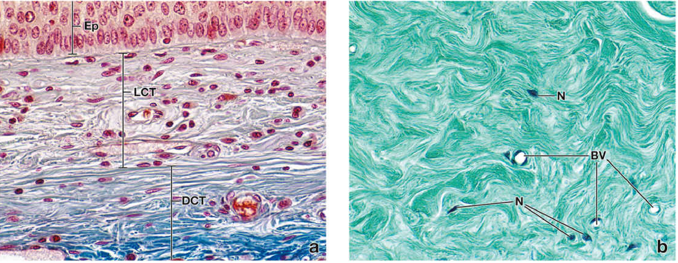
Figure: Mallory-Azan stained section of the epiglottis showing the contrast between Loose Connective Tissue (LCT) - with many cells of varying nuclear shapes and loosely arranged fibers - and Dense Connective Tissue (DCT) below it with thickly packed collagen bundles and sparse fibroblast nuclei (N). x540. - Histology: A Text and Atlas, p. 301
Key Points Summary
- Connective tissue is characterized by its ECM rather than its cells.
- The primary cell is the fibroblast, which secretes collagen and maintains the matrix.
- Loose connective tissue acts as a packing and support tissue beneath epithelia (example: lamina propria of the intestine).
- Dense regular connective tissue provides powerful tensile strength (example: Achilles tendon).
- Specialized connective tissues (bone, blood, cartilage) have highly modified matrices serving specific functions.
This is a shared conversation. Sign in to Orris to start your own chat.