
Give detailed examination of hip with tests and inference
Reading File
Finding Sources
Finding Sources
Finding Sources
Searching PubMed
"hip" AND "physical examination"
Searching the Web
hip physical examination FADIR FABER test interpretation clinical guideline
Reading File
Reading File
Reading File
Searching Images
clinical hip examination special tests FADIR FABER Trendelenburg diagram
Reading File
Reading File
Finding Sources
Finding Sources
Reading File
Examination of the hip
Examine both hips, compare sides throughout, and assess the lumbar spine, sacroiliac joints, abdomen, groin, knee, and neurovascular status because hip pain is often referred. - Bailey and Love's Short Practice of Surgery, p. 510
1. Preparation and gait
Expose from the lower abdomen to knees. Ask about the site of pain first:
- Groin/anterior hip pain: usually intra-articular hip pathology, such as osteoarthritis (OA), femoroacetabular impingement (FAI), labral disease, synovitis.
- Lateral pain: greater trochanteric pain syndrome, gluteus medius/minimus tendinopathy or tear.
- Buttock/posterior pain: may be lumbar spine, sacroiliac joint, deep gluteal pathology, or referred pain.
Observe the patient walking, turning, and sitting/standing.
| Finding | Inference |
|---|---|
| Antalgic gait: shortened stance phase on painful side | Painful hip, knee, or lower-limb disorder |
| Trendelenburg lurch: trunk leans toward stance side | Compensation for weak or painful hip abductors |
| Circumduction gait | Stiff hip, leg-length discrepancy, or neurological weakness |
| Toe-out posture | Hip external-rotation contracture, effusion, or acute intra-articular irritation |
| Toe-in posture | Increased femoral anteversion, internal-rotation contracture |
2. Look
Inspect from front, side, and behind with the patient standing, then lying supine.
Look for:
- Scars, sinuses, erythema, swelling, bruising
- Muscle wasting, especially gluteal and quadriceps wasting
- Pelvic tilt, scoliosis, lumbar lordosis
- Limb posture: flexion, adduction, abduction, internal or external rotation deformity
- Apparent or true limb shortening
- Heel raise or walking aid
Inference
| Sign | Likely significance |
|---|---|
| Flexion-adduction deformity with apparent shortening | Advanced OA, chronic hip disease, spasticity |
| Fixed external rotation and shortening after trauma | Femoral neck or intertrochanteric fracture until proven otherwise |
| Swelling, redness, warmth with marked pain | Infection, inflammatory arthritis, acute synovitis |
| Sinus/scar | Previous surgery, tuberculosis, chronic osteomyelitis, or infection |
| Gluteal wasting | Chronic hip pathology, abductor weakness, superior gluteal nerve lesion |
3. Feel
Palpate with the patient supine and then lateral/prone as necessary. Compare sides.
Bony landmarks
- Anterior superior iliac spine (ASIS)
- Iliac crest
- Pubic tubercle
- Greater trochanter
- Ischial tuberosity
Soft tissues and groin
- Inguinal ligament and femoral pulse
- Inguinal lymph nodes
- Inguinal canal for hernia
- Hip flexor and adductor origin
- Greater trochanter, gluteal tendons, iliotibial band
- Sacroiliac joint and lumbar spine when indicated
Inference
| Site/finding | Inference |
|---|---|
| Point tenderness over greater trochanter | Greater trochanteric pain syndrome, trochanteric bursitis, gluteal tendinopathy |
| Tenderness over adductor origin or rectus femoris | Muscle strain or avulsion injury |
| Groin mass/cough impulse | Inguinal or femoral hernia |
| Deep anterior groin pain with movement, little superficial tenderness | Intra-articular hip source |
| Warmth and diffuse painful restriction | Synovitis, inflammatory arthritis, septic arthritis |
Tenderness directly over the greater trochanter particularly supports trochanteric bursitis or abductor enthesopathy. - Bailey and Love's Short Practice of Surgery, p. 510
4. Assess limb length
Apparent shortening
Measure from umbilicus to medial malleolus.
- May result from pelvic obliquity, scoliosis, fixed adduction/abduction deformity, or flexion deformity.
True shortening
Square the pelvis first, then measure from ASIS to medial malleolus.
- True shortening suggests femoral neck shortening, proximal femoral deformity, hip dislocation, destructive hip disease, or previous surgery.
Additional clue: in fixed adduction, the ipsilateral limb appears short; in fixed abduction, it appears long because of pelvic tilt.
5. Movements
Assess active then passive movement. Stabilize the opposite ASIS with one hand to ensure pelvic movement is not mistaken for hip movement. Compare sides. - Bailey and Love's Short Practice of Surgery, p. 510
Record pain, range, end-feel, clicking, and muscle spasm.
| Movement | Typical range | Method | Important inference |
|---|---|---|---|
| Flexion | 100-135° | Supine, flex knee and bring thigh to abdomen | Reduced in OA, effusion, flexion contracture |
| Extension | 15-30° | Best tested prone | Limited in flexion contracture |
| Abduction | 0-40° | Supine, pelvis fixed | Early limitation in OA; painful in abductor pathology |
| Adduction | 0-30° | Move leg across midline | Restriction may indicate capsular stiffness |
| Internal rotation | 0-40° | Supine with hip extended and then flexed to 90° | Often earliest and most useful restricted movement in OA or FAI |
| External rotation | 0-60° | As above | Excessive rotation may occur with capsular laxity; painful limitation may indicate intra-articular disease |
Normal ranges vary with age and habitus. Internal rotation and abduction are often the first movements lost in hip OA. - Firestein & Kelley's Textbook of Rheumatology, p. 948
Important patterns
| Pattern | Inference |
|---|---|
| Painful restriction of internal rotation, then abduction | Hip OA, FAI, synovitis |
| Pain at extremes of passive rotation | Synovitis or intra-articular inflammation |
| Severe pain even on gentle log roll | Highly irritable hip: consider septic arthritis, inflammatory arthritis, stress fracture, osteonecrosis, or advanced OA |
| Reproducible click/catch/lock during ROM | Labral tear or intra-articular loose body |
| Pain-free passive ROM but pain on resisted movement | Muscle/tendon pathology rather than joint pathology |
Special tests
A. Log-roll test
Method: Patient lies supine with the hip extended and relaxed. Gently roll the whole leg internally and externally.
Positive: Pain, excessive external rotation, or marked guarding.
Inference:
- Pain suggests an intra-articular hip lesion or acute hip irritability.
- Excessive external rotation may suggest capsular laxity.
- This is useful because it places little stress on structures outside the hip joint.
B. Thomas test
Purpose: Detects a fixed flexion deformity.
Method
- Patient lies supine.
- Flex the opposite hip fully to flatten lumbar lordosis and fix the pelvis.
- Allow the tested leg to relax toward the couch.
Positive: The tested thigh rises from the couch or cannot reach neutral extension. The angle between thigh and couch estimates the flexion contracture.
Inference: Fixed flexion deformity due to chronic hip disease, OA, inflammatory arthropathy, psoas tightness, or longstanding pain-related posture.
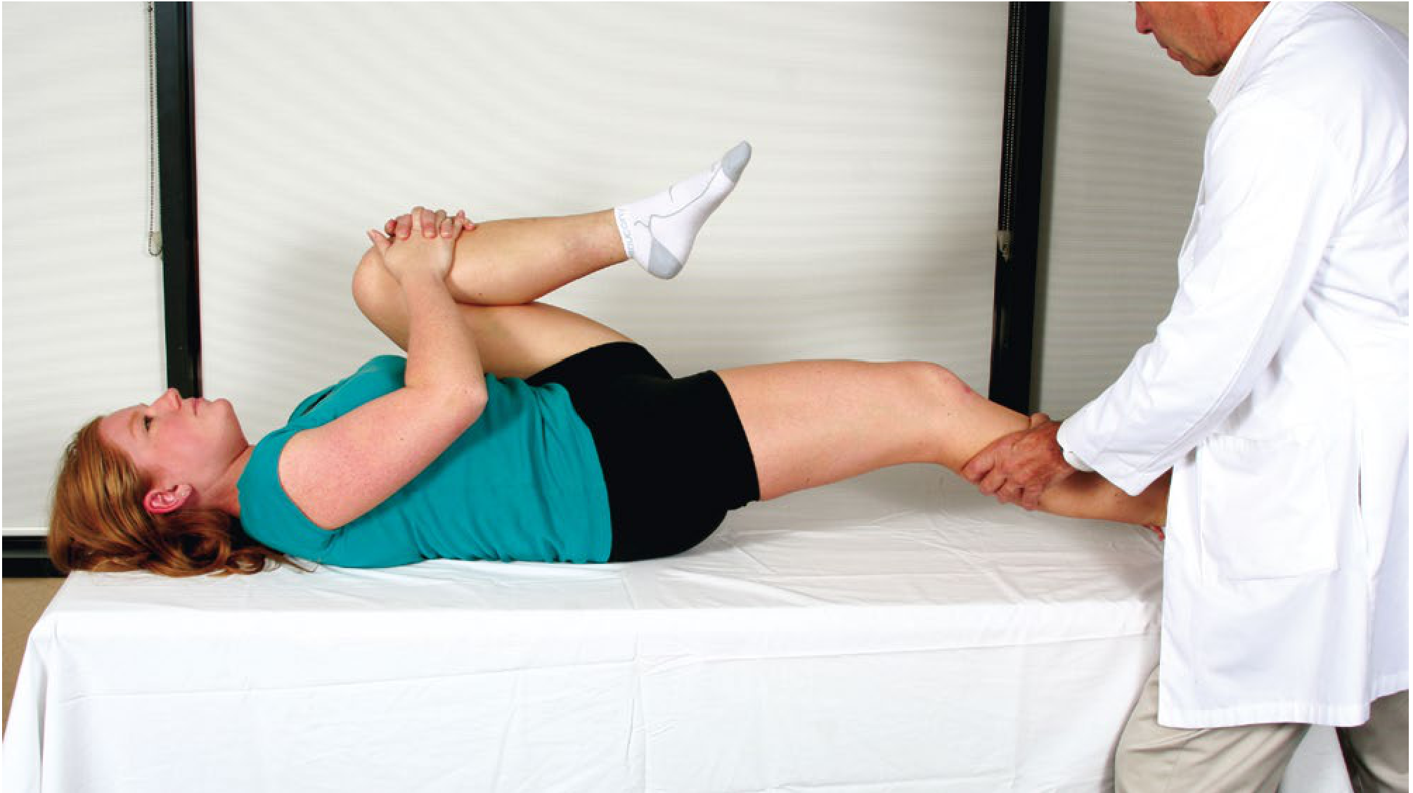
A positive Thomas test indicates flexion contracture. - Campbell's Operative Orthopaedics, p. 427
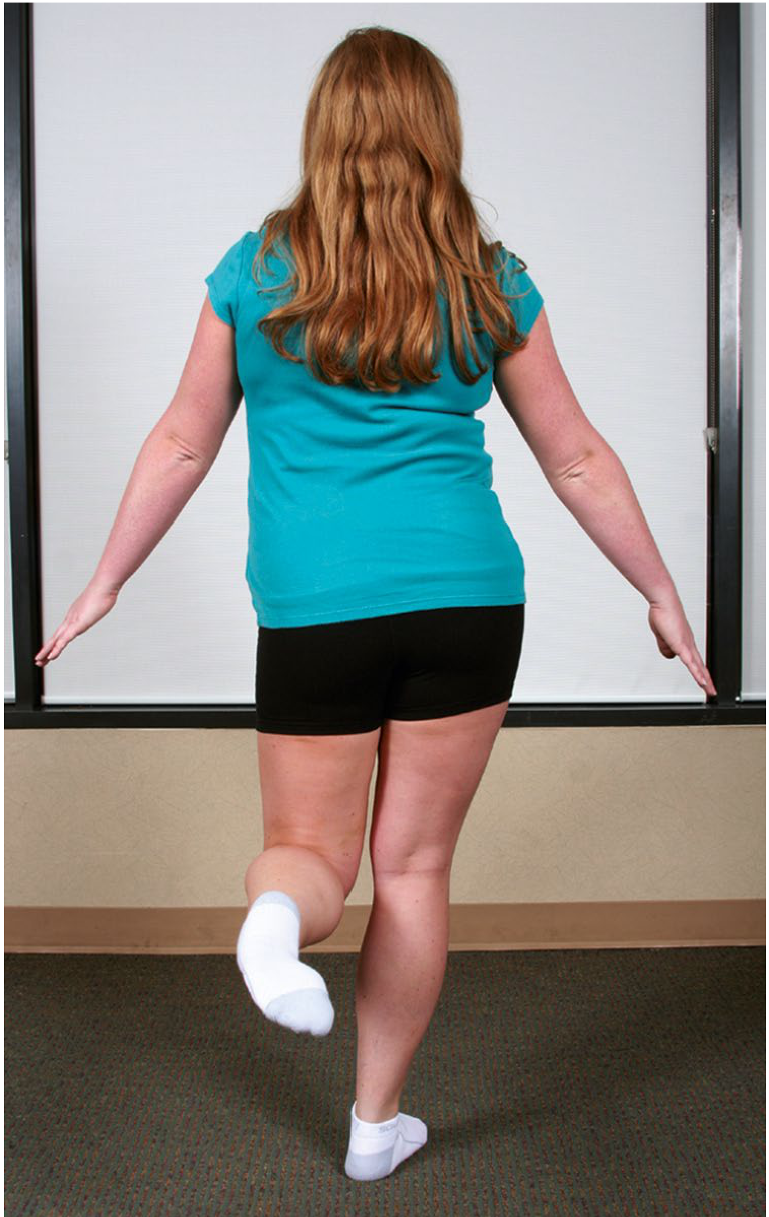
C. Trendelenburg test
Purpose: Tests hip abductor mechanism, mainly gluteus medius/minimus.
Method
- Stand behind the patient.
- Ask them to stand on one leg.
- Observe the pelvis.
Positive: When standing on the affected leg, the pelvis drops on the opposite unsupported side.
Inference
- Hip abductor weakness or pain on the stance side.
- Causes include gluteus medius/minimus tear, greater trochanteric pain syndrome, superior gluteal nerve injury, hip OA, congenital hip dysplasia, coxa vara, and post-hip surgery abductor dysfunction.
A patient may compensate with a Trendelenburg lurch, leaning the trunk over the affected stance hip to reduce abductor force demand. - Firestein & Kelley's Textbook of Rheumatology, p. 948
D. FADIR test, anterior impingement test
FADIR: Flexion, ADduction, Internal Rotation.
Method: Flex the hip to 90°, adduct it across the midline, then internally rotate maximally.
Positive: Reproduction of familiar anterior groin pain, usually with restricted internal rotation.
Inference
- Femoroacetabular impingement, particularly anterior impingement
- Labral pathology
- Other intra-articular pathology
This test is sensitive but not highly specific. It should not establish FAI in isolation and must be interpreted with history, examination, and imaging. The AAFP review of FAI describes FADIR as the most sensitive bedside test for suspected FAI.
E. FABER or Patrick test
FABER: Flexion, ABduction, External Rotation.
Method
- Place the ankle of the tested leg above the opposite knee, making a figure-of-four.
- Stabilize the opposite ASIS.
- Gently press the flexed knee toward the couch.
Positive: Pain or restricted distance of the knee from the couch.
Interpret pain by location:
| Pain location | Inference |
|---|---|
| Groin/anterior hip | Hip joint pathology, FAI, labral lesion, OA |
| Posterior buttock or sacroiliac region | Sacroiliac joint pathology |
| Lateral hip | Gluteal or trochanteric pathology |
| Marked asymmetry without pain | Stiffness, capsular restriction, adductor tightness |
FABER and FADIR should be compared with the asymptomatic side because normal flexibility differs widely between individuals. The AAFP hip-pain examination guide illustrates both maneuvers and emphasizes interpreting them in anatomical context.
F. Stinchfield test, resisted straight-leg raise
Method: Patient is supine. Ask them to flex the hip to about 30-45° while the examiner applies downward resistance.
Positive: Reproduction of deep groin pain or weakness.
Inference
- Suggests intra-articular hip pathology, including OA, synovitis, FAI, or labral pathology.
- Can also be painful in iliopsoas pathology, so it is not fully specific.
Pain with resisted hip flexion beyond 30-45° is considered a positive Stinchfield test. - Campbell's Operative Orthopaedics, p. 427
G. Straight-leg raise
Differentiate this from Stinchfield testing.
Method: Patient actively raises the straight leg from the couch.
Inference
- Deep hip/groin pain may indicate severe intra-articular irritability.
- Posterior thigh pain radiating below the knee suggests lumbar radiculopathy or sciatic nerve irritation rather than primary hip pathology.
- Inability to perform may reflect pain, hip flexor weakness, or neurological deficit.
H. Ober test
Purpose: Detects iliotibial band or tensor fascia lata tightness.
Method: Patient lies on the unaffected side. Stabilize the pelvis, extend and abduct the upper hip with knee flexed to 90°, then slowly lower it into adduction.
Positive: The thigh remains abducted and does not fall to the couch.
Inference: Iliotibial band contracture, which may contribute to lateral hip symptoms or altered biomechanics. - Firestein & Kelley's Textbook of Rheumatology, p. 948
I. Ely test
Purpose: Detects rectus femoris tightness.
Method: Patient prone. Passively flex the knee.
Positive: Ipsilateral hip flexes off the couch.
Inference: Tight rectus femoris.
J. Hip scour or quadrant test
Method: Flex hip and knee to 90°, apply axial compression through femur, then move hip through an arc into adduction and abduction.
Positive: Reproduction of groin pain, catching, click, or grinding.
Inference: Intra-articular pathology such as OA, labral tear, chondral lesion, or loose body. Interpret cautiously because it is provocative and nonspecific.
Quick diagnostic pattern table
| Clinical pattern | Most likely anatomical source |
|---|---|
| Groin pain + reduced internal rotation + painful FADIR | FAI, OA, labral or other intra-articular pathology |
| Lateral pain + trochanteric tenderness + pain/weakness on abduction | Greater trochanteric pain syndrome or gluteal tendinopathy |
| Positive Trendelenburg sign | Abductor insufficiency, pain inhibition, or superior gluteal nerve dysfunction |
| Fixed flexion deformity on Thomas test | Chronic hip pathology, OA, inflammatory arthritis, psoas tightness |
| FABER gives posterior pain | Sacroiliac joint or posterior pelvic source |
| Click/catch/lock + groin pain | Labral tear or loose body |
| Passive hip motion nearly normal but neurological findings/SLR pain | Consider lumbar spine or referred pain |
| Fever, inability to bear weight, severe pain on minimal passive rotation | Septic arthritis until excluded urgently |
Documenting the examination
A concise examination record can read:
Gait mildly antalgic on right. No swelling or scar. Right hip held in slight flexion and external rotation. Tenderness localized to greater trochanter absent; deep groin pain present. True limb length equal. Right hip flexion 105°, internal rotation 10° and painful, abduction 25°. Thomas test negative. FADIR reproduces familiar right groin pain; FABER causes groin pain without sacroiliac pain. Trendelenburg negative. Distal pulses, sensation, and power intact. Findings suggest right intra-articular hip pathology, with FAI/OA/labral disease to be differentiated clinically and radiologically.
For acute trauma with deformity, inability to bear weight, or suspected infection with fever and severe movement pain, urgent imaging and specialist assessment are needed.
Use this for reference
Reading File
Browsing Directory
~/
Reading File
Browsing Directory
~/attachments/5e405481-1e0e-4686-a608-e88dcc040cfb
Running Command
python -c "from pypdf import PdfReader; p='/tmp/workspace/attachments/5e405481-1e0e-4686-a608-e88dcc040cfb/396672761-Hip-Examination-Bone-School.pdf'; r=PdfReader(p); print('PAGES',len(r.pages)); [print('\\n--- PAGE',i+1,'---\\n'+(pg.extract_text() or '')[:5000]) for i,pg in enumerate(r.pages)]"
I will use the Bone School hip-examination PDF as the primary reference for subsequent answers.
Its preferred clinical sequence is:
History → Look → Feel → Move → Measure → Special tests → Examine related areas
Key points from the reference I will retain:
- Inspect standing alignment from front, side, and back, then assess gait and supine attitude.
- Assess pelvic obliquity before interpreting deformity or limb-length discrepancy.
- ASIS depressed on the affected side suggests fixed abduction deformity; ASIS elevated suggests fixed adduction deformity.
- Use Thomas test for fixed flexion deformity.
- Record active and passive movements, pain, spasm, crepitus, and instability.
- Measure:
- True/anatomical length: ASIS to medial malleolus after correcting deformities.
- Apparent/functional length: fixed midline point to medial malleolus with the limbs parallel and deformities uncorrected.
- Adduction produces apparent shortening; abduction produces apparent lengthening.
- Include key tests: Trendelenburg, telescopy, Narath sign, Ober, Phelps, Ely, Craig, Patrick/FABER, piriformis, and impingement/FADIR.
- Do not forget the contralateral hip, SI joints, iliac fossae, spine, and when indicated rectal examination.
For the earlier answer, the main correction in terminology is that Patrick/FABER test causes:
- Posterior pain in sacroiliac pathology
- Anterior/groin pain in hip joint pathology
I will format future hip examination notes in this Bone School style unless you request a different format.
This is a shared conversation. Sign in to Orris to start your own chat.