
SLE pathogenesis
I'll start by reading the medical library skill, then search for SLE pathogenesis content in parallel.
Reading File
Now let me search the library for SLE pathogenesis content and images in parallel.
Finding Sources
Finding Sources
Searching Images
SLE systemic lupus erythematosus pathogenesis mechanism diagram
Searching PubMed
"systemic lupus erythematosus" pathogenesis
Good - I found the key textbooks. Let me now read the dedicated SLE sections from Firestein & Kelley's Rheumatology and the Cellular/Molecular Immunology book in parallel.
Finding Sources
Searching Images
lupus autoantibody antinuclear antibody immune complex pathogenesis
Let me now read the main SLE pathogenesis sections from Firestein & Kelley's Rheumatology and the Cellular/Molecular Immunology textbook in parallel.
Finding Sources
Reading File
Now let me read the core pathogenesis content from Firestein & Kelley's Rheumatology (the main SLE chapter):
Reading File
Reading File
Excellent - I found detailed pathogenesis content and diagram images. Let me read more of the core SLE pathogenesis blocks and fetch the key diagrams:
Reading File
Reading File
Loading Image
Excellent diagram. Now let me read the dedicated SLE pathogenesis chapter from Firestein & Kelley's block 5 (which has 11 paragraphs - likely the core pathogenesis chapter):
Reading File
Reading File
Let me now read the main SLE pathogenesis chapter block 20 which had the main pathogenesis references, and also look for the dedicated SLE pathogenesis chapter in the rheumatology text:
Reading File
Finding Sources
Let me find and read the main SLE chapter from Firestein & Kelley's block 20 (which has the main pathogenesis section), and also search for the Rheumatology 2022 SLE content:
Finding Sources
The block10 entry for "Lupus Erythematosus" has no paragraph line ranges but is likely the main chapter introduction. Let me read that and also the SLE chapter in the Rheumatology 2-Volume Set (Elsevier 2022):
Reading File
Reading File
Let me now find the core SLE pathogenesis chapter in the Firestein & Kelley text (block20 has the key SLE clinical chapter):
Reading File
Let me search for the specific SLE pathogenesis section using paragraphs_fts to find the core immunological mechanism content:
Finding Sources
Finding Sources
Excellent! Found the key section "Pathogenesis of Systemic Lupus Erythematosus" in Cellular and Molecular Immunology textbook and the Key Points/Mechanisms section in Rheumatology 2022. Let me read these now:
Reading File
Reading File
I now have comprehensive content from multiple authoritative sources. Let me read a bit more of the Rheumatology 2022 pathogenesis section to get the complete picture on innate/adaptive mechanisms:
Reading File
I now have very comprehensive, multi-source content. Let me compile the full answer.
Pathogenesis of Systemic Lupus Erythematosus (SLE)
SLE is the prototypic human immune complex disease - a chronic, multisystem autoimmune condition driven by a breakdown of self-tolerance. Its pathogenesis is multifactorial, involving genetic susceptibility, epigenetic changes, environmental triggers, and dysregulation of both innate and adaptive immunity.
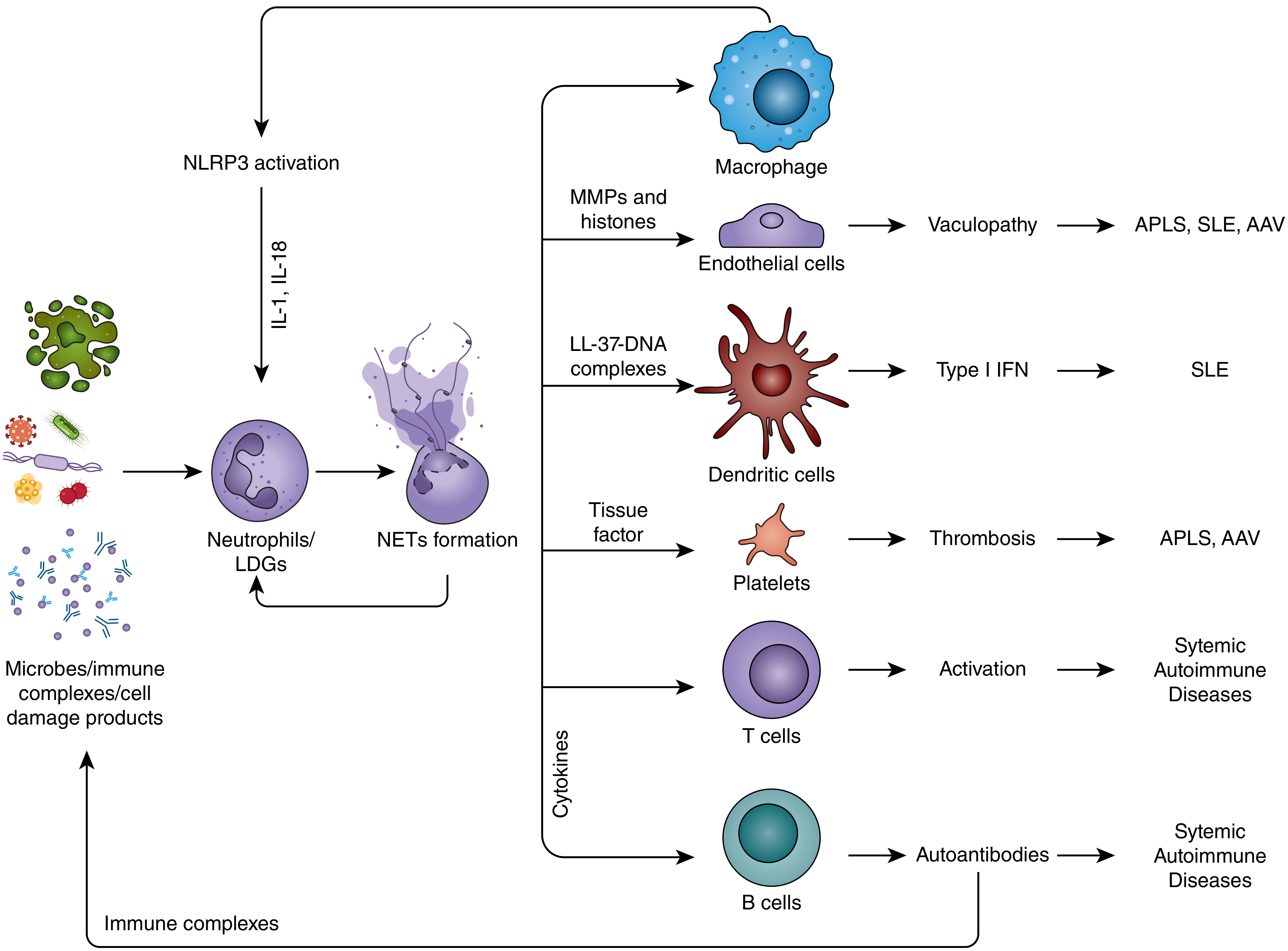
Fig. 11.7 - Neutrophils/LDGs and NETs in systemic autoimmune diseases. NET products (LL-37-DNA complexes) drive type I IFN production by dendritic cells, while cytokines activate T and B cells, causing the autoantibodies and vasculopathy characteristic of SLE. - Firestein & Kelley's Textbook of Rheumatology
1. Disease Phases
SLE develops as a multistep process:
- Genetic/epigenetic susceptibility phase - inherited risk
- Subclinical immune dysregulation - early immune abnormalities
- Asymptomatic autoimmunity - autoantibodies appear (on average 3.4 years before diagnosis; ANA, anti-Ro, anti-La, antiphospholipid antibodies first; anti-Sm and anti-RNP later; anti-dsDNA ~2.7 years before diagnosis)
- Clinical SLE - triggered by additional environmental hits
- Relapsing-remitting course - with accrual of organ damage over years
- Firestein & Kelley's Textbook of Rheumatology, p. 237; Rheumatology 2-Volume Set (Elsevier 2022)
2. Genetic Susceptibility
-
HLA associations: HLA-DR2 and HLA-DR3 carry an odds ratio of 2-3; both together ~5x risk
-
Complement deficiencies: C1q, C2, or C4 deficiency in ~5% of SLE patients - these impair clearance of immune complexes and apoptotic cells, and impair B cell tolerance
-
FcγRIIB polymorphism: reduced inhibitory signaling on B cells, allowing unchecked activation
-
GWAS genes: IRF3, IRF5, IRF7, STAT4, PTPN22, BLK, TNFAIP3, TREX1, DNase I
-
Monogenic lupus: rare mutations in complement components, DNase I, TREX1 (encoding a 3'-5' DNA exonuclease), SAMHD1 - all cause inability to degrade self-DNA/RNA
-
Sex: 10:1 female-to-male ratio; sex hormones (estrogen) influence immune responses; X-linked genes relevant (TLR7 is X-linked - its duplication causes lupus in mouse models)
-
Concordance: ~25-50% in monozygotic twins vs ~5% in dizygotic - strong genetic but not purely genetic basis
-
Cellular and Molecular Immunology (Abbas), p. 1273-1274; Rheumatology 2022
3. Environmental Triggers
| Trigger | Mechanism |
|---|---|
| UV light | Induces apoptosis of keratinocytes → release of nuclear antigens; oxidizes DNA; causes DNA breaks |
| Infections (EBV, others) | Molecular mimicry; TLR activation by nucleic acid-containing complexes; induction of type I IFN |
| Drugs (procainamide, hydralazine, isoniazid) | Altered DNA methylation → drug-induced lupus |
| Ionizing radiation | Alteration of self-antigens (enzymatic cleavage, phosphorylation) |
| Microbiome dysbiosis | Enterococcus gallinarum translocation from gut to lymphoid organs activates TLR7, induces Th17/Tfh cells, triggers anti-dsDNA/RNA antibodies |
- Rheumatology 2022 (Elsevier), Table 139.1
4. Central Initiating Mechanism: Impaired Clearance of Apoptotic Cells
This is the central initiating event in SLE pathogenesis:
-
UV light and other stressors cause apoptosis of cells
-
Normally, apoptotic cells are rapidly phagocytosed by macrophages
-
In SLE, clearance is defective: reduced complement (C1q opsonizes apoptotic debris), reduced DNase I, reduced CRP, macrophage dysfunction
-
Nuclear material (DNA, histones, ribonucleoproteins) from uncleared apoptotic cells becomes accessible as self-antigens
-
These nucleic acid-containing complexes activate TLR7 (RNA) and TLR9 (DNA) on B cells and plasmacytoid dendritic cells (pDCs)
-
Result: break in B cell tolerance and massive production of antinuclear autoantibodies
-
Cellular and Molecular Immunology, p. 1274; Rheumatology 2022
5. The Type I Interferon Axis
This is the hallmark of SLE:
-
Plasmacytoid DCs (pDCs) are the major source of IFN-α
-
TLR7/TLR9 activation by nucleic acid-containing immune complexes triggers pDC IFN-α production
-
The IFN-α gene signature (upregulation of IFN-stimulated genes in peripheral blood) is found in 50-80% of SLE patients and correlates with disease activity
-
IFN-α drives:
- Dendritic cell maturation and enhanced antigen presentation
- B cell class switching and autoantibody production
- T cell activation and effector differentiation
- Upregulation of BLyS/BAFF (B cell survival factor)
-
In the presymptomatic phase: IFN-α activity rises ~2 years before clinical diagnosis
-
Key feedback loop: autoantibody-antigen immune complexes are taken up by pDCs via FcγRIIa, reactivating TLR9/7 and driving more IFN-α - a self-amplifying cycle
-
Cellular and Molecular Immunology, p. 1274-1275; Rheumatology 2022, p. 477
6. B Cell Dysregulation and Autoantibody Production
- SLE B cells have a lower activation threshold, defective tolerance checkpoints (both central and peripheral), and increased survival signals
- Key autoantibodies and their pathogenic roles:
| Autoantibody | Target | Pathogenic role |
|---|---|---|
| Anti-dsDNA | Double-stranded DNA | Immune complex deposition → glomerulonephritis; correlates with disease activity |
| Anti-Sm | snRNP (spliceosome) | Specific for SLE; part of immune complexes |
| Anti-Ro/SSA | RNA-binding protein | Neonatal lupus, photosensitivity, SCLE |
| Anti-La/SSB | RNA-binding protein | Associated with anti-Ro |
| Anti-histone | Histones | Drug-induced lupus |
| Antiphospholipid (aPL) | Phospholipids/β2-GPI | Thrombosis, pregnancy loss (antiphospholipid syndrome) |
| Anti-RBC / anti-platelet | Erythrocytes, platelets | Hemolytic anemia, thrombocytopenia |
-
Immune complex deposition in glomeruli, joints, and skin triggers complement activation → inflammation and tissue damage
-
BLyS/BAFF is elevated in SLE and promotes B cell survival and class switching; belimumab (anti-BLyS) is FDA-approved for SLE
-
Cellular and Molecular Immunology; Rheumatology 2022, p. 2900-2901
7. T Cell Abnormalities
SLE T cells show several intrinsic defects:
-
Reduced IL-2 production: CD4+ T cells produce less IL-2, which normally maintains regulatory T cells (Tregs); Treg dysfunction allows unchecked autoreactive responses
-
Lower activation threshold: abnormal TCR signaling via substitution of FcεRIγ for CD3ζ chain, enhanced calcium mobilization
-
Tfh (T follicular helper) cell expansion: drives germinal center reactions and high-affinity autoantibody maturation
-
Th17 expansion: IL-17-producing cells are increased, especially in patients with renal involvement; IL-17 promotes neutrophil recruitment and tissue inflammation
-
CD8+ T cell exhaustion: impaired cytotoxic T cell function
-
Abnormal DNA methylation in T cells: hypomethylation of immune genes (ITGAL/CD11a, perforin, CD70) causes overexpression and autoreactivity
-
Rheumatology 2022, Key Points; Firestein & Kelley's
8. Innate Immune Dysregulation
Neutrophils and Low-Density Granulocytes (LDGs)
- SLE patients have a distinct population of LDGs (low-density granulocytes) in peripheral blood
- LDGs have a striking type I IFN gene signature and produce far more IFN-α than normal neutrophils
- LDGs and activated neutrophils form NETs (Neutrophil Extracellular Traps):
- NETs consist of DNA decorated with antimicrobial proteins (LL-37, histones, elastase, MPO)
- LL-37-DNA complexes are potent activators of pDCs → massive IFN-α release
- NETs expose modified self-antigens, stimulating autoantibody production
- NETs cause direct vascular endothelial damage → vasculopathy in SLE
- Impaired NET clearance (due to anti-NET antibodies, DNase I deficiency) perpetuates the cycle
Dendritic Cells
- Plasmacytoid DCs are the main IFN-α producers
- TLR7/9 activation by RNA- and DNA-containing immune complexes triggers IFN-α
- Conventional DCs (myeloid DCs) are reduced in circulation (trafficked to lymph nodes) but are potent antigen presenters
Macrophages
- Impaired efferocytosis (phagocytosis of apoptotic cells) - "tingible body" macrophages in germinal centers are reduced
- Macrophage activation contributes to cytokine storm in severe SLE
Basophils
-
Basophils infiltrate lymph nodes in SLE patients with an activated phenotype (upregulated CD63, CD203c, CXCR4)
-
They produce IL-4 which promotes autoantibody production by B cells and Th17 differentiation
-
Basophil depletion reduces disease in murine models
-
Firestein & Kelley's Rheumatology, pp. 237-238 (NETs/LDG section)
9. Complement System in Tissue Damage
- Immune complex deposition in glomeruli → classical pathway complement activation
- C3b and C4b opsonize complexes; C5a recruits neutrophils
- Paradox: complement deficiency (C1q, C2, C4) predisposes to SLE, yet complement activation mediates tissue damage in established disease
- C1q normally opsonizes apoptotic cells for clearance; its deficiency leads to accumulation of autoantigen and autoimmunity
- Once immune complexes form, complement-mediated inflammation drives nephritis and vasculitis
10. Epigenetic Dysregulation
-
DNA hypomethylation in SLE CD4+ T cells - affects genes including ITGAL (CD11a/CD18), perforin, CD70, and a widespread IFN-hypomethylation signature in CD4+, CD19+, and CD14+ cells
-
Histone modifications: altered acetylation and methylation patterns
-
MicroRNAs: miR-21 and miR-148a downregulate DNMT1, promoting hypomethylation
-
Drugs like procainamide and hydralazine inhibit DNMT1 → drug-induced lupus via demethylation
-
Firestein & Kelley's Rheumatology, p. 4227
11. Cytokine Network
| Cytokine | Role in SLE |
|---|---|
| IFN-α (type I IFN) | Master driver; activates all immune arms; correlates with disease activity |
| BLyS/BAFF | B cell survival/activation; target of belimumab |
| IL-6 | B cell maturation, acute phase response; elevated in active SLE |
| IL-12/IL-23 | Promote Th1/Th17 differentiation; genetic variants in IL-12 pathway linked to SLE |
| IL-17 | Promotes neutrophil recruitment; elevated in renal SLE |
| IL-10 | Pro-B cell, anti-T cell/APC; complex role |
| TNF-α | Can be pro- or anti-inflammatory; paradoxically anti-TNF therapy can trigger lupus-like disease |
| APRIL/BAFF | B cell survival; both elevated in SLE |
Summary: Integrated Pathogenic Loop
Genetic susceptibility + Environmental triggers
         ↓
Impaired apoptotic cell clearance → Exposure of nuclear antigens
         ↓
TLR7/9 activation (by RNA/DNA complexes) on pDCs and B cells
         ↓
IFN-α surge (type I IFN signature) + NETosis by LDGs
         ↓
B cell activation → Antinuclear autoantibody production
T cell activation → Tfh expansion → Germinal center hyperactivity
         ↓
Immune complex formation (anti-dsDNA + DNA; anti-Sm + snRNP, etc.)
         ↓
Deposition in kidneys, skin, joints, vessels
         ↓
Complement activation → Inflammation → Organ damage
         ↓
Tissue damage releases more nuclear antigens → Amplification loop
Key sources:
- Cellular and Molecular Immunology (Abbas), pp. 1273-1275
- Rheumatology, 2-Volume Set (Elsevier 2022), Chapter 139, pp. 2761-2924
- Firestein & Kelley's Textbook of Rheumatology, pp. 237-238, 4227-4246
Recent evidence (2024-2025): A 2025 systematic review (PMID 39917309) details organ-based B cell heterogeneity in SLE, and a 2025 systematic review on thrombosis mechanisms (PMID 40086157) highlights roles of antiphospholipid antibodies and complement in SLE-associated thrombosis - both consistent with the framework above.
This is a shared conversation. Sign in to Orris to start your own chat.