Define stable angina and unstable angina.

Reading File
Finding Sources
Finding Sources
Reading File
Reading File
Reading File
Reading File
Reading File
Here is a thorough definition and comparison of stable and unstable angina, drawn from multiple authoritative textbooks.

Angina Pectoris: Stable vs. Unstable

What is Angina Pectoris?

Angina pectoris literally means "chest pain." It is the clinical manifestation of myocardial ischemia - an imbalance between the oxygen requirements of the heart and the oxygen delivered to it via the coronary arteries. This imbalance can occur through increased demand, diminished delivery, or both. The underlying cause is most commonly atherosclerotic coronary artery disease (CAD). - Swanson's Family Medicine Review, p. 119

Stable Angina

Definition: Stable angina (also called chronic stable angina or effort angina) is chest pain that occurs predictably when myocardial oxygen demand increases - typically during physical exertion, emotional stress, or exposure to cold - and is promptly relieved by rest or nitroglycerin (usually within 1-5 minutes).
Pathophysiology:
  • Fixed atherosclerotic plaques in epicardial coronary arteries limit blood flow.
  • At rest, flow is sufficient; during increased demand (exercise, tachycardia), flow cannot increase proportionately.
  • The resulting ischemia causes accumulation of acidic metabolites, triggering pain.
  • Coronary flow reserve is frequently impaired due to endothelial dysfunction, so ischemia may occur even at lower levels of demand.
Key features:
  • Predictable, reproducible pattern
  • Triggered by exertion or emotional stress
  • Relieved by rest within minutes or by sublingual nitroglycerin
  • Retrosternal chest tightness, pressure, or burning; may radiate to left shoulder, arm, or jaw
  • ECG may show ST-segment depression during episodes
  • Stable over weeks to months
Prognosis: Patients with chronic stable angina are at lower risk than those with unstable angina and can generally be monitored and treated as outpatients. - Swanson's Family Medicine Review, p. 119

Unstable Angina

Definition: Unstable angina is an acute coronary syndrome (ACS) characterized by angina that occurs at rest, is new in onset, or is escalating in severity, frequency, or duration compared to a prior stable pattern. It is not relieved by rest or nitroglycerin and represents a medical emergency. - Katzung's Basic and Clinical Pharmacology, 16th Ed., p. 302; Lippincott Illustrated Reviews: Pharmacology, p. 424
Three main presentations (ACC/AHA):
  1. Angina at rest - pain occurring without any precipitating exertion
  2. New-onset angina - angina appearing for the first time, at least CCS Class III severity
  3. Increasing (crescendo) angina - previously stable angina that becomes more frequent, more severe, more prolonged, or occurs at lower levels of exertion
Pathophysiology:
  • Caused by rupture or fissuring of an atherosclerotic plaque, leading to formation of a labile, partially occlusive thrombus at the plaque site.
  • This reduces coronary flow acutely without completely occluding the vessel (complete occlusion typically causes STEMI).
  • Platelet aggregation and thrombosis play a central role.
  • Inflammation is also implicated - patients on TNF-alpha inhibitors appear to have lower MI risk.
Distinguishing unstable angina from NSTEMI: The presentation of unstable angina and NSTEMI (non-ST elevation myocardial infarction) is clinically identical. The distinction is made by cardiac biomarkers: in NSTEMI, troponins and other markers of myocardial necrosis are elevated; in unstable angina, they are not elevated (no myocyte death has occurred yet). - Swanson's Family Medicine Review, p. 119
Prognosis: Unstable angina carries a high risk of MI and death and is treated as a medical emergency. The 30-day mortality of ACS (unstable angina/NSTEMI) is between 10-20% despite treatment. - Swanson's Family Medicine Review, p. 119

Comparison Table

FeatureStable AnginaUnstable Angina
TriggerExertion, stress, coldRest, minimal exertion, or unprovoked
PatternPredictable, reproducibleNew, increasing, or at rest
DurationShort (1-5 min)Longer (>20 min)
ReliefRest or nitroglycerinNOT relieved by rest or nitrates
PathologyFixed plaque, demand-supply mismatchPlaque rupture + acute thrombus
TroponinsNormalNormal (elevated = NSTEMI)
ECGST depression during episodesMay show ST changes at rest
ACS?NoYes - part of ACS spectrum
UrgencyOutpatient managementMedical emergency
RiskLowerHigh (MI, death)

Key Takeaway

Both types share the same underlying mechanism - myocardial ischemia from CAD - but differ critically in their pathophysiology. Stable angina is a demand problem (fixed plaque limiting flow during stress). Unstable angina is a supply problem (acute plaque rupture and thrombosis reducing flow even at rest). This distinction drives completely different management strategies and urgency levels.
As Lippincott Illustrated Reviews: Pharmacology summarizes: "Stable angina is promptly relieved by rest or nitroglycerin, whereas unstable angina occurs with increased frequency, duration, and intensity and is not relieved by rest or nitroglycerin. Unstable angina is an acute coronary syndrome requiring acute medical attention to prevent progression to MI and death."

Define patent ductus arteriosus.

Finding Sources
Finding Sources
Searching Images

patent ductus arteriosus diagram fetal circulation
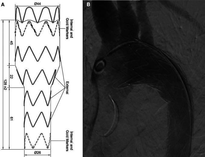
Educational medical graphic illustrating Thoracic Endovascular Aortic Repair (TEVAR) for Patent Ductus Arteriosus (PDA). (A) Schematic diagram of a custom-made tapered stent graft, showing the structural architecture with zig-zag external struts and radio-opaque internal gold markers at the proximal and distal ends. Dimensions are specified: proximal diameter of 44mm, distal diameter of 26mm, and a total length of 128mm with segmented lengths of 45mm, 22mm, and 61mm. (B) Intraoperative aortography following TEVAR deployment. The digital subtraction angiography (DSA) image confirms the successful exclusion of the PDA from the aortic circulation. The stent graft is visible within the thoracic aorta, demonstrating a mesh-like metallic density. No endoleaks are observed, and the aortic arch anatomy, including proximal branch vessels, is partially visible. This visual illustrates endovascular treatment strategies for complex adult congenital heart disease or aortic pathologies, focusing on custom device design and post-procedural diagnostic imaging confirmation.
Educational medical graphic illustrating Thoracic Endovascular Aortic Repair (TEVAR) for Patent Ductus Arteriosus (PDA). (A) Schematic diagram of a custom-made tapered stent graft, showing the structural architecture with zig-zag external struts and radio-opaque internal gold markers at the proximal and distal ends. Dimensions are specified: proximal diameter of 44mm, distal diameter of 26mm, and a total length of 128mm with segmented lengths of 45mm, 22mm, and 61mm. (B) Intraoperative aortography following TEVAR deployment. The digital subtraction angiography (DSA) image confirms the successful exclusion of the PDA from the aortic circulation. The stent graft is visible within the thoracic aorta, demonstrating a mesh-like metallic density. No endoleaks are observed, and the aortic arch anatomy, including proximal branch vessels, is partially visible. This visual illustrates endovascular treatment strategies for complex adult congenital heart disease or aortic pathologies, focusing on custom device design and post-procedural diagnostic imaging confirmation.

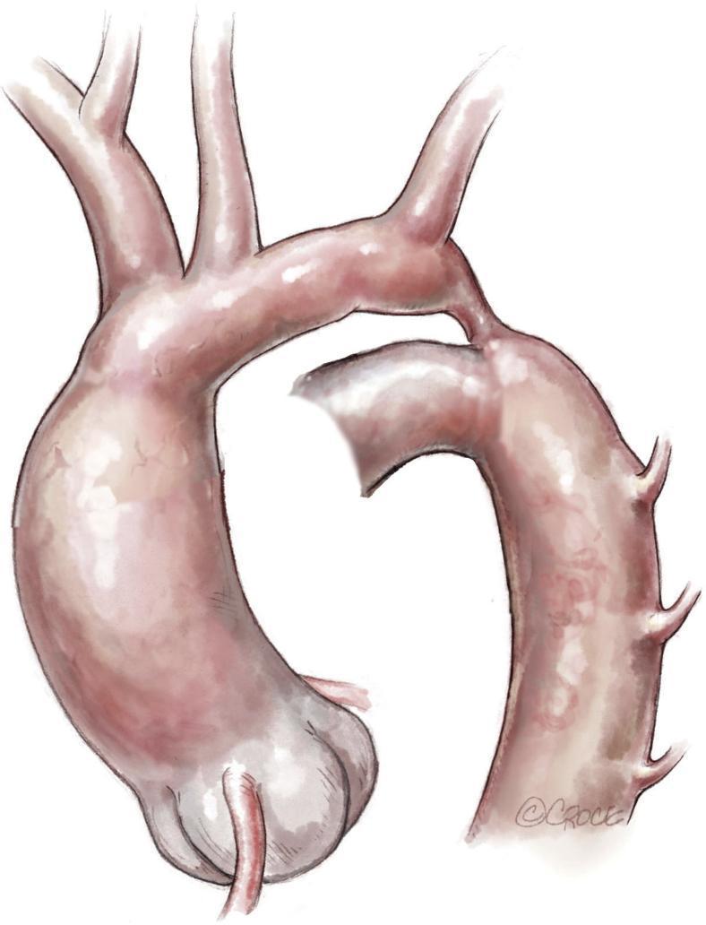
This medical illustration depicts a neonatal aortic arch with ductus-dependent coarctation. The diagram highlights several critical anatomical features: a hypoplastic and elongated distal aortic arch segment situated between the origins of the left common carotid artery and the left subclavian artery. There is a distinct stenotic isthmus, characterized by a significant narrowing of the aortic lumen just proximal to the descending aorta. A patent ductus arteriosus is shown connecting the pulmonary circulation (not fully pictured) to the descending aorta, serving as the primary conduit for distal systemic perfusion. The drawing also visualizes the brachiocephalic trunk and the intercostal arteries branching from the descending thoracic aorta. This illustration is intended for pediatric cardiology and cardiothoracic surgery education to demonstrate the morphology of complex aortic coarctation requiring surgical intervention, such as extended end-to-end anastomosis.
This medical illustration depicts a neonatal aortic arch with ductus-dependent coarctation. The diagram highlights several critical anatomical features: a hypoplastic and elongated distal aortic arch segment situated between the origins of the left common carotid artery and the left subclavian artery. There is a distinct stenotic isthmus, characterized by a significant narrowing of the aortic lumen just proximal to the descending aorta. A patent ductus arteriosus is shown connecting the pulmonary circulation (not fully pictured) to the descending aorta, serving as the primary conduit for distal systemic perfusion. The drawing also visualizes the brachiocephalic trunk and the intercostal arteries branching from the descending thoracic aorta. This illustration is intended for pediatric cardiology and cardiothoracic surgery education to demonstrate the morphology of complex aortic coarctation requiring surgical intervention, such as extended end-to-end anastomosis.

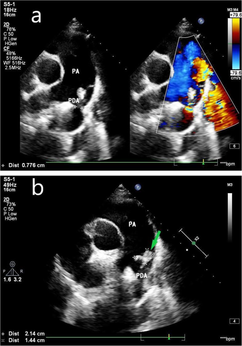
Two-panel fetal echocardiogram at 26 weeks of gestation demonstrating a patent ductus arteriosus (PDA) complicated by infective endocarditis (IE). Panel A features side-by-side 2D grayscale and color Doppler images. The grayscale view identifies the pulmonary artery (PA) and a PDA channel measuring 0.776 cm. The color Doppler demonstrates the left-to-right shunt of the artery through the ductus. Panel B provides a magnified grayscale view of the PDA opening, where a green arrow indicates an irregular, hyperechoic mass. This mass, measuring approximately 2.14 cm by 1.44 cm, represents a vegetation consistent with infective endocarditis. The imaging illustrates the characteristic anatomical connection between the pulmonary artery and the descending aorta in a PDA case, with the added clinical complication of vegetative growth on the vessel wall. This content is suitable for advanced cardiology and maternal-fetal medicine education regarding congenital heart disease and obstetric infections.

Two-panel fetal echocardiogram at 26 weeks of gestation demonstrating a patent ductus arteriosus (PDA) complicated by infective endocarditis (IE). Panel A features side-by-side 2D grayscale and color Doppler images. The grayscale view identifies the pulmonary artery (PA) and a PDA channel measuring 0.776 cm. The color Doppler demonstrates the left-to-right shunt of the artery through the ductus. Panel B provides a magnified grayscale view of the PDA opening, where a green arrow indicates an irregular, hyperechoic mass. This mass, measuring approximately 2.14 cm by 1.44 cm, represents a vegetation consistent with infective endocarditis. The imaging illustrates the characteristic anatomical connection between the pulmonary artery and the descending aorta in a PDA case, with the added clinical complication of vegetative growth on the vessel wall. This content is suitable for advanced cardiology and maternal-fetal medicine education regarding congenital heart disease and obstetric infections.

Reading File
Reading File
Reading File
Reading File
Reading File
Reading File
Here is a comprehensive definition of Patent Ductus Arteriosus, compiled from multiple authoritative textbooks.

Patent Ductus Arteriosus (PDA)

Definition

Patent ductus arteriosus (PDA) is a congenital cardiovascular defect in which the ductus arteriosus - a normal fetal blood vessel that connects the main pulmonary artery (at its bifurcation) to the descending aorta, just distal to the origin of the left subclavian artery - fails to close after birth, resulting in persistent abnormal communication between the two great vessels. PDA accounts for approximately 7% of all congenital heart lesions, and occurs in about 1 in every 2000 births in term infants. - Robbins & Kumar Basic Pathology, p. 261; Schwartz's Principles of Surgery 11th Ed., p. 787

Embryology and Normal Fetal Role

The ductus arteriosus is derived from the sixth aortic arch. In fetal life, it serves a critical physiologic role: it shunts blood from the pulmonary artery to the aorta (a right-to-left shunt), bypassing the fluid-filled, non-respiratory fetal lungs which have a high pulmonary vascular resistance. Ductal flow in fetal life is considerable - approximately 60% of the combined ventricular output. - Schwartz's Principles of Surgery, p. 787
Patency of the ductus during fetal life is actively maintained by locally produced prostaglandin E2 (PGE2) and prostaglandin I2 (PGI2), which induce relaxation of the ductal smooth muscle. - Schwartz's Principles of Surgery, p. 787

Normal Postnatal Closure

At birth, three events trigger ductal closure:
  1. Increased arterial oxygen tension in the neonate's blood - the primary stimulus causing smooth muscle contraction, occurring within 10-15 hours of birth.
  2. Decreased pulmonary vascular resistance as the lungs expand and fill with air.
  3. Falling levels of PGE2 and PGI2 - increased pulmonary blood flow metabolizes these prostaglandins, and removal of the placenta eliminates a major source of them.
Additionally, histamines, catecholamines, bradykinin, and acetylcholine all promote ductal contraction. Functional closure occurs within 1-2 days in healthy term infants; anatomic closure by fibrosis produces the ligamentum arteriosum within the first few months of life. - Robbins & Kumar Basic Pathology, p. 261; Schwartz's Principles of Surgery, p. 787

Pathology: Failure to Close

In PDA, the ductus fails to involute - either through delayed closure (prolonged patency) or persistent failure to close (persistent patency). The embryologic basis is a failure of the muscular wall of the ductus to contract adequately after birth. Failure of TGF-β induction after birth has been suggested as one mechanism. - The Developing Human (Clinically Oriented Embryology), p. 881
Risk factors for PDA include:
  • Prematurity - the most important risk factor; PDAs are found in ~75% of infants at 28-30 weeks gestation; virtually all preterm neonates ≤28 weeks with birth weight <1750 g have a PDA in the first 24 hours.
  • Hypoxia (respiratory distress syndrome, surfactant deficiency, high altitude)
  • Maternal rubella infection during early pregnancy
  • Associated congenital cardiac defects (coarctation of aorta, transposition of great arteries, pulmonary stenosis/atresia)
  • Female sex (2:1 female-to-male ratio)

Hemodynamics: Left-to-Right Shunt

After birth, as pulmonary vascular resistance falls (over 8-10 weeks postnatally), the pressure gradient reverses: aortic pressure now exceeds pulmonary artery pressure, so blood shunts left-to-right (aorta → pulmonary artery) - the opposite direction of fetal flow. This is the hallmark of PDA. - Goldman-Cecil Medicine, p. 1057; Gray's Anatomy for Students, p. 189
Hemodynamic consequences:
  • Increased pulmonary blood flow → pulmonary congestion, increased work of breathing
  • Left ventricular volume overload → left atrial and left ventricular dilation
  • Lower aortic diastolic pressure (blood "runs off" into pulmonary artery during diastole) → wide pulse pressure, bounding pulses
  • Right ventricular strain from elevated pulmonary pressures
  • Risk of pulmonary hypertension if untreated - can eventually lead to Eisenmenger syndrome (reversal to right-to-left shunt with cyanosis), occurring in ~5% of isolated PDA cases

Clinical Features

FeatureDescription
MurmurHarsh, continuous "machinery murmur" at the left first/second intercostal space below the clavicle - heard throughout systole and diastole
PulseWide pulse pressure; bounding, hyperkinetic pulses
PrecordiumHyperdynamic; left ventricular heave
CyanosisAbsent in simple PDA (only present if Eisenmenger develops)
Symptoms (large PDA)Dyspnea, tachypnea, feeding difficulties, tachycardia, exercise intolerance
Small PDAOften asymptomatic, but risk of infective endocarditis (~0.45%/year after second decade)
  • Goldman-Cecil Medicine, p. 1066; Schwartz's Principles of Surgery, p. 787-788; Gray's Anatomy for Students, p. 178-182

Investigations

  • Chest X-ray: Increased pulmonary vascularity, cardiomegaly, prominent pulmonary trunk, dilated left atrium and left ventricle; calcification of PDA in older patients
  • ECG: Left ventricular strain, left atrial enlargement, possible right ventricular hypertrophy
  • Echocardiography with Doppler: Reliably demonstrates ductal patency and estimates shunt size - the primary diagnostic tool
  • Cardiac catheterization: Reserved for cases where pulmonary hypertension and vascular resistance need quantification before closure

Treatment

ApproachDetails
Pharmacological (premature infants)Indomethacin or ibuprofen (COX inhibitors) - inhibit prostaglandin synthesis, promoting ductal closure. Effective in premature neonates; generally ineffective in term infants.
Catheter-based closureTransluminal occlusive devices (e.g., Rashkind double-umbrella, Gianturco coils, Amplatzer ductal occluder) - widely used in suitable patients
Surgical ligationPosterior lateral thoracotomy with ligation/clipping; care must be taken to avoid the left recurrent laryngeal nerve (injury → hoarseness) and the thoracic duct (injury → chylothorax)
The presence of a persistent PDA is sufficient indication for closure given the elevated mortality risk and risk of endocarditis. The estimated death rate for infants with isolated, untreated PDA is approximately 30%, with congestive heart failure as the leading cause. - Schwartz's Principles of Surgery, p. 788

Key Anatomical Relationships

The ductus arteriosus connects:
  • Proximally: Main pulmonary artery (at its bifurcation) / left pulmonary artery
  • Distally: Descending aorta, just distal to the left subclavian artery
After closure, its fibrous remnant is the ligamentum arteriosum, which connects the left pulmonary artery to the aortic arch. - Gray's Anatomy for Students, p. 182; Robbins & Kumar, p. 261
This is a shared conversation. Sign in to Orris to start your own chat.