Explain visual pathway
Reading File
Finding Sources
Reading File
Reading File
Reading File
Running Command
Loading Image
Loading Image
Excellent - I have comprehensive content from both the attached PDF (Chapter 145 of your sensory physiology textbook) and Gray's Anatomy. Here is the full, detailed explanation:
Visual Pathway
The visual pathway transmits visual information from the photoreceptors in the retina all the way to the primary visual cortex in the occipital lobe, with several relay stations in between.
1. Overview of the Pathway
The sequence is:
Photoreceptors (Rods & Cones) → Bipolar Cells → Ganglion Cells → Optic Nerve → Optic Chiasm → Optic Tract → Lateral Geniculate Body (LGB) → Optic Radiation → Primary Visual Cortex (Area 17)
2. From Retina to Optic Nerve
- Visual sensations arise in rods and cones (first-order receptors). These synapse with bipolar cells, which in turn synapse with ganglion cells.
- The axons of all ganglion cells converge at the optic disc (blind spot - no photoreceptors here), exit the eyeball, acquire a myelin sheath (from oligodendrocytes), and are covered by cranial meninges - making the optic nerve a true CNS structure.
- Each optic nerve carries approximately 1 million fibers from one retina.
3. The Optic Chiasm - The Partial Decussation
At the optic chiasm (just anterior to the infundibular stalk):
- Fibers from the nasal (medial) half of each retina cross to the contralateral optic tract.
- Fibers from the temporal (lateral) half of each retina remain ipsilateral and enter the ipsilateral optic tract.
Why does this matter? Each temporal hemiretina sees the same half of the visual field as the nasal hemiretina of the opposite eye. This partial crossing ensures that all information about each visual hemifield is processed by the contralateral hemisphere. For example, objects in the left visual field project to the right visual cortex.
4. Optic Tract
After the chiasm, the fibers travel as the optic tract, which courses around the midbrain, carrying:
- Temporal retinal fibers from the ipsilateral eye
- Nasal retinal fibers from the contralateral eye
Together, these carry information about the contralateral visual hemifield.
5. Lateral Geniculate Body (LGB) - The Thalamic Relay
The optic tract terminates (mostly) in the LGB of the thalamus. The LGB is the primary thalamic relay for vision.
Laminar Organization
The LGB has 6 well-defined layers (numbered 1-6, ventral to dorsal):
| Layers | Cell Type | Cell Name | Input From |
|---|---|---|---|
| 1, 2 | Large cells | Magnocellular | M ganglion cells (movement, depth) |
| 3, 4, 5, 6 | Small cells | Parvocellular | P ganglion cells (color, fine detail) |
Eye-Specific Laminar Input
Each eye projects to specific layers:
- Contralateral (nasal) retina → Layers 1, 4, 6
- Ipsilateral (temporal) retina → Layers 2, 3, 5
This means each eye's input is kept separate within the LGB, even though both eyes contribute to the same hemifield representation.
Retinotopic Map
There is a precise point-for-point (retinotopic) map of the retina in each LGB layer - adjacent retinal points activate adjacent LGB cells.
Collateral Branches from Optic Tract
A small portion of optic tract fibers do not go to the LGB. Instead they project to:
- Pretectal area + Superior colliculus → pupillary light reflex
- Suprachiasmatic nucleus → circadian rhythm regulation
- Pulvinar → attentional/integration functions
6. Optic Radiation (Geniculocalcarine Tract)
Axons from LGB neurons fan out as the optic radiation, also called the geniculostriate tract or geniculocalcarine tract.
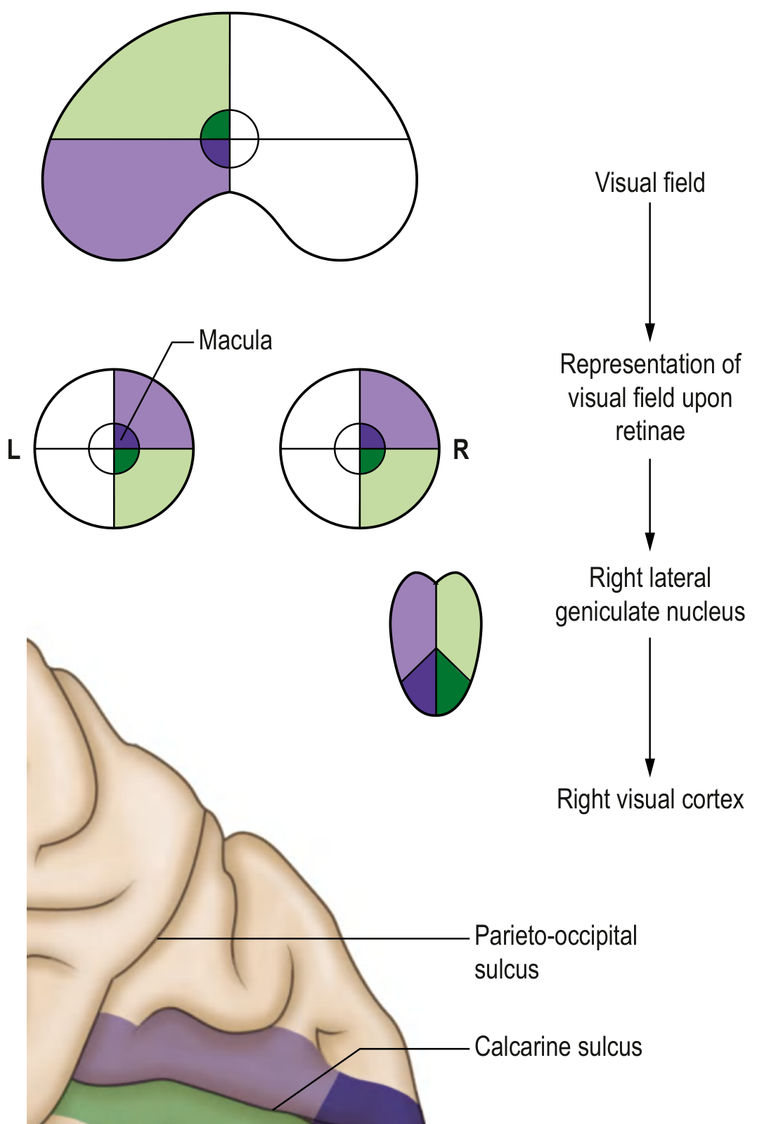
Representation of the visual field at various points along the visual pathway - Gray's Anatomy for Students
The fibers take two routes:
| Division | Retinal Origin | Loop Taken | Cortical Destination |
|---|---|---|---|
| Upper (dorsal) fibers | Upper retina (lower visual field) | Straight back | Upper lip of calcarine fissure |
| Lower (ventral) fibers | Lower retina (upper visual field) | Meyer's loop around inferior horn of lateral ventricle | Lower lip of calcarine fissure |
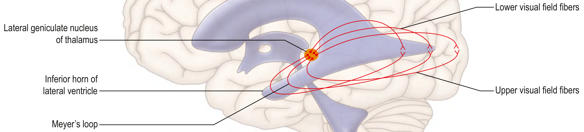
Thalamocortical fibers projecting to the primary visual cortex - Gray's Anatomy for Students
Meyer's loop is clinically important: a temporal lobe lesion can damage only the lower fibers of the optic radiation, causing a superior quadrantanopia ("pie in the sky" defect).
7. Primary Visual Cortex (V1 / Striate Cortex / Area 17)
Located on the banks of the calcarine fissure on the medial surface of the occipital lobe.
- Also called the striate cortex because of a visible stripe of white matter (line of Gennari).
- The macular fibers (from the central retina/fovea) project to the posterior pole of the occipital lobe and have a disproportionately large cortical representation (cortical magnification factor).
- Peripheral retinal fibers terminate anteriorly in the calcarine fissure.
Cortical Layers and Cell Types
The visual cortex, like other cortex, has 6 layers. Magno- and parvocellular LGB fibers project into layer 4.
| Cell Type | Stimulus Preference |
|---|---|
| Simple cells (layer 4) | Respond to bars/edges at a specific orientation AND position |
| Complex cells | Respond to moving bars of specific orientation (movement & velocity) |
| Hypercomplex cells | Respond to moving bars of specific orientation AND specific length/endpoint |
The cortex also has orientation columns (cells in a vertical column share preferred orientation) and ocular dominance columns (alternating input from each eye).
8. Visual Association Areas
Beyond V1, visual information is processed in:
| Area | Brodmann's # | Function |
|---|---|---|
| V1 (Primary) | Area 17 | Basic visual processing |
| V2/V3 (Prestriate) | Area 18, 19 | Form, texture, shape, depth, location, object recognition |
| Frontal eye field | Area 8 | Saccadic eye movements |
| Superior colliculus | Midbrain | Reflexive orienting movements |
There are two main cortical processing streams leaving V1:
- Dorsal stream ("where/how" pathway) → parietal cortex - spatial location, motion
- Ventral stream ("what" pathway) → temporal cortex - object recognition, color, form
9. Visual Field Defects at Different Levels
This is the most clinically tested aspect of the visual pathway:
| Site of Lesion | Visual Field Defect |
|---|---|
| One optic nerve (e.g., right) | Blindness in right eye (monocular) |
| Optic chiasm (central) | Bitemporal hemianopia (tunnel vision) |
| One optic tract (e.g., right) | Left homonymous hemianopia |
| Temporal lobe (Meyer's loop, right) | Left superior quadrantanopia ("pie in the sky") |
| Parietal lobe optic radiation (right) | Left inferior quadrantanopia |
| Right geniculocalcarine tract | Left homonymous hemianopia with macular sparing |
| Occipital (striate) cortex (right) | Left homonymous hemianopia with macular sparing |
Macular sparing occurs because: (i) macular fibers have a separate, large representation in the cortex, and (ii) the occipital pole has dual blood supply (middle + posterior cerebral arteries), so restricted infarcts spare the macula.
Summary Diagram of the Full Pathway
RETINA
  Rods/Cones → Bipolar Cells → Ganglion Cells
        ↓
  OPTIC DISC → OPTIC NERVE
        ↓
  OPTIC CHIASM
  [nasal fibers CROSS; temporal fibers STAY]
        ↓
  OPTIC TRACT
  [carries contralateral hemifield info]
        ↓                           ↓ (collaterals)
  LATERAL GENICULATE BODY     Pretectal area → Pupillary reflex
  (6 layers, retinotopic)     Superior colliculus → Eye movements
        ↓                     Suprachiasmatic n. → Circadian rhythm
  OPTIC RADIATION
  [Upper fibers: straight]
  [Lower fibers: Meyer's loop]
        ↓
  PRIMARY VISUAL CORTEX (V1, Area 17)
  Calcarine fissure, occipital lobe
        ↓
  VISUAL ASSOCIATION AREAS (18, 19)
  + Dorsal (where) & Ventral (what) streams
Sources:
- Sensory Physiology (attached PDF) - Chapters 145 & 146: Visual Pathway and Visual Cortex
- Gray's Anatomy for Students, p. 1346-1347 - Central Visual Pathway
- Principles of Neural Science (Kandel, 6th ed.), p. 547 - Geniculostriate Pathway
This is a shared conversation. Sign in to Orris to start your own chat.