
Wound healing
wound healing phases stages diagram
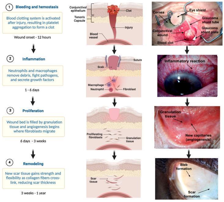
This infographic and clinical photograph series illustrates the four stages of ocular wound healing following glaucoma filtration surgery (e.g., shunt implantation). The diagram is organized into four horizontal rows representing the chronological phases: (1) Bleeding and Hemostasis (0–12 hours), showing platelet aggregation and a visible blood clot at the conjunctival wound site alongside surgical elements like the glaucoma shunt tube and plate. (2) Inflammation (1–6 days), characterized by neutrophil and macrophage recruitment and a clinical appearance of diffuse episcleral/conjunctival injection (redness). (3) Proliferation (6 days–3 weeks), showing fibroblast migration, collagen deposition, and angiogenesis (new capillary formation) clinical visible as fleshy granulation tissue. (4) Remodeling (3 weeks–1 year), depicting scar tissue maturation and the formation of a functional surgical bleb. The figure combines anatomical cross-sections of the conjunctiva and Tenon's capsule with real-world clinical images to demonstrate the physiological transition from acute injury to long-term surgical outcome in ophthalmology.
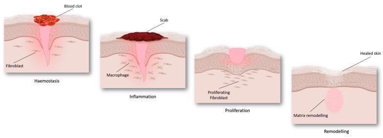
An anatomical pathophysiology diagram illustrating the four sequential stages of cutaneous wound healing: haemostasis, inflammation, proliferation, and remodeling. The illustration depicts a cross-section of the skin layers (epidermis and dermis). During 'Haemostasis', a red blood clot forms on the surface above a funnel-shaped injury, with fibroblasts visible in the surrounding dermis. In the 'Inflammation' stage, a dark-colored scab replaces the initial clot, and macrophages are identified in the underlying tissue. The 'Proliferation' stage demonstrates the formation of pinkish granulation tissue filling the wound gap, accompanied by 'proliferating fibroblasts' migrating into the site. Finally, the 'Remodeling' stage shows 'healed skin' with a restored epidermal surface and an underlying zone of 'matrix remodeling' replacing the previous injury site. The diagram serves as an educational summary of tissue repair mechanisms and cellular involvement at different phases of recovery.
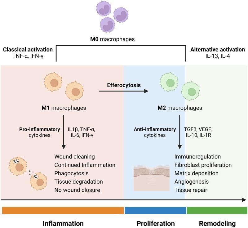
A pathophysiology diagram illustrating macrophage polarization during the stages of wound healing. The process begins with M0 macrophages (undifferentiated) which undergo 'Classical activation' via TNF-α and IFN-γ to become M1 macrophages. This pro-inflammatory phenotype predominates during the 'Inflammation' phase, secreting cytokines (IL-1β, TNF-α, IL-6, IFN-γ) and performing phagocytosis, tissue degradation, and wound cleaning. A transition via efferocytosis leads to 'Alternative activation' stimulated by IL-13 and IL-4, resulting in M2 macrophages. The M2 phenotype characterizes the 'Proliferation' and 'Remodeling' phases, producing anti-inflammatory and growth factors (TGFβ, VEGF, IL-10, IL-1R). These actions promote immunoregulation, fibroblast proliferation, matrix deposition, and angiogenesis, ultimately leading to successful tissue repair. The diagram uses color-coded backgrounds (orange for inflammation, blue for proliferation, green for remodeling) to link cellular phenotypes to clinical wound progression.
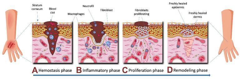
A medical pathophysiology diagram illustrating the four sequential phases of human skin wound healing: Hemostasis (A), Inflammation (B), Proliferation (C), and Remodeling (D). The cross-sectional views depict the stratum corneum, epidermis, and dermis. In the Hemostasis phase (A), a large blood clot fills the deep wound tissue with early macrophage presence. The Inflammatory phase (B) shows a persisting clot with an influx of neutrophils, macrophages, and early fibroblasts. The Proliferation phase (C) illustrates significant fibroblast activity and cellular multiplication beneath a shrinking scab as the epidermal gap begins to close. The final Remodeling phase (D) depicts a freshly healed epidermis and dermis with organized tissue structure and the absence of the initial clot and inflammatory cells. This illustration is an educational resource for understanding dermatological repair mechanisms and cellular recruitment during tissue injury.
"wound healing"[MeSH Terms] AND review
Wound Healing
- Schwartz's Principles of Surgery, 11th ed.
Phases of Wound Healing
1. Hemostasis (Immediate - minutes to hours)
- Vasoconstriction occurs first (mediated by norepinephrine, thromboxane A2, prostaglandin F2α).
- Platelet aggregation and degranulation release alpha-granule contents: fibrinogen, fibronectin, von Willebrand factor, Factor V, platelet-derived growth factor (PDGF), and TGF-β.
- Fibrin clot forms, acting as a scaffold for inflammatory cell recruitment.
- Platelets also release serotonin and histamine, causing vasodilation to allow leukocyte extravasation.
2. Inflammatory Phase (hours to ~7 days)
- First cells to arrive, recruited by IL-1, TNF-α, IL-8, PDGF, and bacterial products
- Phagocytose debris and bacteria; release reactive oxygen species (respiratory burst) and proteases (collagenase, elastase)
- Do not contribute to structural repair; their primary role is bactericidal cleansing
- Derived from blood monocytes; arrive after neutrophils
- The most important cell in wound healing - wounds depleted of macrophages heal poorly
- Functions: phagocytosis, debridement, orchestration of repair through cytokine secretion
- Secrete PDGF, TGF-β, TGF-α, FGF, VEGF, IL-1, TNF-α - the key growth factors driving the next phase
- Macrophage polarization: M1 (pro-inflammatory, early) → M2 (anti-inflammatory, pro-repair, late)
3. Proliferative Phase (~days 4-21)
- Fibroblasts migrate into the wound, proliferate, and begin synthesizing collagen (Types I and III) and extracellular matrix (ECM)
- Initially, Type III collagen predominates (weaker); later replaced by Type I
- Fibroblasts also produce fibronectin, hyaluronic acid, and proteoglycans
- The combination of new vessels, fibroblasts, and loose ECM forms granulation tissue - the hallmark of proliferation
- Granulation tissue appears as pink-red, moist, beefy tissue; it is highly vascular and fragile
- Stimulated by VEGF, FGF, and hypoxia
- Endothelial cells proliferate and migrate into the wound bed
- New capillaries are essential for oxygen and nutrient delivery
- Keratinocytes at wound margins begin migrating across the wound surface within hours of injury
- They dissolve the fibrin clot ahead using plasminogen activator and collagenase
- Keratinocyte migration is stimulated by EGF, TGF-α, and KGF
- Re-epithelialization rate is ~50% faster in a moist wound environment (a principle established by Galen and scientifically validated in the 1960s)
- Begins approximately day 4-5
- Fibroblasts differentiate into myofibroblasts (contain alpha-smooth muscle actin), which actively contract the wound margins
- Contraction reduces wound surface area, particularly significant in secondary healing wounds
- Over-contraction causes contractures (problematic at joints, eyelids, and the neck)
4. Maturation and Remodeling (~day 21 onwards, up to 2 years)
- Collagen is continuously synthesized and degraded by matrix metalloproteinases (MMPs) - collagenase (MMP-1), gelatinase (MMP-2, MMP-9)
- Type III collagen is replaced by Type I collagen, which has a more organized cross-linked structure
- The collagen fibers are reoriented along lines of tension
- Vascularity decreases; the wound becomes paler
- Maximum tensile strength of a healed wound is only ~80% of unwounded skin, reached at ~60 days
- A fresh wound at 1 week has only ~3% tensile strength; at 3 weeks ~20%; at 3 months ~80%
Key Growth Factors in Wound Healing
| Growth Factor | Source | Key Functions |
|---|---|---|
| PDGF | Platelets, macrophages | Chemotaxis of fibroblasts; stimulates collagen synthesis and angiogenesis |
| TGF-β | Platelets, macrophages, T cells | Stimulates matrix synthesis (fibronectin, collagen); TGF-β3 inhibits scar |
| VEGF | Macrophages, fibroblasts | Primary driver of angiogenesis |
| EGF | Platelets, macrophages | Stimulates proliferation of all epithelial cells |
| FGF | Fibroblasts, endothelium | Angiogenesis, mesoderm mitogenesis |
| IGF-1/2 | Platelets, liver | Promote protein and ECM synthesis |
| IL-1 | Macrophages, leukocytes | Pro-inflammatory; stimulates repithelialization |
Types of Wound Healing
| Type | Description |
|---|---|
| Primary intention | Wound edges are reapproximated soon after injury (sutured, stapled, glued). Minimal scarring. Used for clean surgical incisions. |
| Secondary intention | Wound left open; heals by granulation tissue, contraction, and epithelialization. Used for infected/contaminated wounds or large defects. More scarring. |
| Tertiary (delayed primary) | Wound initially left open, then closed after 3-5 days once infection risk is reduced. Best of both methods. |
Collagen in Wound Healing
- Type I - predominant mature dermal collagen; thick, organized fibers; high tensile strength
- Type III - early wound collagen; thin, disorganized; present in embryonic and early granulation tissue
- Collagen synthesis requires Vitamin C (hydroxylation of proline and lysine residues), oxygen, iron, and alpha-ketoglutarate
- Net collagen in a wound = synthesis - degradation (by MMPs)
Factors Affecting Wound Healing
Local Factors
- Infection: Bacterial counts >10⁵ organisms/gram of tissue impair healing; biofilm is a major barrier in chronic wounds
- Blood supply / ischemia: Oxygen is required for collagen synthesis, epithelialization, and bactericidal activity
- Wound moisture: Moist wounds epithelialize faster than dry wounds
- Foreign bodies / necrotic tissue: Impair healing; must be debrided
- Mechanical forces: Excessive tension leads to wide scars
Systemic Factors
| Factor | Effect |
|---|---|
| Diabetes mellitus | Impaired leukocyte function, neuropathy, ischemia (microvascular disease), glycosylation of collagen |
| Malnutrition / hypoalbuminemia | Reduced collagen synthesis; protein stores are critical |
| Vitamin C deficiency (scurvy) | Failure of collagen cross-linking; wound dehiscence |
| Zinc deficiency | Impaired epithelialization and fibroblast proliferation |
| Steroids / immunosuppression | Inhibit inflammation and fibroblast proliferation; thin wounds |
| Chemotherapy / radiation | Damage fibroblasts, endothelium; obliterative endarteritis |
| Aging | Decreased collagen synthesis, reduced macrophage response, slower epithelialization |
| Hypothyroidism / uremia | Impair collagen synthesis and fibroblast function |
| Jaundice | Bile salts impair fibroblast proliferation |
Abnormal Wound Healing
Hypertrophic Scars
- Raised, erythematous scars that remain within the wound boundaries
- Caused by excess collagen deposition
- Often regress over time
- Treatment: topical silicone, intralesional corticosteroids, compression therapy, surgical excision
Keloids
- Extend beyond the original wound margins - distinguish from hypertrophic scars
- More common in darker skin phototypes (Fitzpatrick types IV-VI)
- Predilection: earlobes, deltoid, presternal, face
- Do not spontaneously regress
- Treatment: repeated excision + adjuvant radiotherapy; intralesional steroids
Chronic Non-Healing Wounds
- Stuck in a prolonged inflammatory phase
- Common causes: venous ulcers, diabetic foot ulcers, pressure ulcers, arterial insufficiency
- Characterized by: elevated MMPs, decreased growth factors, senescent fibroblasts, persistent biofilm
- Management: debridement, offloading, moist dressing, treat underlying cause, advanced therapies (bioengineered skin, NPWT)
Wound Dehiscence
- Re-opening of closed wound, typically 5-8 days postoperatively
- Risk factors: infection, poor technique, malnutrition, steroid use, obesity
Wound Classification (Surgical)
| Class | Definition | Infection Risk |
|---|---|---|
| Class I - Clean | Uninfected, no inflammation, no GI/GU tract entry | ~2% |
| Class II - Clean-contaminated | GI or GU tract entered under controlled conditions | ~10% |
| Class III - Contaminated | Open wounds, major break in sterile technique | ~20% |
| Class IV - Dirty/infected | Old wounds with devitalized tissue or pus | ~40% |
Fetal Wound Healing
- High levels of TGF-β3 (anti-scarring isoform) and low TGF-β1/β2
- High hyaluronic acid content in fetal ECM
- Reduced inflammatory response
- Regenerative rather than reparative healing - this is the basis of research into scarless wound therapies
Summary - Key Points
- Wound healing occurs in four overlapping phases: hemostasis → inflammation → proliferation → remodeling
- The macrophage is the most critical cell in wound healing
- Collagen: Type III early → Type I late; maximum strength ~80% of normal
- Epithelialization is 50% faster in moist environments
- Tensile strength never returns to 100% of pre-injury levels
- Keloids extend beyond wound margins; hypertrophic scars do not
- Vitamin C and zinc are essential micronutrients for healing
- Diabetes and ischemia are the most clinically important impairers of healing