
Achlasia cardia
achalasia cardia barium swallow bird beak esophagus
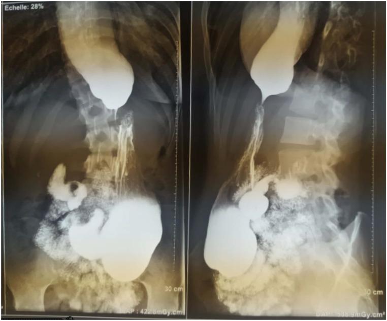
This diagnostic image consists of two views of a barium swallow x-ray radiography, showing the upper gastrointestinal tract in an anteroposterior and lateral perspective. The esophagus is markedly dilated (megaesophagus) with significant retention of the radiopaque barium contrast medium. At the gastroesophageal junction, there is a smooth, tapered narrowing of the distal esophagus, creating the classic 'bird's beak' appearance characteristic of achalasia. This narrowing represents the failure of the lower esophageal sphincter (LES) to relax. In the stomach, the barium presents a 'snowflake-falling' pattern, indicating irregular distribution and altered gastric filling. These findings are primary indicators of esophageal motility disorders, specifically achalasia, where the primary educational focus is on identifying the combination of proximal dilation and distal muscular obstruction at the cardia. This clinical imaging is essential for students and residents to recognize stages of Achalasia Cardia (AC) based on esophageal diameter and morphology.
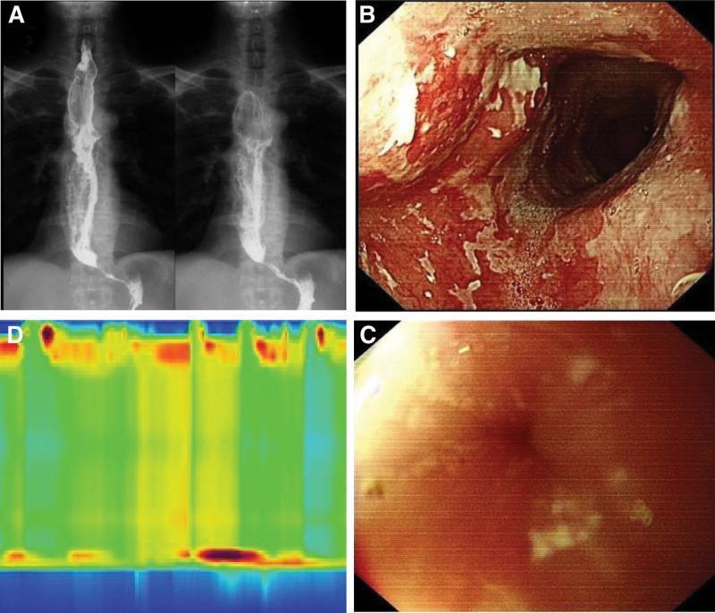
This composite diagnostic image illustrates the clinical findings of achalasia through multiple modalities. (A) Barium swallow (upper GI angiography) displays a significantly dilated esophageal lumen with a classic 'bird’s beak' sign, characterized by smooth, symmetric tapering at the gastroesophageal junction. (B, C) Endoscopic views (EGD) demonstrate stagnant food residue and extensive white mucosal plaques in the mid-esophagus, suggestive of secondary candidiasis or stasis-induced changes. View C specifically shows a tightly closed cardia that provided resistance to the endoscope. (D) A high-resolution esophageal manometry (HRM) topographic plot reveals aperistalsis in the esophageal body (low-pressure/green zones) and impaired relaxation of the lower esophageal sphincter (LES), indicated by persistent high-pressure bands at the distal end. Together, these images teach the classic triad of achalasia: esophageal dilation, failure of LES relaxation, and loss of peristalsis, useful for gastroenterology and radiology trainees.
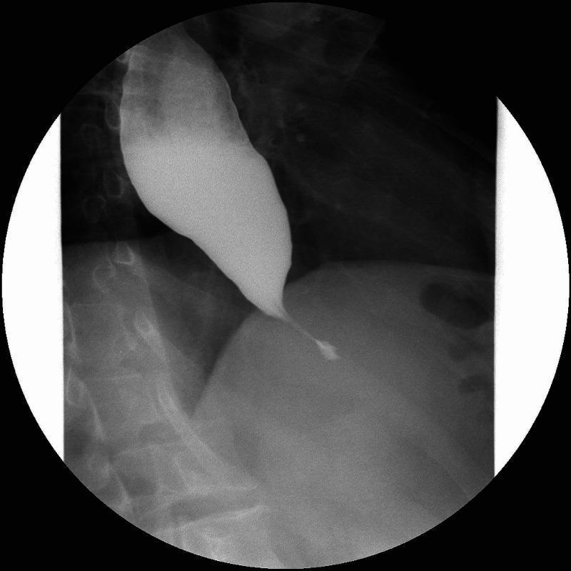
This diagnostic image is a contrast esophagogram (barium swallow) of the upper gastrointestinal tract. The radiograph reveals a significantly dilated esophagus filled with radiopaque barium contrast. There is a classic 'bird's beak' appearance at the distal esophagus, characterized by a smooth, symmetric, and tapered narrowing as the lumen approaches the gastroesophageal junction. This radiographic finding is pathognomonic for achalasia, reflecting the failure of the lower esophageal sphincter (LES) to relax and the loss of esophageal peristalsis. The contrast outlines a clear transition from the proximal megaesophagus to the functional obstruction at the cardia. This image is a primary educational tool for illustrating the diagnostic features of esophageal motility disorders in gastroenterology and radiology. It demonstrates the clinical significance of barium studies in evaluating patients presenting with symptoms such as dysphagia, regurgitation, and weight loss.
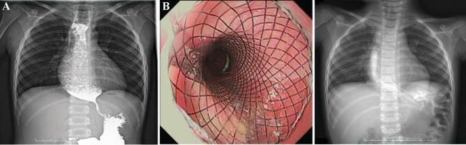
A three-panel figure documenting the diagnosis and management of achalasia of the cardia using a self-expandable metal stent (SEMS). Panel A: A preoperative upper gastrointestinal barium swallow radiograph (X-ray) showing classic features of achalasia. There is marked proximal esophageal dilation with a smooth, tapered narrowing at the gastroesophageal junction, creating a pathognomonic 'bird-beak' or 'beak-like' appearance, resulting in barium retention. Panel B: An endoscopic view showing a fully expanded, mesh-like metallic stent positioned within the esophageal lumen. The stent's lattice structure is in close apposition to the pink, slightly inflamed esophageal mucosa, ensuring patency. Panel C: Postoperative barium swallow radiograph showing the esophagus following stent placement. The contrast medium now passes freely through the previously constricted cardia into the stomach, indicating successful relief of the obstruction. This series illustrates the clinical transition from mechanical obstruction to restored luminal patency in a pediatric patient with refractory esophageal stricture.
achalasia cardia treatment
Achalasia Cardia
Definition
- Impaired or absent relaxation of the lower esophageal sphincter (LES) with swallowing
- Aperistalsis of the smooth muscle esophagus
Epidemiology
- Incidence: ~1 per 100,000 population per year
- Prevalence: ~10 per 100,000
- Affects males and females equally
- Bimodal age distribution: peaks in 20-40 years and again after 60 years
Pathophysiology
Core Defect
Inhibitory Neuron Loss
- Deglutitive inhibition (LES relaxation)
- Sequenced propagation of esophageal peristalsis
Excitatory Neuron Involvement
Etiology of Ganglion Cell Loss
- Myenteric plexus infiltrate shows cytotoxic T cells (resting and activated)
- Antibodies against myenteric neurons detected in patient sera (especially with specific HLA alleles)
- Suspected trigger: chronic latent HSV-1 infection (HSV-1 also found in LES of non-achalasic organ donors, suggesting genetic predisposition is required)
Chicago Classification (High-Resolution Manometry Subtypes)
| Type | HRM Finding | Features |
|---|---|---|
| Type I (Classic) | Aperistalsis, no pressurization | Advanced disease; dilated esophagus |
| Type II | Aperistalsis + panesophageal pressurization | Intermediate disease |
| Type III (Spastic) | Premature/spastic distal contractions | Unique pathogenesis - plexus inflammation without destruction; no LES relaxation |
Clinical Features
- Dysphagia - present in ALL patients for solids; majority also have liquid dysphagia. Gradual onset, often present for years before diagnosis. Eventually plateaus.
- Regurgitation - nonbilious, non-acid, mixed with saliva. Classic: food eaten hours or days earlier is regurgitated. Worsens with recumbency.
- Chest pain - in ~2/3 of patients early in disease course. Related to esophageal spasm (possibly longitudinal muscle). May spontaneously improve over time.
- Weight loss
- Hiccups
- Halitosis (from fermentation of retained food)
- Paradoxical "heartburn" - actually due to bacterial fermentation of retained food causing esophageal acidification, NOT true GER
- Aspiration pneumonia (up to 10% with advanced disease)
- Rarely: airway compromise/stridor from a dilated esophagus compressing the membranous trachea
Diagnosis
1. Barium Swallow
2. High-Resolution Manometry (HRM) - Gold Standard
- Absent or markedly impaired LES relaxation (elevated integrated relaxation pressure, IRP)
- Aperistalsis in the esophageal body
- Allows subtype classification (Types I, II, III)
3. Endoscopy
- Required in all patients to exclude pseudoachalasia (malignancy at the cardia)
- May show: dilated esophagus, retained food/saliva, "pop" when endoscope passes through the tight cardia
- Endoscopic view shows tightly closed cardia with resistance to scope passage
4. Other
- CT scan/EUS: to rule out infiltrative/extrinsic causes
- Timed barium esophagram: useful for monitoring treatment response
Differential Diagnosis
- Pseudoachalasia - malignancy (gastric, lung, pancreatic), infiltrative disease, or post-surgical distortion mimicking achalasia. Distinguished by shorter symptom duration, age >50, marked weight loss, CT/EUS findings.
- Chagas disease - T. cruzi infection destroying myenteric ganglion cells. Produces identical picture but with additional visceral involvement (megacolon, megaduodenum, cardiomyopathy). Diagnosed by serology/PCR.
- Distal esophageal spasm (DES) - differs in that LES relaxation is preserved (except in spastic achalasia/Type III)
- Other: scleroderma, EoE, GERD with stricture
Treatment
1. Pharmacologic (Temporizing Only)
- Nitrates (sublingual isosorbide dinitrate) and calcium channel blockers (nifedipine 30-40 mg/day before meals) - reduce LES pressure; limited efficacy, significant side effects (headache, flushing, orthostasis)
- Sildenafil (50 mg) - blocks PDE-5, increases cGMP from NO; peaks at 15-20 min, lasts <1 hour. Limited by cost and side effects.
- Best reserved as temporizing measures; not definitive treatment.
2. Botulinum Toxin Injection (Endoscopic)
- 80-100 units injected into the LES in 4 quadrants (intrasphincteric)
- Mechanism: irreversibly inhibits ACh release from cholinergic terminals, reducing neurogenic LES pressure
- ~66% improvement in dysphagia at 6 months; minimal efficacy at 1 year
- Repeat injections possible but cause fibrosis, limiting long-term use
- Best reserved for elderly/frail patients who are poor surgical candidates
3. Pneumatic Dilation
- Requires distension of LES to ≥3 cm to partially disrupt circular sphincter muscle
- Uses Rigiflex balloons (3.0, 3.5, 4.0 cm) over a guidewire, positioned fluoroscopically
- Outpatient procedure under conscious sedation
- Efficacy: ~85-90% overall; type II achalasia responds best (up to 100% in the European RCT)
- Main risk: esophageal perforation (~1%)
- May require repeat dilations
4. Surgical Heller Myotomy (Laparoscopic)
- Longitudinal division of the LES circular muscle (cardiomyotomy)
- Usually combined with partial fundoplication (Dor or Toupet) to prevent post-op GERD
- Efficacy: ~90%; effective across all subtypes
- Type III achalasia responds better to myotomy (86%) than to pneumatic dilation (40%)
- Risk of perforation: ~1% (comparable to pneumatic dilation)
5. POEM (Per-Oral Endoscopic Myotomy)
- Endoscopic approach creating a submucosal tunnel to perform myotomy
- Particularly effective for Type III (spastic) achalasia due to ability to extend the myotomy proximally
- Efficacy comparable to Heller myotomy; avoids external incisions
- Post-POEM GERD is a concern (no anti-reflux procedure done)
- Recent meta-analysis (PMID 40399616) comparing POEM vs Heller myotomy outcomes confirms comparable efficacy
Treatment Choice by Subtype:
| Subtype | Preferred Treatment |
|---|---|
| Type I | Heller myotomy or POEM |
| Type II | Pneumatic dilation (first-line) or Heller/POEM |
| Type III | POEM or Heller myotomy (dilation less effective) |
Complications
| Complication | Notes |
|---|---|
| Aspiration pneumonia | From nocturnal regurgitation |
| Esophageal candidiasis | From stasis of food/secretions |
| Esophageal carcinoma | Long-term risk; ~16x increased risk of squamous cell carcinoma |
| Megaesophagus | End-stage progressive dilation |
| Airway compromise | Dilated esophagus compressing trachea (rare) |
| Post-treatment GERD | Especially after myotomy without fundoplication |
Key Points Summary
- Achalasia = failure of LES relaxation + aperistalsis, due to loss of inhibitory myenteric neurons (NO/VIP)
- Gold standard diagnosis: high-resolution manometry
- Classic radiology: "bird's beak" on barium swallow
- HRM subtypes (I, II, III) guide therapy choice
- Definitive treatment: pneumatic dilation or Heller myotomy/POEM (~90% efficacy)
- Botulinum toxin: for poor surgical candidates only
- All patients need endoscopy to exclude pseudoachalasia/malignancy
Sources: Sleisenger & Fordtran's Gastrointestinal and Liver Disease, pp. 729-738; Clinical Gastrointestinal Endoscopy, 3e; Current Surgical Therapy, 14e