Respiratory tract epithelialm
Reading File
Finding Sources
Finding Sources
Reading File
Reading File
Reading File
Reading File
Reading File
Finding Sources
Loading Image
Loading Image
Loading Image
Here is a comprehensive overview of the epithelium of the respiratory tract, from nose to alveoli, based on Histology: A Text and Atlas and Junqueira's Basic Histology.
Respiratory Tract Epithelium
The lining epithelium changes progressively from the nasal vestibule to the alveoli, transitioning from stratified → pseudostratified → simple → squamous as the airways narrow.
1. Nasal Vestibule
- Stratified squamous epithelium — a direct continuation of facial skin
- Contains vibrissae (hairs) that trap large particulate matter
- Sebaceous glands assist in particle entrapment
- Posteriorly transitions into pseudostratified epithelium
2. Respiratory Region (Nasal Cavity, Trachea, Bronchi)
The dominant epithelium of the conducting airways is pseudostratified ciliated columnar epithelium (also called "respiratory epithelium"). It lines the nasal cavity, nasopharynx, larynx (most of it), trachea, bronchi, and larger bronchioles.
Five cell types make up this epithelium:
| Cell Type | Features | % of Cells |
|---|---|---|
| Ciliated columnar cells | ~250 cilia/cell; extend full thickness; mucociliary escalator | ~70% (most abundant) |
| Goblet cells | Mucus-secreting; interspersed among ciliated cells; increase during chronic irritation | 5–15% |
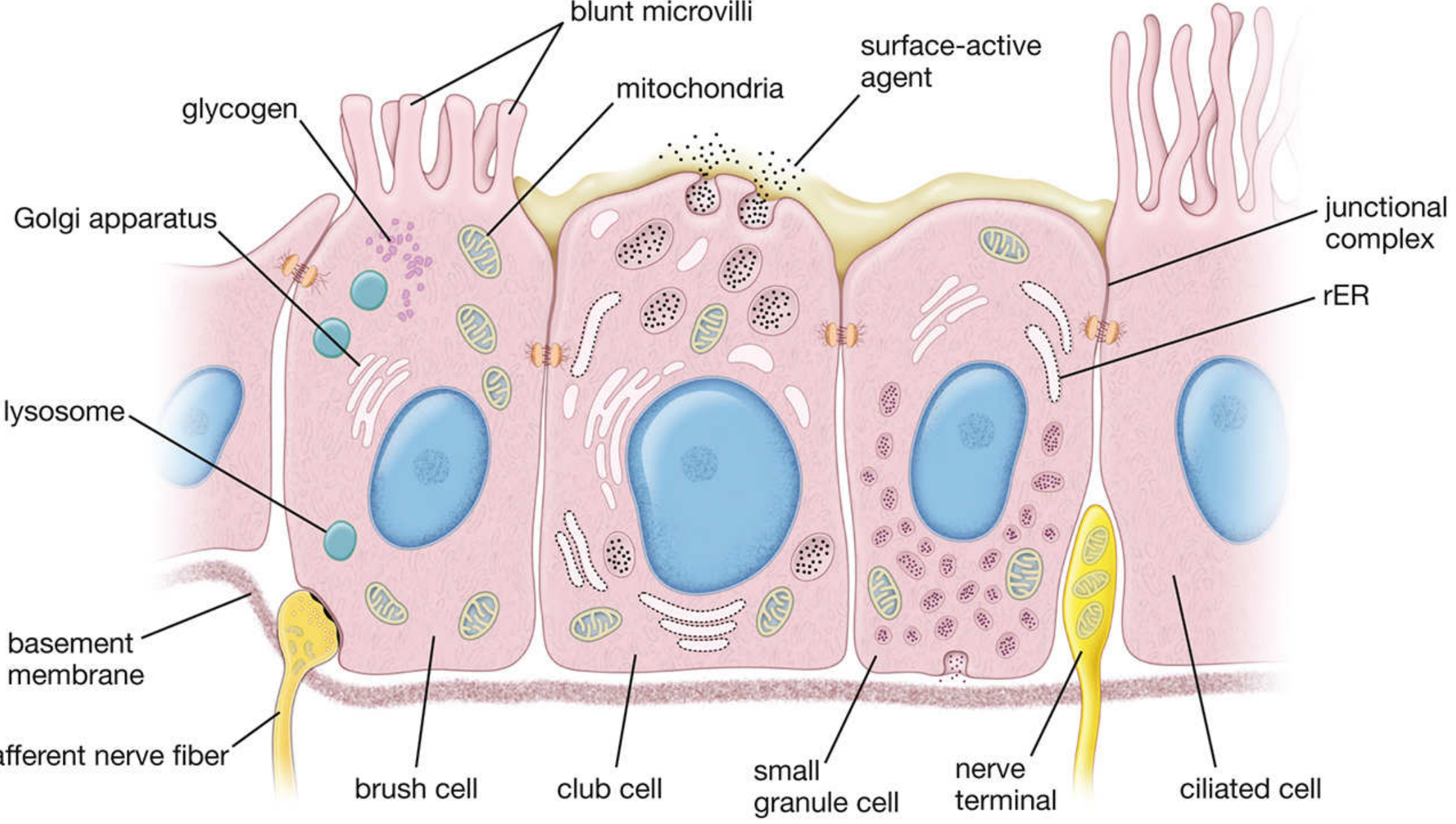
| Brush cells | Blunt microvilli; chemosensory/general sensory; synaptic contact with afferent nerve endings | Few |
| Small granule cells (Kulchitsky cells) | Secretory granules; endocrine cells of diffuse neuroendocrine system (DNES) | Few |
| Basal cells | On basement membrane; do not reach surface; stem cells for all other types | Variable |
Electron micrograph of human trachea — showing ciliated cells, goblet cells, and basal cells (×1,800):
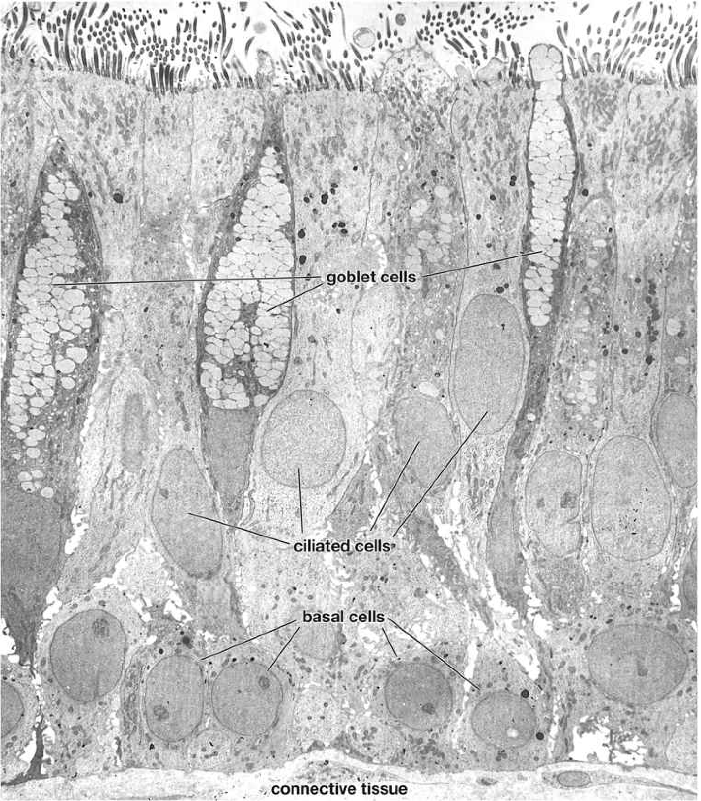
H&E photomicrograph of tracheal wall — epithelium (Ep) with goblet cells (G), prominent basement membrane (BM), lamina propria (LP), and submucosa (SM) with seromucous glands (×400):

Key functional point: The cilia beat in coordinated waves, forming the mucociliary escalator — sweeping the mucous blanket (with trapped particles) upward toward the pharynx for clearance.
3. Terminal Bronchioles
As the airways narrow (<1 mm diameter), the epithelium transitions:
- Simple columnar ciliated → simple cuboidal epithelium
- Goblet cells disappear
- Club cells (formerly Clara cells) become the dominant non-ciliated cell type
- Dome-shaped, non-ciliated, no mucus granules
- Secrete surface-active agents (surfactant-like lipoproteins) to aid airway expansion
- Also have detoxifying enzyme activity (smooth ER-rich)
Diagram of terminal bronchiolar epithelium showing brush cell, club cell, small granule cell, and ciliated cell with their ultrastructural features:
4. Respiratory Bronchioles
- Wall alternates between thick regions (club cells + smooth muscle) and thin regions (alveolar wall-like, for gas exchange)
- Scattered alveoli interrupt the wall — this is where gas exchange begins
5. Alveoli (Alveolar Ducts → Alveolar Sacs → Alveoli)
The alveolar epithelium consists of two cell types (pneumocytes):
| Cell | Type | Features |
|---|---|---|
| Type I alveolar cells (type I pneumocytes) | Extremely thin, squamous | Cover ~95% of alveolar surface; form the air-blood barrier |
| Type II alveolar cells (type II pneumocytes) | Cuboidal; lamellar bodies | Secrete surfactant (reduces surface tension); serve as progenitors for type I cells; innate immune role |
The interalveolar septum — the site of the air-blood barrier — consists of:
- Alveolar epithelium + basal lamina
- Capillary endothelium + basal lamina
- ± Interstitial connective tissue (elastic and reticular fibers)
Summary: Epithelial Transitions Along the Airway
Nasal vestibule → Stratified squamous
Nasal cavity/trachea/bronchi → Pseudostratified ciliated columnar (respiratory epithelium)
Bronchioles → Simple columnar/cuboidal ciliated + club cells
Terminal bronchioles → Mostly club cells (simple cuboidal)
Respiratory bronchioles → Cuboidal (club cells) + interrupted by alveoli
Alveolar ducts/sacs → Squamous (type I) + cuboidal (type II) pneumocytes
Olfactory Epithelium (roof of nasal cavity)
A special region: pseudostratified but contains:
- Bipolar olfactory neurons (chemosensory)
- Supporting (sustentacular) cells
- Basal stem cells
Sources: Histology: A Text and Atlas with Correlated Cell and Molecular Biology (Ross & Pawlina), 9th ed.; Junqueira's Basic Histology, 17th ed.
Grades of
Asking for Preferences
Mets
Asking for Preferences
This is a shared conversation. Sign in to Orris to start your own chat.