
Infective endocarditis latest diagnostic crieterion
"infective endocarditis" AND "diagnostic criteria" AND "Duke criteria"
infective endocarditis 2023 ESC guidelines diagnostic criteria updated Duke criteria
https://pmc.ncbi.nlm.nih.gov/articles/PMC10681650
infective endocarditis Duke criteria diagnostic echocardiography vegetation
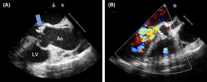
Diagnostic ultrasound imaging featuring transesophageal echocardiography (TEE) for the assessment of infective endocarditis. Panel A displays a grayscale long-axis view of the left ventricle (LV) and aorta (Ao). A blue arrow identifies a prominent, hyperechoic, and motile vegetation measuring approximately 1.5 cm localized on the noncoronary cusp of the aortic valve. Panel B shows the same view with Color Doppler application, demonstrating high-velocity, mosaic, and turbulent retrograde flow from the aorta into the left ventricle, indicating severe aortic regurgitation. The imaging illustrates the cardiac manifestations of systemic infection, highlighting the correlation between valvular vegetations and resultant hemodynamic dysfunction. This visual is intended for medical education regarding the diagnostic criteria for Duke's definitive infective endocarditis and the assessment of valvular pathology.
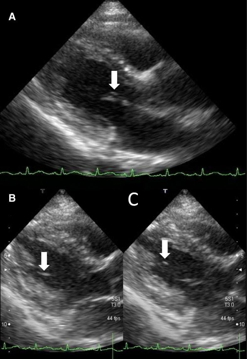
This set of diagnostic images consists of three transthoracic echocardiography (TTE) frames in the parasternal long-axis (PLAX) view, showcasing cardiac pathology associated with infective endocarditis. Panel A displays a 4 x 1 mm echogenic mass (vegetation) attached to the anterior leaflet of the mitral valve, indicated by a white arrow. Panels B and C capture different phases of the cardiac cycle, specifically presystole and end-diastole, highlighting a second, larger vegetation. This second mass is elongated and mobile, measuring approximately 10 x 2 mm, and is located on the chorda tendinea of the mitral valve. The images demonstrate classic ultrasonographic findings of endocardial vegetations, emphasizing their morphology, size, and anatomical attachment sites within the left ventricle. These features are critical for diagnosing infective endocarditis using clinical criteria such as the Modified Duke Criteria. The content is suitable for cardiology and internal medicine education, focusing on valvular heart disease and diagnostic ultrasound interpretation.
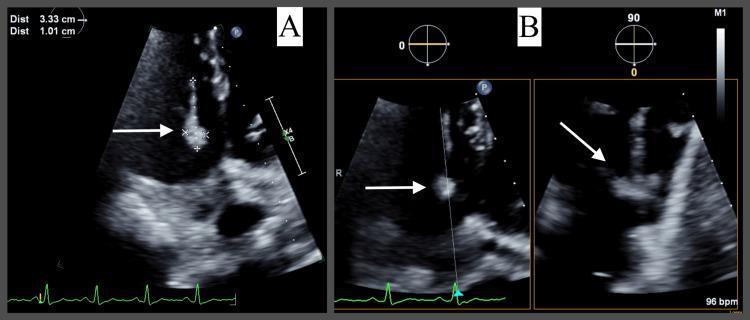
Transthoracic echocardiography (TTE) diagnostic images demonstrating a large, mobile vegetation on the tricuspid valve, characteristic of infective endocarditis. Image A shows a zoomed-in apical four-chamber view where white arrows highlight a hyperechoic mass attached to a tricuspid valve leaflet; on-screen calipers measure the vegetation at 3.33 cm x 1.01 cm. Image B utilizes biplane visualization, presenting simultaneous orthogonal planes to detail the three-dimensional morphology and spatial relationship of the mass within the right heart chambers. The images illustrate key diagnostic criteria for right-sided endocarditis, focusing on mass size, echogenicity, and attachment to valvular structures. This content is relevant for cardiology and infectious disease education, specifically for teaching the echocardiographic identification of major Duke criteria in clinical practice.
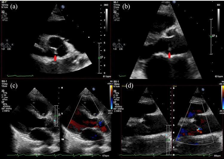
Transthoracic echocardiography (TTE) panel illustrating diagnostic findings of infective endocarditis. (a, b) Parasternal long-axis views in 2D mode showing a distinct, irregular echogenic mass (vegetation) attached to the aortic valve leaflet, indicated by red arrows. The vegetation displays independent motion and different acoustic properties compared to the valve tissue. (c, d) Parasternal long-axis views utilizing color flow Doppler to assess valvular function. These images demonstrate a mosaic-colored retrograde jet originating from the aortic valve and extending into the left ventricular outflow tract during diastole, diagnostic of mild aortic regurgitation. This educational visual facilitates the identification of major modified Duke criteria through imaging evidence of valvular vegetations and secondary valvular incompetence (regurgitation) in the clinical context of suspected cardiac infection.
Infective Endocarditis: 2023 Diagnostic Criteria
Background
Classification of IE Cases
| Classification | Clinical Criteria Required |
|---|---|
| Definite | 2 major criteria OR 1 major + 3 minor OR 5 minor criteria |
| Possible | 1 major + 1 minor OR 3 minor criteria |
| Rejected | Firm alternate diagnosis; resolution with antibiotics ≤4 days; no pathologic evidence at surgery with antibiotics ≤4 days; does not meet possible criteria |
Pathologic Criteria (sufficient alone for Definite IE)
- Microorganisms identified by culture or histology in a vegetation, explanted prosthetic valve, ascending aortic graft, CIED, or embolus
- Histopathologic findings of active endocarditis in cardiac tissue, vegetation, explanted valve, CIED, or embolus
I. MAJOR CRITERIA
A. Microbiologic Major Criteria (NEW updates in bold)
- Typical organisms (commonly cause IE) isolated from 2 or more separate blood culture sets
- Typical organisms include: Viridans streptococci, Streptococcus gallolyticus (bovis), HACEK group organisms, Staphylococcus aureus, community-acquired enterococci (without primary focus), and now Enterococcus faecalis regardless of acquisition source (NEW - previously excluded ~30% of E. faecalis definite IE)
- Non-typical organisms (occasionally/rarely cause IE) isolated from 3 or more separate blood culture sets
- Note: Timing and separate venipuncture requirements removed in 2023 (previously required 12-hour interval)
- Positive PCR or nucleic acid-based technique from blood for Coxiella burnetii, Bartonella spp., or Tropheryma whipplei (NEW)
- C. burnetii: antiphase I IgG antibody titer >1:800, or isolated from a single blood culture
- Bartonella spp.: IgG titer >1:800 by indirect immunofluorescence assay (enzyme immunoassay also acceptable as NEW addition)
- Amplicon/metagenomic sequencing positive for C. burnetii, Bartonella, or T. whipplei from blood (NEW)
B. Imaging Major Criteria (NEW additions)
- Vegetation, valvular/leaflet perforation, valvular/leaflet aneurysm, abscess, pseudoaneurysm, or intracardiac fistula
- Significant new valvular regurgitation (worsening/changing of pre-existing not sufficient)
- New partial dehiscence of prosthetic valve (compared with previous imaging)
- Cardiac CT is now equivalent to echocardiography as a major imaging criterion (NEW)
- Abnormal metabolic activity involving a native or prosthetic valve, ascending aortic graft (with valve involvement), intracardiac device leads, or other prosthetic material
- For prosthetic valves implanted >3 months ago or native valves: qualifies as Major Criterion
- For valves implanted <3 months ago: counts as Minor Criterion only (inflammation vs. infection difficult to distinguish)
C. Surgical Major Criterion (NEW CATEGORY):
- Evidence of IE observed by direct intraoperative inspection during cardiac surgery, in the absence of major microbiologic or imaging criteria, and in the absence of pathologic criteria
II. MINOR CRITERIA
A. Predisposing Conditions (expanded)
- Previous history of IE (NEW)
- Prosthetic valve or previous valve repair
- Congenital heart disease (moderate-to-high risk)
- More than mild regurgitation or stenosis of any etiology
- Endovascular cardiac implantable electronic device (CIED) (NEW)
- Transcatheter valve implant/repair (NEW)
- Hypertrophic obstructive cardiomyopathy
- Injection drug use
B. Fever
- Temperature ≥38.0°C (100.4°F)
C. Vascular Phenomena (expanded)
- Clinical or radiographic evidence of: arterial emboli, septic pulmonary infarcts, cerebral abscess, splenic abscess (NEW), mycotic aneurysm, intracranial hemorrhage, conjunctival hemorrhages, Janeway lesions, purulent purpura, thromboembolic phenomena
- Spondylodiscitis (NEW in 2023 ESC criteria)
D. Immunologic Phenomena
- Positive rheumatoid factor, Osler's nodes, Roth's spots, immune complex-mediated glomerulonephritis
E. Microbiological Evidence (not meeting Major Criteria)
- Positive blood cultures for an organism consistent with IE but not meeting major criteria requirements
- NEW: Positive culture, PCR, or nucleic acid-based test (amplicon/metagenomic sequencing, in situ hybridization) for an organism consistent with IE from a sterile body site other than cardiac tissue, prosthesis, or embolus
Key Changes from 2000 Modified Duke Criteria: Summary Table
| Domain | Old (2000) | New 2023 |
|---|---|---|
| E. faecalis | Non-typical (3+ cultures needed) | Typical organism (2+ cultures sufficient) |
| Timing of blood cultures | 12 h apart required | Removed |
| Cardiac CT | Not included | Major criterion (= echo) |
| FDG PET/CT | Not included | Major criterion |
| Intraoperative inspection | Not included | New Major criterion |
| PCR/sequencing | Not included | Major criterion for C. burnetii, Bartonella, T. whipplei |
| Metagenomic sequencing | Not included | Minor (for other organisms); Major for above 3 |
| Splenic/cerebral abscess | Not included | New minor vascular criterion |
| Prior IE, CIED, transcatheter valve | Not included | New minor predisposing criteria |
Echocardiographic Findings (illustrative)
Performance Comparison
| Criteria Version | Sensitivity | Specificity |
|---|---|---|
| 2000 Modified Duke | 74.9% | 94.9% |
| 2015 ESC | 80.0% | 93.9% |
| 2023 Duke-ISCVID | 84.2% | 93.9% |
| 2023 ESC | 82.7% | 92.3% |
- Textbook of Clinical Echocardiography, Table 14.1 (2023 Duke Criteria)
- Harrison's Principles of Internal Medicine 22E, Further Reading