
Ventilator associated pneumonia MD Anaesthesia pg exam 30 marks answer
Reading File
Now let me search the medical library for comprehensive VAP content in parallel.
Finding Sources
Finding Sources
Searching PubMed
"ventilator-associated pneumonia"[MeSH Terms] AND prevention
Excellent results. Let me now read the most relevant sources in parallel - Miller's Anesthesia (primary for MD Anaesthesia exam) and Barash Clinical Anesthesia, plus the comprehensive Frameworks textbook.
Reading File
Reading File
Reading File
Excellent content gathered. Let me read more of the Barash Clinical Anesthesia content and the Goldman-Cecil for complete treatment/management details.
Reading File
Reading File
Reading File
Let me fetch the flowchart image and also get pathogenesis/diagnosis content from Goldman-Cecil and Harrison's.
Loading Image
Reading File
Finding Sources
Now let me get the Harrison's content on pathogenesis and prevention, and also the Goldman-Cecil antibiotic table.
Reading File
Reading File
I now have comprehensive content from Harrison's (2025), Barash Clinical Anesthesia (9e), Miller's Anesthesia (10e), and Goldman-Cecil Medicine. Let me compile the complete 30-mark MD Anaesthesia exam answer.
Reading File
Reading File
I now have comprehensive information from multiple authoritative sources. Let me write the complete 30-mark MD Anaesthesia examination answer.
Ventilator-Associated Pneumonia (VAP)
MD Anaesthesia PG Examination - 30 Marks Answer
1. DEFINITION
Ventilator-associated pneumonia (VAP) is defined as a new lower respiratory tract infection developing in a patient who has been on mechanical ventilation for 48 hours or more via an endotracheal or tracheostomy tube, and which was not present or incubating at the time ventilation was initiated.
- Early-onset VAP: occurs within the first 48-72 hours of intubation
- Late-onset VAP: occurs after 72 hours (>5 days of hospitalization in some classifications)
The CDC/NHSN uses a broader "Ventilator-Associated Event (VAE)" surveillance framework - requiring at least 4 days of mechanical ventilation - which avoids subjective radiologic interpretation.
(Harrison's Principles of Internal Medicine 22E; Barash Clinical Anesthesia 9e)
2. INCIDENCE AND EPIDEMIOLOGY
- VAP occurs in up to 10-40% of intubated patients; it is the most frequent ICU-acquired infection and most frequent ventilator-associated complication
- Peak incidence: 5-9 days after intubation; cumulative risk is proportional to duration of ventilation
- Incidence using NHSN criteria: <4% per mechanical ventilation episode (reflecting improved prevention practices)
- VAP accounts for approximately 35% of all hospital-acquired pneumonia cases
- With COVID-19, incidence has again increased in mechanically ventilated patients
- On any given ICU day, ~10% of patients have pneumonia - VAP in the overwhelming majority
(Harrison's 22E; Frameworks for Internal Medicine; Barash 9e)
3. PATHOGENESIS
Three factors are critical:
1. Colonization of the oropharynx with pathogenic microorganisms
2. Aspiration of these organisms from the oropharynx into the lower respiratory tract
3. Compromise of normal host defense mechanisms (impaired cough reflex, ciliary dysfunction, altered mucosal immunity)
Specific Mechanisms:
| Mechanism | Details |
|---|---|
| Microaspiration | Almost all intubated patients experience microaspiration; pooling of secretions above the ETT cuff leaks past the cuff |
| ETT biofilm | Bacteria form biofilm on the inner surface of the endotracheal tube - reservoir for persistent/recurrent infection |
| Colonization progression | Colony counts increase to high levels days before clinical pneumonia manifests |
| Impaired airway protection | ETT prevents normal cough reflex and mucociliary clearance |
| Aspiration of gastric contents | Acid suppression raises gastric pH, allowing bacterial overgrowth and colonization |
| Sinusitis | Bacterial sinusitis (especially with nasal tubes) predisposes to VAP via microaspiration of infected secretions |
| Hematogenous spread | Less common route - organisms seed the lungs from distant infection sites |
(Harrison's 22E, p.1073; Goldman-Cecil Medicine; Barash 9e)
4. MICROBIOLOGY
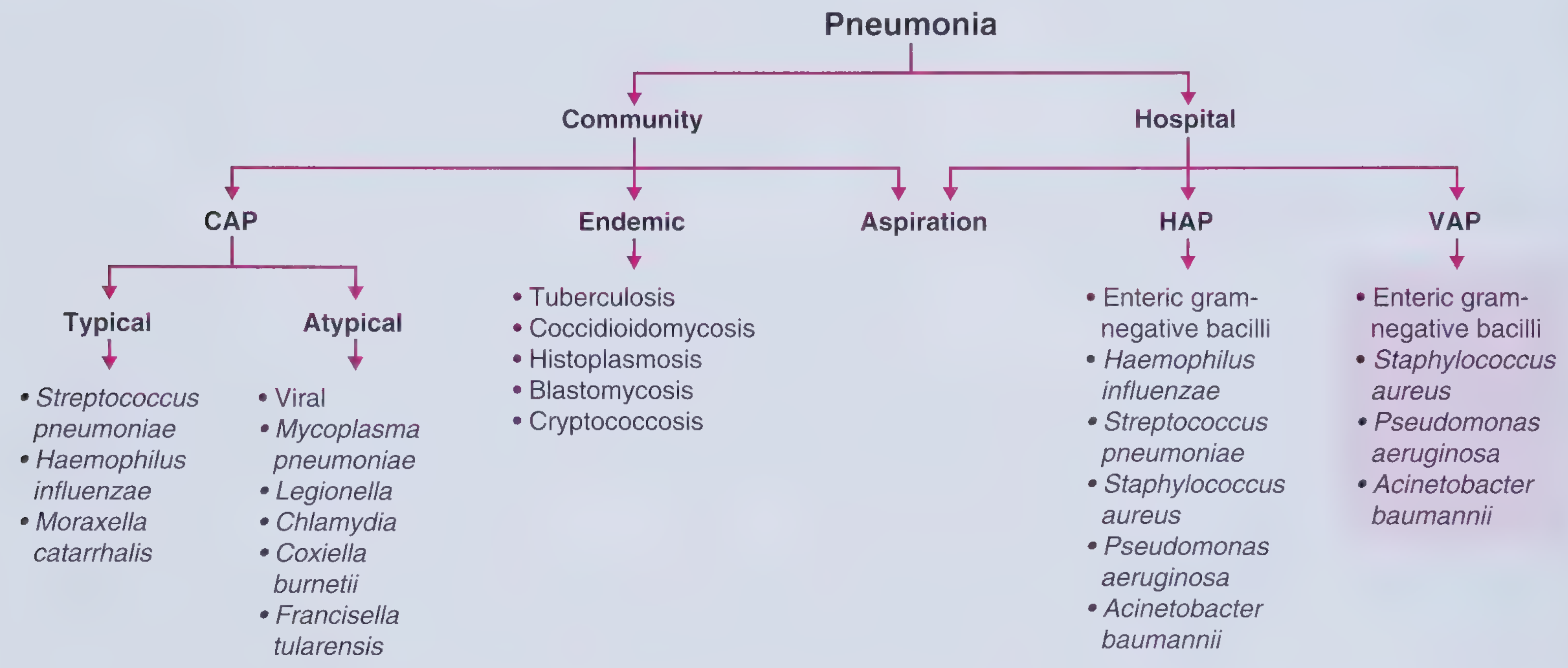
Non-MDR (Core) Pathogens - typically Early-Onset VAP:
- Streptococcus pneumoniae
- Haemophilus influenzae
- Methicillin-sensitive Staphylococcus aureus (MSSA)
- Antibiotic-sensitive Enterobacteriaceae (E. coli, Klebsiella pneumoniae, Proteus spp., Enterobacter spp., Serratia marcescens)
MDR Pathogens - typically Late-Onset VAP:
- Pseudomonas aeruginosa (most important; dual-drug coverage often required)
- Methicillin-resistant Staphylococcus aureus (MRSA)
- Acinetobacter baumannii (may be susceptible only to polymyxins)
- ESBL-producing strains
- Carbapenem-resistant Enterobacteriaceae
- Legionella pneumophila, Burkholderia cepacia
Key distinction: Early-onset VAP (< 48-72 h) is generally caused by antibiotic-sensitive organisms; late-onset VAP (>72 h, especially with prior antibiotic use) is associated with MDR pathogens and carries higher mortality.
(Harrison's 22E Table 131-6; Frameworks for Internal Medicine)
5. RISK FACTORS
Patient Factors:
- Advanced age, immunocompromised state, malnutrition
- COPD, ARDS, high APACHE-II score
- Reduced level of consciousness
Treatment/Procedure-Related Factors:
- Duration of mechanical ventilation (most important risk factor)
- Supine positioning
- Nasogastric/nasotracheal tubes (sinusitis predisposition)
- Continuous IV sedation (reduces ability to clear secretions)
- Prior intravenous antibiotic use within 90 days (strongest risk factor for MDR)
- Presence of septic shock; ARDS before VAP onset
- Acute renal replacement therapy before VAP
For MDR Pathogens specifically:
- Prior IV antibiotic use within 90 days
- Presence of septic shock
- ARDS before VAP
- ≥5 days of hospitalization before VAP onset
- Acute renal replacement therapy before VAP
(Frameworks for Internal Medicine; Harrison's 22E)
6. CLINICAL FEATURES AND DIAGNOSIS
Clinical Criteria:
VAP is diagnosed when ALL of the following are present:
- New or progressive infiltrate on chest radiograph
- Plus at least 2 of the following:
- Fever (>38.5°C) or hypothermia (<36°C)
- Leukocytosis (>10,000/mm³) or leukopenia (<4,000/mm³)
- Purulent tracheobronchial secretions (change in color/consistency)
- Deterioration in oxygenation (↑FiO₂ requirement, ↑PEEP)
Important caveat: Radiographic findings alone are insufficient - pulmonary edema, atelectasis, ARDS, and pulmonary contusion can mimic VAP on CXR.
Clinical Pulmonary Infection Score (CPIS):
A composite score using temperature, WBC, tracheal secretions, PaO₂/FiO₂, CXR, and culture results. Score >6 supports VAP diagnosis.
Microbiologic Diagnosis:
Non-invasive (preferred):
- Endotracheal aspirate (quantitative culture: threshold ≥10⁶ CFU/mL)
- Blood cultures (positive in ~15% of VAP cases)
Invasive:
- Bronchoalveolar lavage (BAL): threshold ≥10⁴ CFU/mL; intracellular organisms seen in >5% of cells = positive
- Protected Specimen Brush (PSB): threshold ≥10³ CFU/mL
- Blind bronchial sampling as alternative if bronchoscopy unavailable
IDSA guideline preference: Tracheal aspirate over BAL (though conflicting evidence - BAL appropriate for selected patients).
(Barash 9e; Harrison's 22E; Goldman-Cecil Medicine)
7. PREVENTION - THE VAP BUNDLE
The IHI (Institute for Healthcare Improvement) "100,000 Lives Campaign" (2005) popularized the VAP bundle approach. When implemented collectively, bundle components show synergistic benefit greater than any single intervention.
Core Bundle Elements:
| Intervention | Evidence Basis |
|---|---|
| Head-of-bed elevation 30-45° | Reduces microaspiration; simplest, no-cost measure |
| Daily sedation vacation | Reduces ventilator days, allows extubation readiness assessment |
| Daily assessment for extubation readiness | Minimizes duration of mechanical ventilation |
| Strict hand hygiene | Role-modeling increases compliance from 20% to 94% |
| Oral hygiene with chlorhexidine | Reduces VAP incidence by ~30% (effect strongest in cardiac surgery patients; blinded studies less clear) |
| Subglottic secretion drainage (SSD) | ETTs with subglottic suctioning ports - meta-analysis supports reduction in VAP incidence and shorter ICU/hospital stay |
| Avoidance of nasal tubes | Prevents sinusitis and secondary VAP |
| Selective Digestive Decontamination (SDD) | Non-absorbable antimicrobials reduce VAP; controversial due to risk of resistance selection |
Additional Prevention Measures:
- Avoid unnecessary sedation - use lowest effective dose
- Early mobilization - reduces ventilator days
- Silver-coated endotracheal tubes - NOT proven beneficial despite theoretical rationale
- Sucralfate vs. acid-suppression: Acid suppression (PPIs/H2 blockers) raises gastric pH → bacterial overgrowth → increased VAP risk; sucralfate maintains acid barrier; reserve acid suppression for high-risk GI bleeding patients
- Noninvasive ventilation (NIV) where possible - eliminates ETT-related risk entirely
- Prophylactic antibiotics: 2 days of IV amoxicillin-clavulanate in post-cardiac-arrest patients on targeted hypothermia reduces early VAP by ~45%
(Goldman-Cecil Medicine; Miller's Anesthesia 10e; Barash 9e; Harrison's 22E)
8. TREATMENT
General Principles:
- Do not delay treatment - start antibiotics after culture specimens are collected; delay increases mortality
- De-escalation strategy: Start broad, then narrow spectrum based on culture results at 48-72 hours
- Antibiotic selection based on local antibiogram (hospital-specific resistance patterns)
- Duration: 7-8 days (vs. longer courses) is recommended for most VAP - shorter courses reduce resistance without increasing failure rates
Early-Onset VAP (without MDR risk factors):
Narrow-spectrum, single-agent therapy:
| Antibiotic | Dose |
|---|---|
| Ceftriaxone | 1-2 g IV every 12-24 hours |
| Levofloxacin | 750 mg IV every 24 hours |
| Ciprofloxacin | 400 mg IV every 8 hours |
| Ampicillin-sulbactam | 1.5-3 g IV/IM every 6 hours |
| Ertapenem | 1 g IV/IM once daily |
Late-Onset VAP / MDR Risk Factors:
Broad-spectrum, dual-agent therapy covering MDR gram-negatives AND MRSA:
For gram-negatives (choose one):
- Antipseudomonal cephalosporin: Ceftazidime 2 g IV q8h, or Cefepime 1-2 g q8-12h; newer: ceftazidime/avibactam 2.5 g q8h, ceftolozane/tazobactam 3 g q8h
- Antipseudomonal carbapenem: Meropenem 1 g q8h, Imipenem 500 mg q6h
- Piperacillin-tazobactam 4.5 g IV q6h
For MRSA coverage (add one):
- Vancomycin 15-20 mg/kg IV q8-12h (monitor troughs; target 10-15 mcg/mL)
- Linezolid 600 mg IV/PO q12h - preferred over vancomycin in renal insufficiency and high-MIC MRSA isolates (15% more efficacious than adjusted-dose vancomycin)
Indications for MRSA Coverage:
- Prior IV antibiotic use within 90 days
- ICU where >10-20% of S. aureus isolates are methicillin-resistant
- Unit where MRSA prevalence is unknown
- Known MRSA carrier
- Severely ill patient at high risk of mortality
For Acinetobacter baumannii:
- Carbapenem or ampicillin-sulbactam for susceptible strains
- Polymyxin B or Colistin for pan-resistant strains
- Inhaled colistin may be added as adjunct
For Pseudomonas aeruginosa:
- Combination therapy recommended (40-50% failure rate with any single regimen)
- Consider prolonged/continuous infusion of beta-lactams to optimize pharmacodynamics
- Biofilm on ETT allows persistence - early ETT change may be considered in refractory cases
(Goldman-Cecil Medicine Table 85-10; Harrison's 22E; Barash 9e)
9. COMPLICATIONS
- Increased mortality: Crude mortality 30-70%; attributable mortality ~5-25% (VAP with COVID-19 has higher attributable mortality)
- Prolonged mechanical ventilation: Most important consequence - each additional day on ventilator increases risk of further complications
- Increased ICU and hospital length of stay
- Necrotizing pneumonia (P. aeruginosa, S. aureus): pulmonary hemorrhage, empyema
- Long-term: bronchiectasis, parenchymal scarring, recurrent pneumonia
- Nutritional depletion - catabolic state worsens outcomes
- Treatment failure - especially with MRSA (40% clinical failure with standard vancomycin) and Pseudomonas (40-50% failure regardless of regimen)
10. PROGNOSIS AND MONITORING
- Clinical improvement (especially oxygenation) expected within 48-72 hours of appropriate therapy
- Chest radiographic findings often worsen initially - less reliable than clinical/oxygenation parameters for response assessment
- Procalcitonin has a role in guiding duration of therapy alongside clinical response
- Serial quantitative cultures clarify microbiologic response
Biomarkers used in monitoring VAP:
- Procalcitonin (PCT) - guides antibiotic de-escalation and duration
- C-reactive protein (CRP)
- Serial CPIS scoring
SUMMARY TABLE
| Feature | Details |
|---|---|
| Definition | Pneumonia developing ≥48 h after endotracheal intubation |
| Incidence | Up to 40% of intubated patients; peak at 5-9 days |
| Mortality | Crude 30-70%; attributable 5-25% |
| Early-onset organisms | S. pneumoniae, H. influenzae, MSSA, sensitive gram-negatives |
| Late-onset organisms | MRSA, P. aeruginosa, Acinetobacter, ESBL-producers |
| Diagnosis | New CXR infiltrate + fever/WBC change + purulent secretions + micro cultures |
| Gold standard culture | Endotracheal aspirate (≥10⁶ CFU/mL) or BAL (≥10⁴ CFU/mL) |
| Prevention bundle | Head elevation 30-45°, daily sedation vacation, extubation readiness, hand hygiene, CHX oral care, subglottic suctioning |
| Early-onset treatment | Ceftriaxone or levofloxacin (monotherapy) |
| Late-onset treatment | Antipseudomonal beta-lactam + vancomycin/linezolid |
| De-escalation | At 48-72 h based on culture results |
| Duration | 7-8 days for most cases |
References: Miller's Anesthesia 10e (p.11450-11452); Barash, Cullen & Stoelting's Clinical Anesthesia 9e (p.4862-4865); Harrison's Principles of Internal Medicine 22E (2025, p.1072-1077); Goldman-Cecil Medicine (Table 85-10); Frameworks for Internal Medicine
This is a shared conversation. Sign in to Orris to start your own chat.