
Perthes disease for 10marks
"Legg-Calve-Perthes Disease"[MeSH Terms]
Perthes disease radiograph femoral head fragmentation
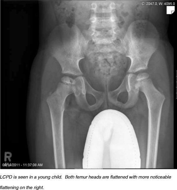
This diagnostic image is an anteroposterior (AP) radiograph of the pelvis and proximal femurs in a pediatric patient. The radiograph demonstrates bilateral pathological changes in the femoral heads consistent with Legg-Calv)-Perthes disease (LCPD). There is significant bilateral flattening and fragmentation of the femoral epiphyses (coxa plana), with more pronounced deformity and articular surface collapse on the right side compared to the left. The right femoral head shows increased radiodensity (sclerosis) and broadening of the femoral neck (coxa magna). The joint spaces appear preserved, but the femoral heads are poorly contained within the acetabula. Gonadal shielding is visible in the midline. This image serves as a clinical example of idiopathic avascular necrosis of the proximal femoral epiphysis in a child, highlighting the asymmetric progression and characteristic morphological alterations used in the radiographic classification and staging of LCPD.
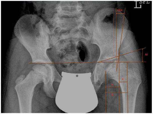
Anteroposterior (AP) pelvic radiograph of a 9-year-old male demonstrating severe Legg-Calv-Perthes disease (LCPD) of the left hip. The image features several quantitative orthopedic measurements used to assess hip dysplasia and femoral head coverage. On the affected left side, the femoral head exhibits significant flattening (coxa plana), fragmentation, and lateralization compared to the normal right side. Key radiographic markers highlighted in orange include the Acetabular Index (AI), which measures the slope of the acetabular roof; Wiberg’s Center-Edge Angle (CEA), indicating the degree of lateral femoral head coverage; and Reimer’s Index (RI), denoted by the horizontal segments A (covered width), A' (uncovered width), and B (total head width). The radiograph illustrates marked hip joint incongruence and subluxation. Anatomical landmarks such as the pelvic teardrops and ischial tuberosities are visible, serving as reference points for the horizontal inter-teardrop line. This diagnostic image is used to quantify the severity of LCPD and plan preoperative surgical interventions like pelvic osteotomy.
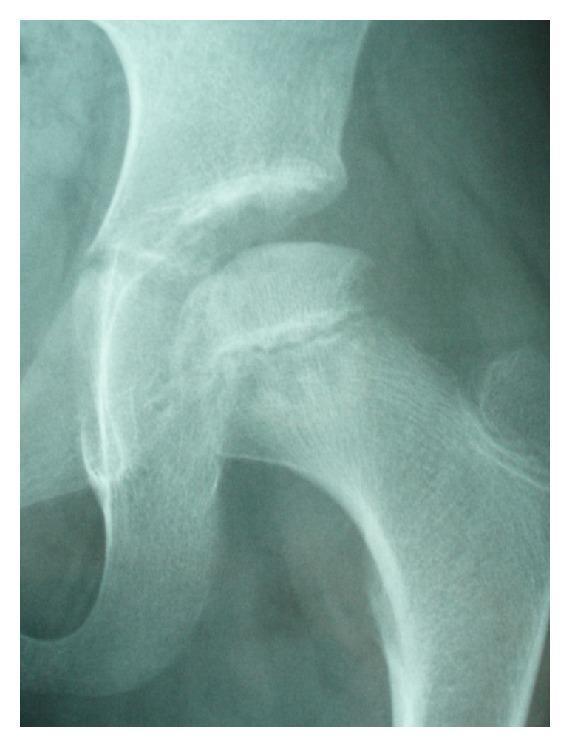
A diagnostic radiograph (X-ray) focusing on the left hip joint of a pediatric patient. The image shows the anatomical relationship between the acetabulum and the proximal femur. Key pathological features include visible bone resorption within the femoral capital epiphysis, characterized by areas of reduced radiopacity and irregular trabecular density compared to the adjacent femoral neck. The epiphysis appears somewhat flattened with a linear lucency, suggesting the fragmentation phase of Legg-Calv)-Perthes disease (LCPD). The joint space is visualized, and the growth plate (physis) is still evident, consistent with a young patient. This image serves as a clinical demonstration of the progression from transient synovitis to ischemic necrosis of the femoral head. It is intended for intermediate to advanced medical learners focusing on pediatric orthopedics and musculoskeletal radiology.
Legg-Calvé-Perthes Disease (Coxa Plana)
Definition
Epidemiology and risk factors
- It is most common in boys aged 4 to 8 years. They are typically very active and have delayed skeletal maturation. - Miller's Review of Orthopaedics; Bailey & Love 28th Ed
- Boys are affected about 4 to 5 times more often than girls.
- Risk factors are a positive family history, low birth weight and abnormal birth presentation.
- Bilateral disease occurs in 12 to 15% of cases. It is usually asynchronous. If both hips look affected at the same time, exclude multiple epiphyseal dysplasia with a skeletal survey.
Pathology (stages of Waldenström)
- Initial (avascular): the epiphysis is sclerotic and the joint space is widened. X-rays may stay normal for 4 to 6 months.
- Fragmentation: the necrotic bone is resorbed and the head is soft, so collapse occurs. This is the stage used for the lateral pillar classification.
- Reossification: new bone forms. This may last up to 18 months.
- Healed/remodelling: remodelling continues until skeletal maturity.
Clinical features
- Symptoms: a painless or mildly painful limp, often with pain referred to the knee or thigh. Symptoms are usually insidious.
- Signs:
- Trendelenburg or antalgic gait.
- Restricted hip movement, especially abduction and internal rotation.
- Muscle wasting of the thigh, and later limb shortening.
Investigations
- X-ray (AP and frog-lateral):
- Early: a small ossific nucleus, lateral joint space widening and a subchondral fracture (crescent sign).
- Later: sclerosis, fragmentation, flattening (coxa plana), a broad short neck (coxa magna) and a poorly contained head.
- MRI: detects early disease and the extent of the avascular area.
- Bone scan: shows a cold area early.
- Ultrasound: shows effusion.
- Blood tests: to exclude infection and other causes. These include thyroid function, sickle cell screening and a bone survey for epiphyseal dysplasia.
Classification
| Group | Lateral pillar height | Prognosis |
|---|---|---|
| A | Little or no involvement | Uniformly good |
| B | More than 50% maintained | Good if bone age under 6 years; poorer in older children |
| B/C | 50% or less, or narrow (2 to 3 mm), poorly ossified or depressed | Treated like B |
| C | Less than 50% maintained | Poor in all age groups |
- Gage sign (a V-shaped defect at the lateral physis)
- Lateral calcification
- Lateral subluxation
- Metaphyseal cyst
- Horizontal growth plate
Prognostic factors
- A poor prognosis goes with age at onset over 8 years (bone age over 6), female sex, Herring group C, loss of abduction and a non-spherical head.
- The most important determinant is the sphericity of the femoral head at maturity. An aspherical head leads to early osteoarthritis. - Miller's Review, p. 270
- About 13% of patients eventually need hip arthroplasty. The risk is highest when head involvement is severe. - Campbell's Operative Orthopaedics 15th Ed
Management
- Observation and activity restriction.
- Analgesics and NSAIDs, and short-term traction or partial weight bearing for irritability.
- Physiotherapy to maintain abduction and internal rotation.
- Petrie casts or abduction bracing. These have fallen out of favour.
- Femoral varus (derotation) osteotomy.
- Pelvic osteotomy: Salter, triple, Dega or Pemberton.
- Shelf acetabuloplasty, to prevent lateral subluxation and lateral overgrowth.
- Valgus femoral osteotomy for hinge abduction.
- Chiari or shelf osteotomy for hips that can no longer be contained.
- Trochanteric advancement or distal transfer and cheilectomy.
- Total hip arthroplasty in adulthood.
Complications and sequelae
- Coxa magna, coxa plana and coxa breva.
- Hinge abduction and femoroacetabular impingement.
- Leg length discrepancy and trochanteric overgrowth.
- Secondary osteoarthritis. Hips with a dysplastic or retroverted acetabulum and coxa vara can make later arthroplasty harder. - Campbell's Operative Orthopaedics
Differential diagnosis
- Transient synovitis, septic arthritis and juvenile idiopathic arthritis.
- Multiple epiphyseal dysplasia, hypothyroidism, sickle cell disease and Gaucher disease.