
Explain cardiac action potential of conduction cells from it's introduction to its every important point along with suitable diagrams and graphs it's ionic charge during each phase and their common names with stages of repolarization and depolarisation
diagram cardiac pacemaker action potential SA node phases 4 0 3 ionic currents
"cardiac pacemaker" AND "action potential"
cardiac pacemaker action potential phases funny current calcium channels physiology
https://cvphysiology.com/arrhythmias/a004
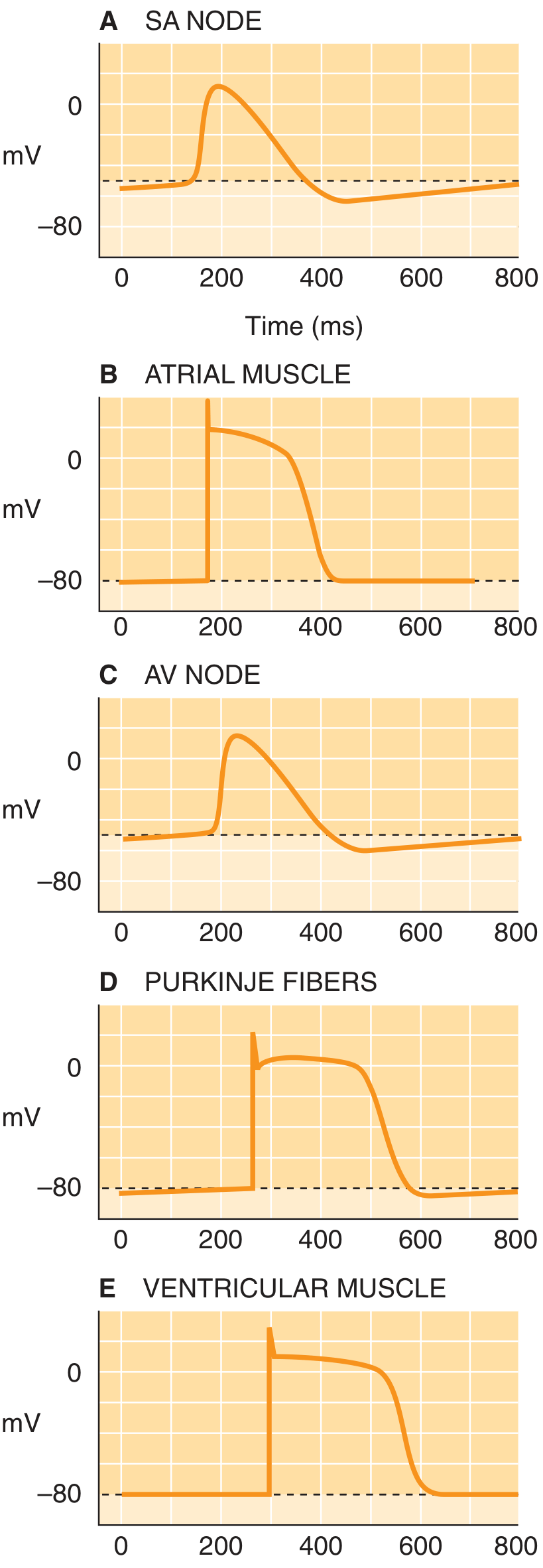
Cardiac action potential in conduction cells
- SA node: normal pacemaker, initiates each heartbeat
- AV node: delays the impulse before it enters ventricles
- His bundle, bundle branches, Purkinje fibers: rapidly transmit the impulse through ventricles
-
Slow-response action potential: SA and AV nodal cells
This is usually what is meant by the action potential of pacemaker or nodal conduction cells. -
Fast-response action potential: His-Purkinje cells
It resembles the ventricular muscle-cell action potential, but Purkinje cells can also show slow spontaneous phase-4 depolarization.
1. Introduction: what is an action potential?
- At a negative voltage, the inside of the cell is more negative than the outside.
- Depolarization means the interior becomes less negative, then positive.
- Repolarization means the interior returns toward negativity.
- Hyperpolarization means it becomes more negative than its usual level.
| Ion movement | Electrical effect inside cell |
|---|---|
| Na⁺ enters | More positive, depolarization |
| Ca²⁺ enters | More positive, depolarization |
| K⁺ leaves | More negative, repolarization |
| Cl⁻ enters | More negative, hyperpolarization |
2. Why nodal cells are special
- do not have a stable resting membrane potential
- have a maximum negative voltage of approximately -60 mV
- slowly depolarize during diastole
- use mainly Ca²⁺ influx, not fast Na⁺ influx, for the phase-0 upstroke
- do not have clear phases 1 and 2
Phase 4 → Phase 0 → Phase 3 → repeat
3. Diagram: SA nodal cell action potential
Membrane
potential
(mV)

 +10 |                         /\
   0 |                        /  \
 -10 |                       /    \
 -20 |                      /      \
 -30 |                     /        \
 -40 |--------------------|          \        Threshold
 -50 |                  /              \
 -60 |______/-----------                  \____
          ↑        ↑                         ↑
       Maximum   Phase 0                  Phase 3
       diastolic depolarization        repolarization
       potential
          \_______________________________/
                    Phase 4
          spontaneous diastolic depolarization
                 or pacemaker potential

             Time →
The phases and their common names
| Phase | Common name | Main ionic event |
|---|---|---|
| 4 | Pacemaker potential / spontaneous diastolic depolarization | Na⁺ and Ca²⁺ enter, while K⁺ efflux falls |
| 0 | Depolarization / upstroke | Ca²⁺ enters through L-type Ca²⁺ channels |
| 3 | Repolarization | K⁺ leaves the cell; Ca²⁺ channels inactivate |
| 1 | Initial repolarization | Absent in nodal cells |
| 2 | Plateau | Absent in nodal cells |
4. Phase 4: pacemaker potential
Other names
- Spontaneous diastolic depolarization
- Prepotential
- Pacemaker potential
- Automatic depolarization
Phase 4

 -60 mV                                     -40 mV
   │                                           │
   │          gradual depolarization           │
   └───────────────╱╱╱╱╱╱╱╱╱╱╱╱╱──────────────┘
       If current       ICa,T          ICa,L begins
Ionic basis of phase 4
A. Funny current, If
- Despite opening when the cell is very negative, If produces a net inward positive current.
- It is predominantly inward Na⁺ movement, with some K⁺ contribution.
- Na⁺ entering the cell makes the interior progressively more positive.
B. Decreased K⁺ efflux
- K⁺ channels progressively close
- less K⁺ leaves the cell
- the outward positive current decreases
- the membrane voltage gradually becomes less negative
C. T-type Ca²⁺ channels
- Ca²⁺ moves into the cell.
- This adds to depolarization.
- These channels help the membrane reach threshold.
D. L-type Ca²⁺ channels begin to open near threshold
Net charge movement during phase 4
Outside                     Membrane                     Inside

Na⁺  ───────────────────────►                         Positive charge enters
Ca²⁺ ───────────────────────►                         Positive charge enters
K⁺   ◄─────────────── less than before                Less positive charge exits

Result: inside becomes gradually less negative.
Importance of phase-4 slope
- Steeper phase-4 slope: threshold reached sooner, heart rate rises.
- Flatter phase-4 slope: threshold reached later, heart rate falls.
5. Phase 0: depolarization or upstroke
Common names
- Upstroke
- Depolarization
- Phase-0 depolarization
Phase 0

-40 mV                   0 to +10 mV
   │                         │
   └───────────────────────╱
                     Ca²⁺ enters
                   through ICa,L
Main ionic movement
Ca²⁺: extracellular fluid ───► nodal cell
Important contrast with ventricular muscle and Purkinje cells
| Cell type | Ion chiefly responsible for phase 0 | Speed of upstroke |
|---|---|---|
| SA node, AV node | Ca²⁺ through L-type channels | Slow |
| Atrial and ventricular muscle | Na⁺ through fast Na⁺ channels | Fast |
| Purkinje fibers | Na⁺ through fast Na⁺ channels | Very fast |
- the nodal phase-0 upstroke is less steep
- nodal conduction is slow
- AV nodal delay occurs
6. Phase 3: repolarization
Common names
- Final repolarization
- Rapid repolarization
- Repolarizing phase
- L-type Ca²⁺ channels inactivate, so inward positive charge falls.
- K⁺ channels open.
- K⁺ leaves the cell.
- The interior becomes negative again.
Phase 3

 +10 mV
   │\
   │ \
   │  \
   │   \___________________ -60 mV
   │
   └── K⁺ exits cell
       Ca²⁺ entry stops
Ionic movement
Inside                           Membrane                  Outside

K⁺  ───────────────────────────────►                    Positive charge leaves
Ca²⁺ entry decreases or stops

Result: the cell interior becomes negative again.
7. Complete ionic-current graph
Phase:       4                   0                  3
             │                   │                  │
Voltage:    -60 mV ──► -40 mV ──► +5 mV ──► -60 mV

If:          ↑ early             ↓                  off
             Na⁺ inward

ICa,T:       ↑ late phase 4      ↓                  off
             Ca²⁺ inward

ICa,L:       begins near          ↑↑↑                closes
             threshold            Ca²⁺ inward

IK:          progressively        low initially      ↑↑↑
             declining                                K⁺ outward
| Current | Full name | Dominant ion | Main phase |
|---|---|---|---|
| If | Funny or pacemaker current | Mainly Na⁺ inward | Early phase 4 |
| ICa,T | T-type calcium current | Ca²⁺ inward | Late phase 4 |
| ICa,L | L-type calcium current | Ca²⁺ inward | Phase 0 |
| IK | Potassium current | K⁺ outward | Phase 3 |
8. Stages of depolarization and repolarization
Depolarization stages
1. Slow diastolic depolarization
- Corresponds to phase 4
- If current begins
- K⁺ efflux decreases
- T-type Ca²⁺ influx assists
- Cell moves from around -60 mV toward threshold
2. Threshold
- Approximately -40 mV
- L-type Ca²⁺ channels activate
3. Rapid depolarization
- Corresponds to phase 0
- Ca²⁺ influx causes the upstroke
- The cell interior becomes positive
Repolarization stage
Final repolarization
- Corresponds to phase 3
- L-type Ca²⁺ channels inactivate
- K⁺ exits
- Membrane returns toward approximately -60 mV
- A new pacemaker cycle begins
9. Why phases 1 and 2 are absent in nodal cells
- Phase 1 is initial repolarization, largely due to transient outward K⁺ current.
- Phase 2 is the plateau, produced by balance between Ca²⁺ entry and K⁺ exit.
Nodal cell:          4 → 0 → 3

Fast-response cell:  4 → 0 → 1 → 2 → 3
10. Comparison: nodal versus Purkinje action potentials
| Feature | SA/AV nodal cell | Purkinje fiber |
|---|---|---|
| Main role | Pacemaking and AV delay | Rapid ventricular conduction |
| Action-potential type | Slow response | Fast response |
| Resting potential | No stable resting potential | Stable, about -90 mV |
| Phase 4 | Spontaneous depolarization | Usually stable, but can slowly depolarize |
| Phase-0 ion | Ca²⁺ influx | Fast Na⁺ influx |
| Phase-0 slope | Slow | Steep and rapid |
| Phases 1 and 2 | Not distinct | Present |
| Conduction velocity | Slow, especially AV node | Fast |
11. Autonomic effects
Sympathetic stimulation
- increases cAMP
- increases If
- increases Ca²⁺ current
- steepens phase 4
- threshold is reached earlier
- increases heart rate: positive chronotropy
- increases AV nodal conduction: positive dromotropy
Steeper phase 4 = more action potentials per minute
Parasympathetic stimulation
- decreases cAMP
- decreases If and L-type Ca²⁺ current
- increases K⁺ conductance through acetylcholine-sensitive K⁺ channels
- makes the cell more negative
- flattens phase 4
- slows SA-node firing and AV-nodal conduction
Flatter phase 4 + more negative maximum diastolic potential
= slower heart rate
12. High-yield clinical correlations
- Ivabradine blocks the funny current, If. It slows phase-4 depolarization and lowers heart rate.
- Beta blockers reduce sympathetic stimulation, flatten phase 4, and slow heart rate and AV nodal conduction.
- Verapamil and diltiazem block L-type Ca²⁺ channels. They slow nodal phase 0 and AV nodal conduction.
- Adenosine increases K⁺ conductance and suppresses Ca²⁺-dependent AV nodal conduction. It can terminate many AV-node-dependent supraventricular tachycardias.
- Hyperkalemia makes membrane potential less negative and can impair excitability and conduction.
One-line summary
SA and AV nodal cells spontaneously depolarize during phase 4 because of If, reduced K⁺ efflux, and Ca²⁺ entry; phase 0 is caused by L-type Ca²⁺ influx; phase 3 is caused by K⁺ efflux. They have phases 4, 0, and 3 only.
Make a comparison table of nodal and ventricular action potentials
"cardiac action potential" AND nodal
nodal versus ventricular cardiac action potential phases ionic basis physiology
| Feature | Nodal action potential | Ventricular action potential |
|---|---|---|
| Main cells | SA node and AV node | Ventricular working myocytes |
| Functional role | Pacemaker generation and AV nodal delay | Forceful ventricular contraction |
| Type | Slow-response action potential | Fast-response action potential |
| Automaticity | Present, especially SA node | Absent normally |
| Stable resting membrane potential | Absent | Present |
| Phase 4 voltage | Maximum diastolic potential about -60 mV | Stable resting membrane potential about -90 mV |
| Phase 4 behavior | Slow spontaneous depolarization | Flat, stable resting phase |
| Name of phase 4 in nodal cells | Pacemaker potential / spontaneous diastolic depolarization | Resting membrane potential |
| Main cause of phase 4 | Funny current If with net Na⁺ entry, declining K⁺ efflux, T-type Ca²⁺ entry, and contribution from intracellular Ca²⁺ cycling | High K⁺ permeability through inward-rectifier K⁺ current, IK1 |
| Threshold | About -40 mV | About -70 mV |
| Phase 0 | Present | Present |
| Phase-0 name | Slow depolarization / upstroke | Rapid depolarization / upstroke |
| Main ion in phase 0 | Ca²⁺ influx through L-type Ca²⁺ channels, ICa,L | Rapid Na⁺ influx through fast voltage-gated Na⁺ channels, INa |
| Phase-0 slope | Slow and less steep | Very rapid and steep |
| Consequence of phase-0 slope | Slow conduction, especially in AV node | Fast conduction through ventricular myocardium |
| Phase 1 | Absent | Present |
| Phase-1 name | Not applicable | Initial rapid repolarization |
| Main event in phase 1 | Not applicable | Transient outward K⁺ efflux, Ito |
| Phase 2 | Absent | Present and prominent |
| Phase-2 name | Not applicable | Plateau phase |
| Main event in phase 2 | Not applicable | Balance between inward Ca²⁺ current through L-type channels and outward K⁺ current |
| Phase 3 | Present | Present |
| Phase-3 name | Repolarization | Final rapid repolarization |
| Main event in phase 3 | L-type Ca²⁺ channels inactivate and K⁺ leaves the cell | Ca²⁺ channels close and outward K⁺ currents dominate, chiefly IKr and IKs |
| Main ion causing repolarization | K⁺ efflux | K⁺ efflux |
| Action-potential phases | 4, 0, 3 only | 4, 0, 1, 2, 3 |
| Plateau | No true plateau | Characteristic long plateau |
| Action-potential duration | Shorter, approximately 100-150 ms, varies with rate and site | Approximately 200-300 ms |
| Refractory period | Relatively long for nodal tissue; conduction is strongly rate-dependent | Long refractory period prevents tetany |
| Response to sympathetic stimulation | Increases If and Ca²⁺ currents, steepens phase 4, increases heart rate and AV conduction | Increases Ca²⁺ current and alters K⁺ currents, increases contractility and usually shortens action-potential duration |
| Response to parasympathetic stimulation | Strong effect: increases K⁺ efflux, decreases If and ICa,L, hyperpolarizes cell and flattens phase 4 | Minimal direct vagal effect on ventricular myocardium |
| Drugs that act prominently | Adenosine, beta blockers, verapamil, diltiazem, ivabradine | Class I Na⁺ channel blockers and Class III K⁺ channel blockers have major effects |
Simplified diagrams
NODAL CELL: slow-response AP

mV
 +10 |                    /\
 -40 |-------------------/  \---------------- threshold
 -60 |______/────────────      \______
          Phase 4       0         3
       pacemaker      Ca²⁺ in    K⁺ out
       If, Ca²⁺ in
VENTRICULAR CELL: fast-response AP

mV
 +20 |              /──────────\
   0 |             /            \
 -90 |____________/              \__________
              0    1     2        3     4
            Na⁺ in  K⁺   Ca²⁺ in    K⁺ out
                         plateau
High-yield memory aid
-
Nodal cells:
Funny Na⁺ in during phase 4 → Ca²⁺ in during phase 0 → K⁺ out during phase 3. -
Ventricular cells:
Na⁺ in during phase 0 → brief K⁺ out during phase 1 → Ca²⁺ in creates phase-2 plateau → K⁺ out during phase 3.
Create a labeled diagram of both action potentials
An accurate, clear educational medical diagram comparing two cardiac action potentials side by side. Left panel: SA/AV nodal pacemaker action potential, vertical axis membrane potential in mV from -70 to +20 and horizontal axis time. Label Phase 4 spontaneous diastolic depolarization or pacemaker potential, funny current If mainly Na+ inward and decreased K+ efflux; label threshold -40 mV; label Phase 0 Ca2+ influx via L-type calcium channels; label Phase 3 K+ efflux and calcium channel inactivation; label maximum diastolic potential -60 mV. Clearly show phases 1 and 2 absent. Right panel: ventricular myocyte action potential, vertical axis membrane potential mV from -100 to +30 and horizontal axis time. Label Phase 4 resting membrane potential -90 mV, Phase 0 rapid Na+ influx, Phase 1 transient K+ efflux, Phase 2 plateau with Ca2+ influx via L-type calcium channels balanced by K+ efflux, and Phase 3 K+ efflux repolarization. Include a concise legend: inward positive ion movement depolarizes; outward K+ movement repolarizes. Use medically conventional colors: blue Na+, orange Ca2+, purple K+, crisp readable typography, no unsupported details.
"cardiac action potential" AND physiology
cardiac nodal versus ventricular action potential phases ion currents physiology
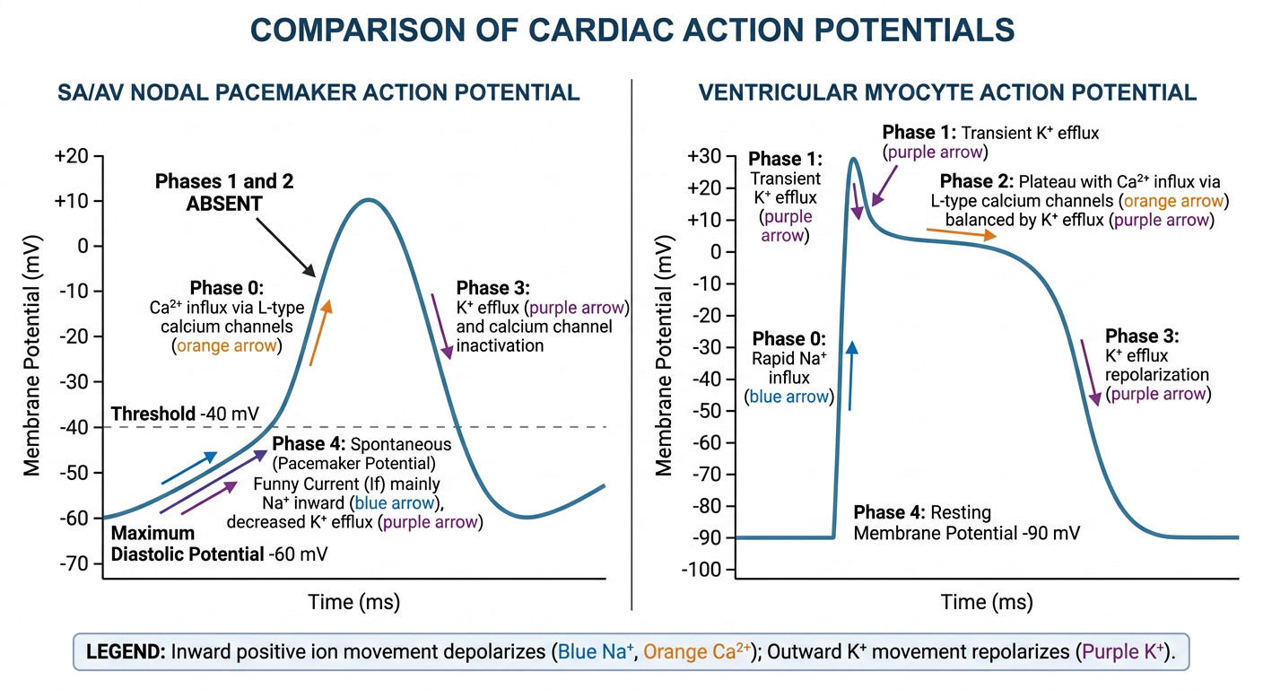
- Nodal cells, SA and AV node: phase 4 is spontaneous pacemaker depolarization, phase 0 depends on L-type Ca²⁺ influx, and phase 3 is due to K⁺ efflux. Phases 1 and 2 are absent.
- Ventricular myocytes: phase 0 is fast Na⁺ influx; phase 1 is brief K⁺ efflux; phase 2 is the Ca²⁺-dependent plateau; and phase 3 is K⁺-mediated repolarization.
Detaild notes on ventricular action potential with proper diagram and complete details
ventricular myocyte cardiac action potential diagram phases 0 1 2 3 4 ionic currents
"ventricular action potential" AND ion channels
ventricular myocyte action potential phases 0 1 2 3 4 ionic currents physiology
Accurate educational medical diagram of a ventricular cardiac myocyte action potential. A large clean graph with membrane potential in mV on vertical axis from -100 to +30 and time in milliseconds on horizontal axis. Clearly label Phase 4 resting membrane potential -90 mV with IK1 potassium current, Phase 0 rapid depolarization with fast Na+ influx INa, Phase 1 initial repolarization with transient outward K+ efflux Ito, Phase 2 plateau with L-type Ca2+ influx ICa,L balanced by delayed rectifier K+ efflux IKr and IKs, Phase 3 final repolarization with K+ efflux IKr IKs and reactivation of IK1. Include arrows for Na+, Ca2+, and K+ movement and concise legible labels. Add a small color legend: blue Na+ inward, orange Ca2+ inward, purple K+ outward. No nodal action potential.
Which section should I develop into detailed notes first?