


b991d931-b4d8-4d08-b04f-ccf6b8248385.jpg
ed77e679-fdb9-4461-b5e8-b9a424ae8f26.jpg
8 old boy complain of this symptoms along with itching and pain . It’s also present in mouth unable to open mouth and eat
analyse_media
analyse_media
Reading File
The clinical picture here is highly characteristic. An 8-year-old with skin lesions on the hands AND mouth involvement with itching/pain - this is classic Hand, Foot and Mouth Disease (HFMD). Let me search the medical library for detailed information.
Searching sources...
Finding Sources
Finding Sources
Searching the Web
Hand foot mouth disease 8 year old child clinical features treatment 2024
Excellent - the diagnosis is confirmed. Let me fetch the detailed textbook content and the clinical images.
Reading File
Reading File
Reading File
I have all the information needed. Let me fetch a key clinical image to include in the response.
Loading Image
Loading Image
I now have a complete picture. Here is a thorough clinical answer:
Diagnosis: Hand, Foot, and Mouth Disease (HFMD)
The clinical picture fits perfectly - an 8-year-old child with:
- Small papulo-vesicular lesions on the palms with itching and pain
- Simultaneous oral lesions causing inability to open mouth and eat
This is classic Hand, Foot, and Mouth Disease (HFMD).
Comparing the Child's Images to Textbook Photos
The child's hand shows small raised erythematous papules/early vesicles on the palmar surface and along the finger creases - matching exactly the lesion pattern below:
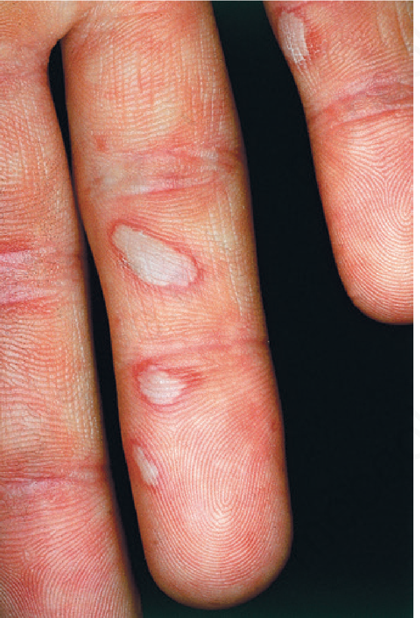
Classic HFMD - gray oval vesicles with red halo running parallel to skin lines on fingers
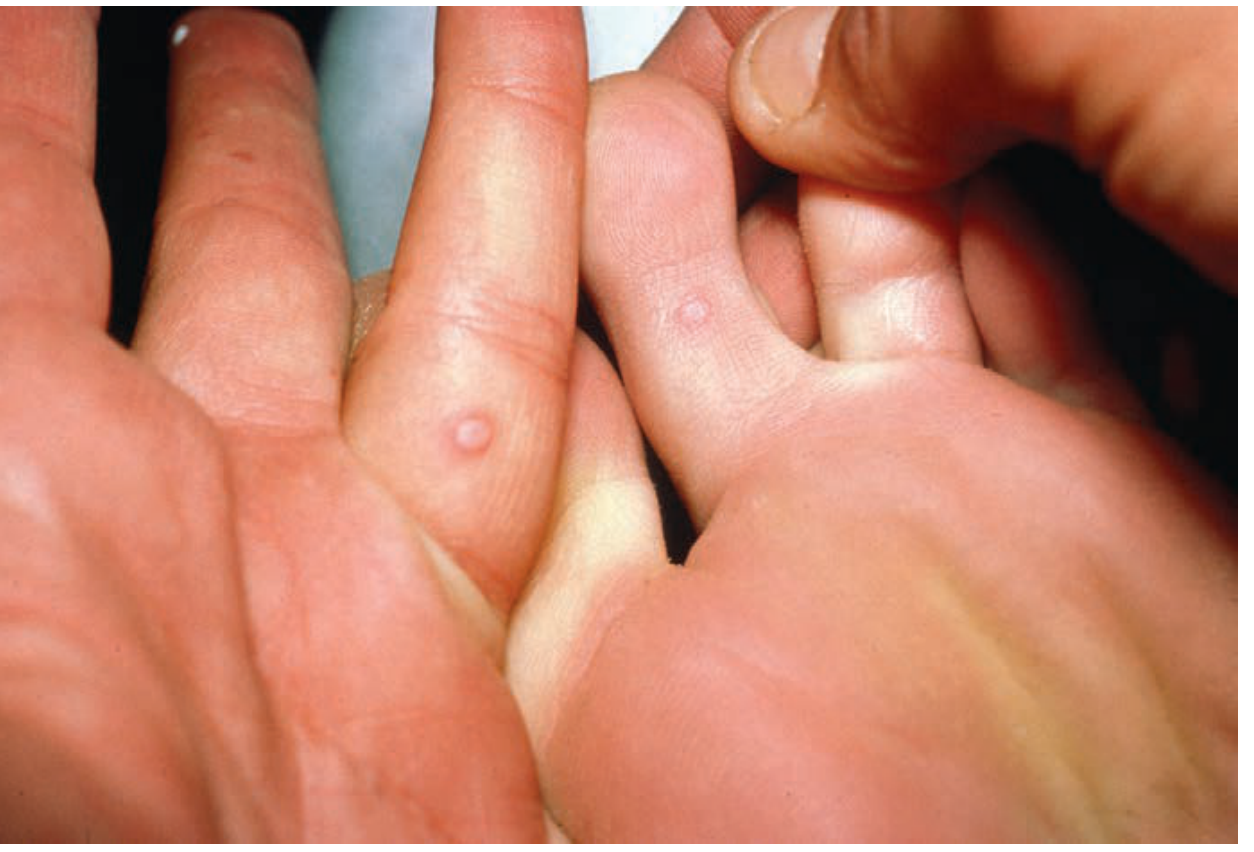
Small dome-shaped papulo-vesicles on the palm - closely matching what is seen in this child
Etiology
- Primary causative agent: Coxsackievirus A16 (most common)
- Also caused by: other Coxsackie A and B viruses, Enterovirus 71 (associated with neurological complications), and increasingly Coxsackievirus A6 (CVA6) which causes more atypical/widespread disease
- Affects children 6 months to 10 years of age primarily, though adults can be infected too
- Andrews' Diseases of the Skin, p. 457
Pathophysiology
The virus enters via the oral/fecal-oral route, replicates in the oropharynx and GI tract, then disseminates hematogenously to skin and mucous membranes. The skin lesions result from intraepidermal blister formation via vacuolar and reticular degeneration of keratinocytes. No inclusion bodies or multinucleated giant cells are present (which helps distinguish it from herpes).
Clinical Features in This Child
| Feature | Classic HFMD |
|---|---|
| Oral ulcers causing pain/difficulty eating | Small (4-8 mm) rapidly ulcerating vesicles on buccal mucosa, tongue, soft palate, gingiva - surrounded by red areola |
| Hand lesions with itching/pain | Asymptomatic to mildly painful red papules → gray 3-7 mm vesicles with red halo, oval/linear, running parallel to skin lines |
| Distribution | Palms > dorsa of fingers; also soles, feet, buttocks |
| Age | 8 years - fits typical range |
The oral lesions are the reason he cannot open his mouth and eat - the ulcers are painful on contact with food/saliva.
When to Suspect Atypical HFMD (CVA6)
If lesions are more widespread (trunk, perioral area, legs, arms), bullous, or severe, suspect Coxsackievirus A6. In children with pre-existing eczema, CVA6 causes "eczema coxsackium" - a severe vesicular and erosive eruption in areas of dermatitis. - Dermatology 2-Volume Set 5e, p. 1716
Serious Complication to Watch For
Enterovirus 71-associated HFMD can cause CNS complications (aseptic meningitis, brainstem encephalitis, acute flaccid paralysis). In the severe EV71 Taiwanese outbreak, 80% of cases with CNS disease had typical HFMD skin lesions. Watch for:
- High fever persisting > 3 days
- Vomiting, headache, neck stiffness
- Myoclonic jerks, limb weakness, altered consciousness
Management
Supportive (no specific antiviral therapy exists):
-
Pain and fever:
- Paracetamol (acetaminophen) for fever and oral pain - helps the child eat/drink
- Ibuprofen for children >6 months (effective for pain relief)
-
Oral hydration:
- Encourage cold, soft foods and fluids (ice cream, yogurt, cold water)
- Avoid acidic/salty/spicy foods which worsen mouth pain
- Monitor for dehydration - the major risk in children who refuse to eat/drink
-
Oral topical relief:
- Topical anesthetic mouthwash (e.g., diluted lidocaine gel in older children) - with caution
- Magic mouthwash (antacid + diphenhydramine + lidocaine mixture) - sometimes used
-
Skin care:
- Topical calamine or mild corticosteroids for itching if severe
- Keep lesions clean to prevent secondary bacterial infection
-
Isolation:
- Keep the child away from school/daycare until fever resolves and lesions crust over (typically 5-7 days)
- HFMD is highly contagious via direct contact, respiratory droplets, and fecal-oral route
-
Hospitalization indications:
- Severe dehydration unable to take oral fluids
- Signs of CNS involvement
- Immunocompromised child
Prognosis
HFMD usually resolves in less than 1 week and is self-limiting. The oral ulcers typically resolve in 3-5 days. A late complication to be aware of is onychomadesis (nail shedding) occurring approximately 1-2 months after the acute illness, especially with CVA6 strains - parents should be reassured this is benign and nails regrow. - Goldman-Cecil Medicine; Dermatology 2-Volume Set 5e
Important note: This is an AI-assisted clinical analysis for educational and reference purposes. The treating clinician should examine the child directly, confirm the diagnosis, check for signs of dehydration and neurological involvement, and tailor management accordingly.
Syp lovocitrine given but syp not reduced
Levocetirizine (an antihistamine) is not expected to reduce the symptoms of HFMD significantly - it only helps with itching mildly. It does nothing for the viral infection, oral pain, or lesions themselves.
Searching sources...
Finding Sources
Finding Sources
Searching the Web
HFMD hand foot mouth disease treatment management children levocetirizine not effective what to give
Reading File
Why Levocetirizine Didn't Work
Syrup Levocetirizine is just an antihistamine - it blocks H1 receptors to reduce allergic itch. In HFMD, the itching is caused by direct viral inflammation of the skin, not by histamine release. So it provides minimal to no benefit and has no effect on the mouth ulcers, pain, or viral course.
This is a very common prescribing mistake in HFMD - antihistamines are not the right drug here.
What Should Actually Be Given
1. Pain + Fever - THE PRIORITY (especially for oral lesions)
The mouth pain is the main problem - the child can't eat or drink because of it.
| Drug | Dose | Route | Frequency |
|---|---|---|---|
| Paracetamol (Acetaminophen) | 15 mg/kg/dose | Oral syrup | Every 4-6 hours |
| Ibuprofen | 10 mg/kg/dose | Oral syrup | Every 6-8 hours |
Ibuprofen is often more effective than paracetamol alone for the oral ulcer pain because of its anti-inflammatory action. Alternate both for better pain control.
2. Topical Oral Analgesia (for mouth ulcers)
This directly targets the problem of painful oral lesions:
- Topical gel for mouth ulcers - Choline salicylate + Cetalkonium chloride gel (e.g., Bonjela) applied to ulcers with a fingertip, 3-4 times/day
- Glycerin + Thymol mouthwash (diluted) - gentle rinse
- Magic mouthwash - a mixture of:
- Liquid antacid (magnesium hydroxide/aluminum hydroxide)
- Diphenhydramine syrup
- Mix 1:1, swish and spit/swallow 3-4x daily
- Rosen's Emergency Medicine specifically recommends topical diphenhydramine + aluminum hydroxide + magnesium hydroxide combination for HFMD oral lesions
Avoid viscous lidocaine in young children due to systemic toxicity risk.
3. Oral Hydration - CRITICAL
- Cold fluids: cold water, chilled milk, ice cream, cold yogurt (cold numbs the ulcers and makes swallowing easier)
- Avoid: orange juice, tomatoes, spicy/salty food - these burn the ulcers
- If the child is not drinking at all for >6-8 hours → IV fluids/hospitalization needed
4. For Itching on Hands
Since levocetirizine failed, the itch is viral in origin. Better options:
- Calamine lotion - applied topically to hand/foot lesions 3-4x daily - provides cooling and mild itch relief
- Topical mild steroid (e.g., 1% hydrocortisone cream) - short course for severe itching on intact skin
5. What NOT to Give
| Drug | Reason to Avoid |
|---|---|
| Antibiotics | Viral disease - no benefit |
| Antiviral (acyclovir) | Not effective against enteroviruses |
| Aspirin | Risk of Reye's syndrome in children |
| Viscous lidocaine | Systemic toxicity risk in children |
| Steroids (systemic) | May worsen viral dissemination |
Red Flags - Refer/Admit Immediately If:
- Child not drinking for >8 hours (dehydration risk)
- High fever > 39°C not responding to antipyretics
- Lethargy, drowsiness, or confusion
- Neck stiffness, vomiting, or seizures (→ CNS involvement from Enterovirus 71)
- Rapid breathing or chest pain
Key Message
HFMD is self-limiting and resolves in 5-7 days without specific treatment. The goal of management is to keep the child comfortable and hydrated during this period. Levocetirizine alone is inadequate - paracetamol + ibuprofen + topical oral analgesia + cold fluids is the correct approach. - Rosen's Emergency Medicine; Andrews' Diseases of the Skin
Syp r b tone junior
"R B Tone Junior" sounds like a multivitamin/tonic syrup for children. Let me look up its exact composition.
Searching sources...
Searching the Web
Syrup RB Tone Junior composition ingredients children tonic India
Syrup R.B. Tone Kid (Junior)
Manufacturer: Medley Pharmaceuticals Ltd.
Composition (per 5 ml)
| Ingredient | Amount |
|---|---|
| Elemental Iron | 80 mg |
| Folic Acid (Vitamin B9) | 200 mcg |
| Vitamin B12 (Cyanocobalamin) | 2 mcg |
(Some formulations also include Vitamin B-complex and Calcium - verify on the actual label.)
What It Is
This is a haematinic / nutritional supplement - it is an iron + folate + B12 syrup used for:
- Iron deficiency anaemia
- Nutritional deficiencies causing weakness and fatigue in children
Is It Relevant for This Child's HFMD?
No - R.B. Tone Junior has NO role in treating HFMD.
This syrup is for anaemia and nutritional support. It will NOT:
- Reduce viral lesions
- Relieve mouth pain
- Treat itching
- Shorten the course of the disease
Why It May Have Been Prescribed
Possibly prescribed as a background nutritional support if the child was found anaemic or malnourished, OR it may have been prescribed as a general "immune booster" - which is not evidence-based for HFMD.
What This Child Actually Needs Right Now
Given the child has HFMD with painful oral ulcers preventing eating:
| Priority | Drug |
|---|---|
| 1st - Oral pain relief | Paracetamol syrup 15 mg/kg every 6 hrs |
| 2nd - Inflammation + pain | Ibuprofen syrup 10 mg/kg every 8 hrs (alternate with paracetamol) |
| 3rd - Mouth ulcer topical | Choline salicylate gel (Bonjela) or antacid + diphenhydramine mouthwash |
| 4th - Skin itch | Calamine lotion topically on hand lesions |
| 5th - Hydration | Cold soft foods, cold water, cold milk - monitor intake closely |
R.B. Tone can be continued if there is an underlying anaemia, but it is not a treatment for HFMD and should not replace the above.
This is a shared conversation. Sign in to Orris to start your own chat.