
Corneal ulcer summarised
corneal ulcer clinical appearance slit lamp
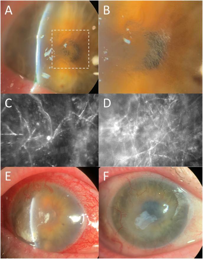
A multi-panel figure illustrating the clinical presentation, diagnostic imaging, and surgical management of a fungal corneal ulcer (Phialophora chinensis keratitis). Panels 1A and 1B (slit lamp photographs) demonstrate a corneal ulcer with surrounding anterior stromal haze, thinning, and characteristic hair-like, filamentous elements visualized on retroillumination. Panels 1C and 1D show in vivo confocal microscopy (IVCM) of the corneal stroma, revealing highly reflective, branching, and interconnected filamentous structures pathognomonic for fungal hyphae. Panel 1E displays clinical progression with worsening conjunctival injection, increased ulcer size, corneal edema, and superficial neovascularization. Panel 1F shows the post-operative appearance following superficial keratectomy and placement of a layered amniotic membrane; visible is the residual membrane graft filling the previously thinned corneal areas. The series highlights the importance of IVCM in diagnosing filamentous fungal keratitis and tracks the disease from initial infection through surgical resolution.
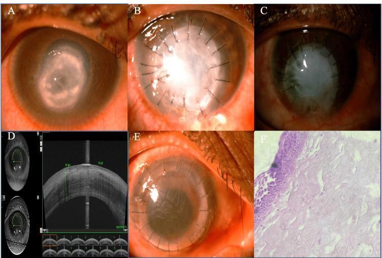
This composite figure illustrates the clinical progression and management of a perforated fungal corneal ulcer through various imaging modalities. (A) Slit-lamp clinical photograph showing a large, central perforated corneal ulcer with a pseudo-cornea, diffuse stromal infiltrates, and an inferiorly shallow anterior chamber. (B) Immediate postoperative appearance following therapeutic penetrating keratoplasty (TPK), displaying a sutured, opaque host tissue graft with circumcorneal injection. (C) One-year follow-up showing a completely opaque graft with inferior conjunctivalization. (D) Anterior segment optical coherence tomography (ASOCT) cross-section demonstrating a thick, epithelialized corneal graft with an apposed graft-host junction. (E) Clinical photograph following a subsequent optical penetrating keratoplasty, showing improved graft clarity and radial sutures. (F) Histopathology (H&E stain, 40x) revealing a stratified squamous non-keratinized five-layer epithelium with underlying edematous stroma. The series highlights the transition from acute infection and perforation to therapeutic stabilization and eventual optical rehabilitation in ophthalmology.
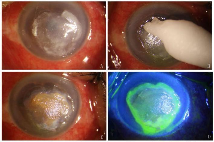
Clinical Slit-lamp Photograph (four panels) illustrating the procedure of Local Application of Silver Nitrate (LASN) for a corneal ulcer. Panel A shows a debrided corneal ulcer appearing as a central whitish, opaque lesion with irregular margins on a congested eye. Panel B demonstrates the application phase, with a silver nitrate cotton stick making contact with the corneal surface. Panel C displays the immediate post-treatment outcome, where the ulcer bed has developed a characteristic brown-to-yellowish precipitate due to the silver nitrate reaction. Panel D shows fluorescein sodium staining under cobalt blue light; intense green fluorescence is localized to the ulcerated area, demarcating the epithelial defect, while the surrounding healthy cornea and conjunctiva remain negative for staining. This sequence illustrates a chemical debridement technique often used in the management of infectious or refractory corneal ulcers to reduce microbial load and promote healing.
"corneal ulcer" management treatment
Corneal Ulcer - Summary
Definition
Pathophysiology
- Desquamation (e.g., exposure keratitis from incomplete lid closure in Bell's palsy)
- Trauma - directly breaches the epithelium and inoculates the cornea
- Direct microbial invasion - particularly with immunosuppression
Causative Organisms
| Category | Organisms |
|---|---|
| Bacteria | Pseudomonas aeruginosa (contact lens users), Streptococcus pneumoniae, Staphylococcus spp., Moraxella spp. |
| Viruses | Herpes simplex (dendritic pattern on fluorescein), Varicella-zoster |
| Fungi | Candida, Aspergillus, Penicillium, Cephalosporium |
| Protozoa | Acanthamoeba (contact lens wearers, exposure to contaminated water) |
- S. pneumoniae and S. aureus: common general bacterial causes
- Pseudomonas: especially virulent; strongly associated with contact lens use
- Fungi/viruses: increasingly common due to widespread topical and systemic immunosuppressant use
Risk Factors
- Contact lens use - the most common cause; risk increases dramatically with extended-wear and sleeping in lenses
- Previous ocular surgery or injury
- Trauma
- History of genital herpes
- Topical or systemic steroids / immunosuppressants
- Exposure keratitis (Bell's palsy)
Clinical Features
- Ocular pain or foreign body sensation
- Photophobia (often consensual due to ciliary spasm)
- Blurred vision (if ulcer is in the central visual axis or if uveitis is present)
- Redness and lid/conjunctival swelling
- Mucopurulent discharge
- Eyelids and conjunctiva: erythematous, possibly with mucopurulent discharge
- Cornea: round or irregular ulcer with a white, hazy base extending into the stroma (WBC infiltration), or heaped-up edges
- Miotic pupil (associated iritis)
- Slit lamp: flare and cells (iritis), occasionally a hypopyon (pus layering in anterior chamber)
- Fluorescein staining: epithelial defect stains with uptake; herpes simplex shows a classic dendritic pattern
Diagnosis
- Clinical - history + slit lamp examination is the cornerstone
- Slit lamp is required for accurate diagnosis (Roberts and Hedges' Clinical Procedures in Emergency Medicine)
- The ophthalmologist typically scrapes the center of the ulcer for culture and sensitivity
- Gram stain from the ulcer base can be obtained
- Do not start antibiotics before culture if an ophthalmologist will see the patient promptly - it makes subsequent organism identification difficult
Management
Immediate Steps
- Emergent ophthalmologic consultation for culture and antibiotic guidance
- If consultation is delayed by hours, initiate topical fluoroquinolones - but ideally obtain cultures first (lightly touch a culture-medium-moistened swab to the ulcer before starting antibiotics)
Antibiotics
- First-line: Topical fluoroquinolone - ciprofloxacin (Ciloxan) or ofloxacin (Ocuflox), 1 drop every hour in the affected eye
- Cultures direct subsequent narrowing of therapy
Adjuncts
- Cycloplegics (e.g., cyclopentolate 1%) - for pain relief from accompanying iritis (ciliary spasm)
- Topical steroids - generally avoided; relatively contraindicated in viral infections; may reduce scarring in some cases but should NOT be started by the emergency physician without ophthalmologic guidance
What NOT to Do
- Do not patch the eye - patching creates a warm, anaerobic environment that promotes rapid Pseudomonas ulceration, corneal melting, and perforation
- Never pressure-patch a contact lens wearer - risk of rapid infection development (Wills Eye Manual)
Follow-up
- Refer to ophthalmologist within 12-24 hours
Complications
| Complication | Notes |
|---|---|
| Corneal scarring | Can permanently impair vision |
| Corneal perforation | Especially with Pseudomonas (rapid, aggressive melting) |
| Anterior/posterior synechiae | From iritis |
| Glaucoma | Secondary to inflammation |
| Cataracts | Long-term complication |
Key Clinical Pearls
- A red eye in a contact lens wearer requires prompt evaluation for corneal ulcer - Textbook of Family Medicine
- Pseudomonas keratitis is particularly virulent; urgent and aggressive treatment is needed to avoid intraocular spread - Goldman-Cecil Medicine
- Herpes simplex keratitis is triggered by any form of corneal stress in patients with prior disease; recognized by its dendritic fluorescein staining pattern
- Acanthamoeba initially presents as a red eye with negative bacterial cultures - often misdiagnosed early
- If the patient is immunocompromised or the ulcer is chronic, consider fungal etiology