
Sickle cell SAM Thalassemia
Reading File
The user asked about three topics: Sickle Cell Disease, SAM (Severe Acute Malnutrition), and Thalassemia. Let me search for all three in parallel.
Finding Sources
Finding Sources
Finding Sources
Now let me read the key sections in parallel - best sources for each topic:
Reading File
Reading File
Finding Sources
Good content so far. Let me now get the sickle cell section from Robbins and more thalassemia + SAM details:
Finding Sources
Reading File
Reading File
Now let me get the Sickle Cell Anemia section from Robbins and fetch key images:
Reading File
Searching Images
sickle cell disease blood smear
I have rich content from all three sections. Let me also grab the SAM section from Park's PSM textbook and search for thalassemia image:
I have comprehensive content from the textbooks. Let me now compile the full answer.
Sickle Cell Disease, SAM, and Thalassemia
1. SICKLE CELL DISEASE (SCD)
Definition & Epidemiology
Sickle cell anemia is the most common familial hemolytic anemia. It is caused by a point mutation in the β-globin gene - a single amino acid substitution (glutamate → valine at position 6). The HbS allele is prevalent where falciparum malaria was endemic: equatorial Africa, parts of India, southern Europe, and the Middle East. In the USA, ~8% of African Americans are HbS carriers; ~1 in 600 have sickle cell anemia.
Pathogenesis
- Normal adult RBC: 96% HbA (α₂β₂), 3% HbA₂, 1% HbF
- In SCD: HbA is completely replaced by HbS; in carriers (sickle trait), ~40-50% is HbS
- On deoxygenation, HbS undergoes conformational change → molecules polymerize via the abnormal valine residue → distort RBC into elongated, crescentic (sickle) shape
- Initial sickling is reversible on reoxygenation, but repeated episodes cause cumulative membrane damage (Ca²⁺ influx, K⁺ and water loss), creating irreversibly sickled cells
Three Key Factors Determining Clinical Severity
- Intracellular levels of non-HbS hemoglobins - HbA and HbF inhibit HbS polymerization. Newborns are protected until HbF falls (~5-6 months). Carriers have HbA that greatly retards sickling.
- MCHC (Mean Corpuscular Hemoglobin Concentration) - higher MCHC = denser cells = more polymerization. Loss of water from RBCs raises MCHC and accelerates sickling.
- Transit time through microcirculation - slower blood flow (e.g., in hypoxic/acidotic states) gives HbS longer to polymerize.
Consequences of Sickling
| Effect | Result |
|---|---|
| Hemolysis | Chronic anemia (Hb ~6-8 g/dL), jaundice, gallstones |
| Vaso-occlusion | Pain crises, stroke, acute chest syndrome, avascular necrosis |
| Organ infarction | Spleen (autosplenectomy → infection risk), kidney, retina |
| Aplastic crises | Parvovirus B19 infection |
Sickle Cell Trait vs. Disease
- Trait (HbAS): ~40% HbS + ~60% HbA → generally asymptomatic, minimal sickling in vivo; rare complications under severe hypoxia
- Disease (HbSS): 100% HbS → full clinical picture
Blood Smear
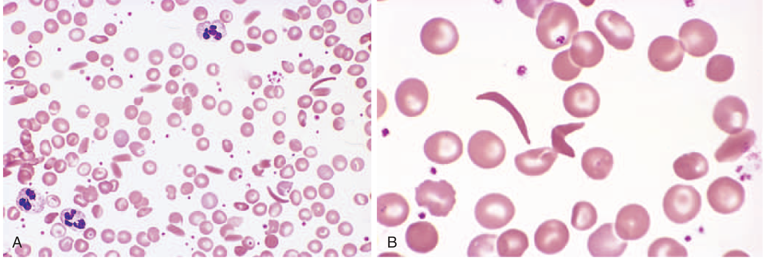
Peripheral blood smear: sickle-shaped and target cells. - Robbins & Kumar Basic Pathology
Management Principles
- HbF induction: Hydroxyurea (↑ HbF, reduces sickling)
- Analgesics for pain crises
- Penicillin prophylaxis + vaccinations (asplenic state)
- Blood transfusions for severe anemia/stroke
- Bone marrow transplant (curative)
- Gene therapy (emerging)
2. SEVERE ACUTE MALNUTRITION (SAM)
Definition (WHO)
SAM = weight-for-height Z-score below -3 SD of WHO median growth standard, OR visible severe wasting, OR presence of nutritional edema. Also: mid-upper arm circumference (MUAC) <115 mm.
~50 million children affected worldwide; responsible for ~45% of deaths in children under 5 in low-resource settings. Severely wasted children are 9 times more likely to die than well-nourished children.
SAM Spectrum: Two Ends
| Feature | Marasmus | Kwashiorkor |
|---|---|---|
| Primary deficit | Calories + protein (severe) | Protein >> calories |
| Protein compartment affected | Somatic (skeletal muscle) | Visceral (liver, plasma proteins) |
| Serum albumin | Normal or slightly reduced | Markedly reduced |
| Edema | Absent | Present (hypoalbuminemia) |
| Subcutaneous fat | Severely depleted | Relatively spared (masked by edema) |
| Body weight | Severely reduced | 60-80% of normal (masked by edema) |
| Appearance | Emaciated, "old man" look; head appears too large | Pot-bellied, puffy face, edematous limbs |
| Liver | Normal | Enlarged, fatty (↓ lipoprotein synthesis) |
| Skin/hair | Normal or dry | "Flaky paint" dermatosis, depigmentation, hair color bands |
| Behavior | Alert but weak | Apathy, listlessness, loss of appetite |
| Cause | Severe food deprivation | Early weaning + carbohydrate-only diet |
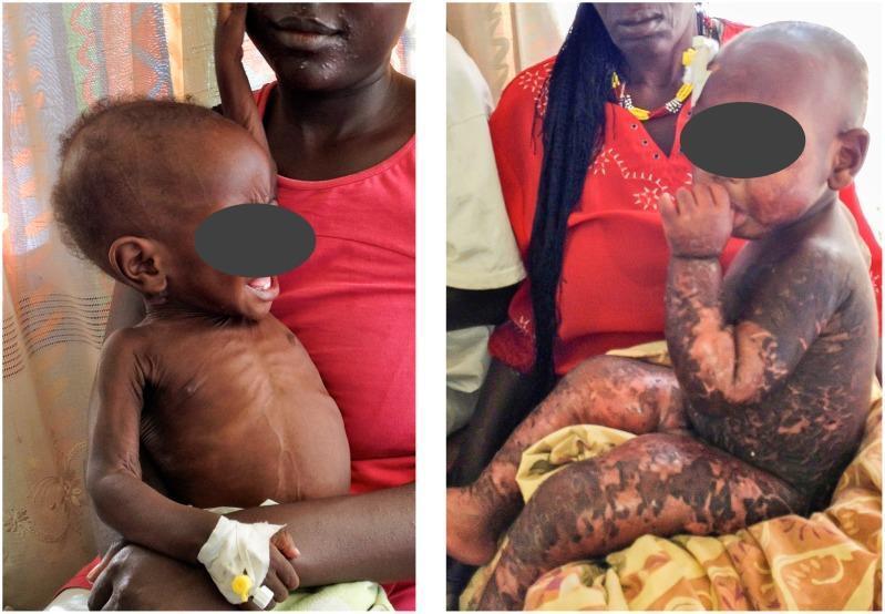
Left: Marasmus (severe wasting, emaciation). Right: Kwashiorkor (oedema, flaky paint dermatosis).
Additional SAM Features (Both Forms)
- Immune deficiency (especially T-cell mediated) → concurrent infections common
- Anemia and multivitamin deficiencies
- Gut microbiome alterations (play a causative role, not just consequence)
- Infections impose additional catabolic stress, worsening malnutrition
Diagnosis
- Weight-for-height Z-score (primary)
- Skinfold thickness (fat stores)
- Midarm circumference (muscle mass): <12.5 cm = severe; 12.5-13.5 cm = mild-moderate; >13.5 cm = normal
- Serum albumin/transferrin (visceral protein compartment)
Prevention (FAO/WHO Framework)
- Breastfeeding promotion
- Supplementary feeding for pregnant/lactating women
- Low-cost weaning foods
- Nutrition education
- Family planning and birth spacing
3. THALASSEMIA
Definition
Thalassemias are inherited disorders caused by mutations in globin genes that decrease synthesis of α- or β-globin chains. The resulting excess unpaired normal chain forms toxic precipitates causing RBC damage and hemolysis. Common in Mediterranean, African, and Asian regions (overlap with malaria-endemic zones) - HbS-like protection against falciparum malaria is hypothesized.
Pathogenesis (Two Mechanisms of Anemia)
- Inadequate HbA formation → small (microcytic), poorly hemoglobinized (hypochromic) RBCs
- Accumulation of unpaired globin chains → toxic precipitates → membrane damage → apoptosis of erythroid precursors in bone marrow = ineffective erythropoiesis
Ineffective erythropoiesis also causes:
- Low hepcidin → ↑ iron absorption → iron overload (even without transfusions)
- Massive extramedullary hematopoiesis → hepatosplenomegaly, skeletal deformities ("chipmunk facies" from frontal bossing in β-thalassemia major)
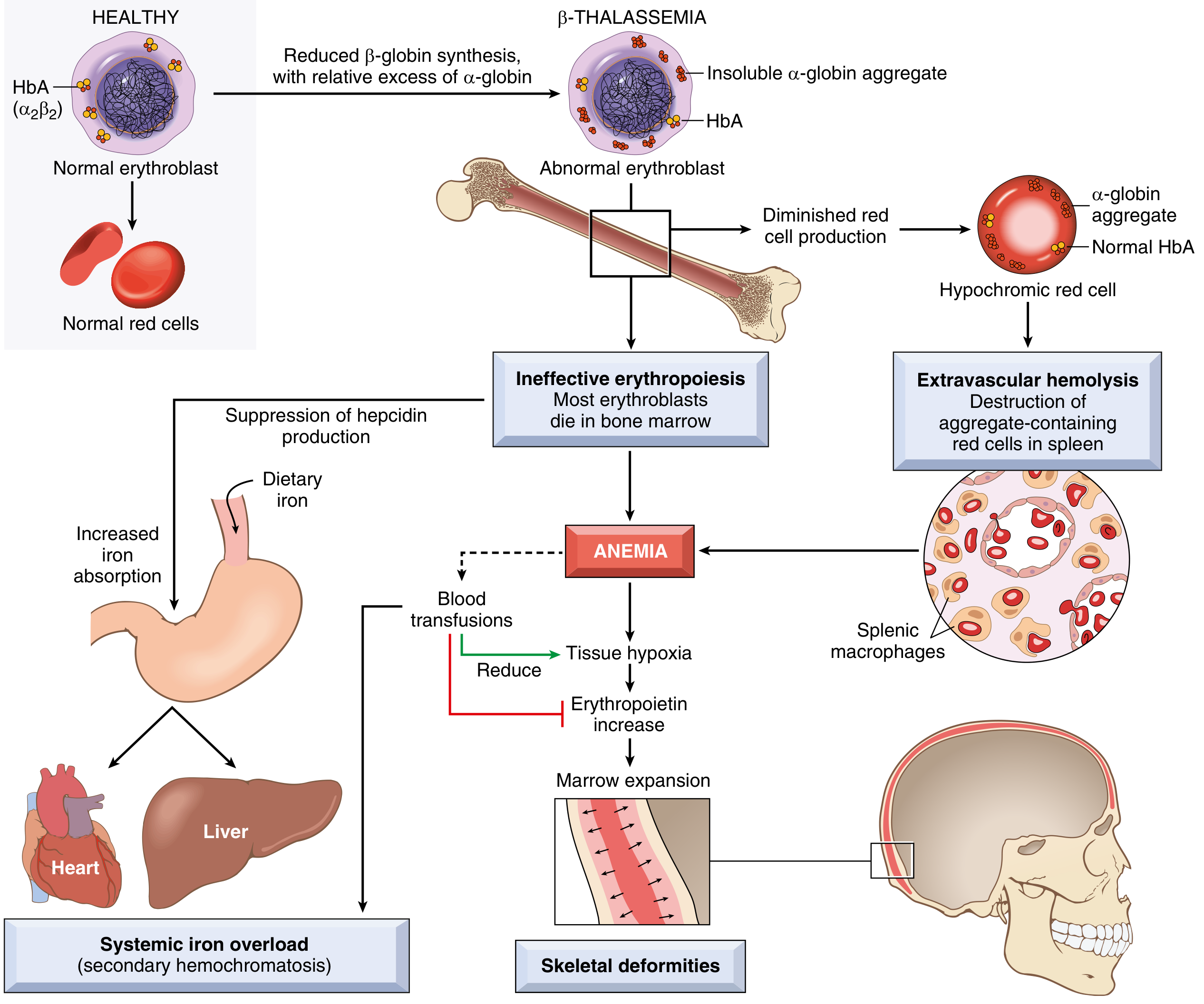
Pathophysiology of β-thalassemia major - Robbins & Kumar Basic Pathology
Classification
β-Thalassemia (mainly point mutations)
| Type | Genotype | Clinical Features |
|---|---|---|
| β-Thal Major (Cooley's anemia) | β⁰/β⁰ or β⁺/β⁰ | Severe anemia; regular transfusions required; iron overload; hepatosplenomegaly; skeletal deformity |
| β-Thal Intermedia | β⁺/β⁺ or β⁺/β⁰ | Moderate anemia; transfusions not regularly required |
| β-Thal Minor (trait) | β/β⁰ or β/β⁺ | Asymptomatic or mild; microcytic hypochromic picture; no significant anemia |
α-Thalassemia (mainly gene deletions - 2 genes per chromosome 16)
| # Genes Deleted | Name | Features |
|---|---|---|
| 1 (-α/αα) | Silent carrier | Completely asymptomatic; no RBC abnormality |
| 2 (--/αα or -α/-α) | α-Thal trait | Mild microcytic hypochromic picture; like β-thal minor |
| 3 (--/-α) | HbH disease | Moderately severe; excess β-chains form HbH (β₄) tetramers; hemolytic anemia |
| 4 (--/--) | Hydrops fetalis | Lethal in utero; excess γ-chains form Hb Bart (γ₄); no oxygen delivery possible |
Key Lab Findings
- Microcytic hypochromic anemia (low MCV, MCH)
- Target cells on peripheral smear
- Elevated HbA₂ (>3.5%) and/or HbF on Hb electrophoresis → diagnostic for β-thal minor
- HbH inclusions (Heinz body-like, with BCB stain) in HbH disease
- No HbA₂ elevation in α-thalassemia
Management
- β-Thal major: Regular blood transfusions (every 3-4 weeks) + iron chelation (deferoxamine, deferasirox, deferiprone) + splenectomy (if hypersplenism), bone marrow transplant (curative), luspatercept (activin receptor ligand trap, reduces ineffective erythropoiesis)
- β-Thal minor: No treatment; genetic counseling
- HbH: Folic acid; transfuse during hemolytic crises; avoid oxidant drugs
- Hydrops fetalis: Intrauterine transfusion (to allow delivery); otherwise fatal
Comparison at a Glance
| Feature | Sickle Cell Disease | Thalassemia | SAM |
|---|---|---|---|
| Type | Hemoglobinopathy | Hemoglobinopathy | Nutritional deficiency |
| Genetic | Point mutation in β-globin (Glu→Val at pos 6) | α/β globin gene deletions/mutations | Not genetic |
| Key mechanism | HbS polymerization on deoxygenation → sickling | Unpaired globin chains → ineffective erythropoiesis | Protein/caloric deficiency → somatic/visceral protein depletion |
| Anemia type | Hemolytic, normocytic | Microcytic hypochromic | Normocytic/mixed (may coexist) |
| Hallmark smear finding | Sickle cells + target cells | Target cells + microcytes | - |
| Key complication | Vaso-occlusion, pain crises, autosplenectomy | Iron overload (transfusion + ineffective erythropoiesis) | Immune deficiency, infection, edema (kwashiorkor) |
| Treatment cornerstone | Hydroxyurea (↑ HbF), transfusions | Transfusions + iron chelation | RUTF (ready-to-use therapeutic food), antibiotics, micronutrients |
| Curative therapy | Bone marrow transplant / gene therapy | Bone marrow transplant | Nutritional rehabilitation |
Sources: Robbins & Kumar Basic Pathology; Park's Textbook of Preventive and Social Medicine
How do you diagnose and manage a pain crisis in sickle cell disease?
A sickle cell pain crisis (vaso-occlusive crisis, VOC) is diagnosed clinically - acute onset of severe pain in a known SCD patient after excluding other causes. Management centers on aggressive hydration, analgesia (opioids titrated to effect), and treating precipitants.
Searching sources...
Finding Sources
Searching PubMed
sickle cell vaso-occlusive crisis management
Finding Sources
Finding Sources
Reading Article
PMID: 42190636
Finding Sources
Finding Sources
Good - found key sections in Rosen's EM and Tintinalli's EM. Let me read both in detail:
Reading File
Reading File
Excellent - very detailed management content from both Tintinalli's and Rosen's EM. Let me get a bit more from the Tintinalli section (it was truncated):
Reading File
Loading Image
I now have comprehensive, detailed content. Let me compile the full answer.
Diagnosis and Management of Sickle Cell Pain Crisis (Vaso-Occlusive Crisis)
What Is a Pain Crisis?
A vaso-occlusive crisis (VOC) is the most common acute complication of sickle cell disease. It occurs when deoxygenated HbS polymerizes → RBCs sickle → obstruct the microvasculature → ischemia and infarction of bone, marrow, and surrounding soft tissue. Pain is typically severe, involves the long bones, back, chest, and abdomen, and is the leading reason SCD patients present to the emergency department.
DIAGNOSIS
Clinical Diagnosis - There Is No Lab Test for Crisis
"No test is available that detects whether a patient is in a crisis - the diagnosis is based largely on clinical presentation." - Rosen's Emergency Medicine
A VOC is a diagnosis of exclusion - you must rule out other causes of pain (infection, ACS, appendicitis, cholecystitis) before attributing pain to VOC alone.
History
- Duration and location of pain
- Fever (low-grade fever can occur in VOC itself; high fever suggests infection)
- Focal swelling or redness
- Precipitating factors: cold exposure, dehydration, infection, exertion, hypoxia, alcohol, high altitude/pressurized aircraft
- Medications already taken
- Prior episodes - is this similar to previous crises? (atypical pain = red flag)
Physical Examination
| System | What to Assess |
|---|---|
| Vital signs | Temperature, pulse oximetry (SpO₂), HR, BP |
| General | Pallor, jaundice, hydration status, respiratory distress |
| Respiratory | Lung sounds (rule out ACS/pneumonia) |
| Abdomen | Tenderness, organomegaly, peritoneal signs |
| MSK | Sites of pain - swelling, warmth, redness over bones/joints |
| CNS | Any focal neurologic signs (stroke) |
WBC >20,000/mm³ with increased bands is NOT typical for simple VOC - suggests infection.
Investigations
| Test | Purpose |
|---|---|
| CBC + differential | Baseline Hb (compare to known baseline); WBC for infection |
| Reticulocyte count | If Hb dropped ≥2 g/dL from baseline - assess aplastic vs hemolytic crisis |
| Chest X-ray | Rule out pneumonia, ACS (new pulmonary infiltrate) |
| Blood cultures + urine culture | If fever present |
| LFTs, bilirubin | Hepatic involvement, cholestasis |
| Renal function + urinalysis | Renal infarction, papillary necrosis |
| Type and screen | If transfusion likely needed |
Reticulocyte count interpretation:
- Retic <3% (or below patient's usual): suggests aplastic crisis (parvovirus B19)
- Retic >12% with nucleated RBCs: suggests rapid hemolysis
Peripheral Blood Smear in SCD
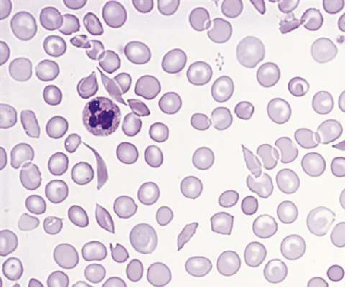
Sickle cells and target cells on peripheral smear - Rosen's Emergency Medicine
MANAGEMENT
General Goals
"Rest, adequate nutrition, hydration, oxygenation, analgesia, transfusion, and therapy for infection - directed toward symptomatic relief and interrupting the cycle of deoxygenated sickling and intravascular sludging." - Rosen's Emergency Medicine
Step 1: Immediate Supportive Measures
| Measure | Details |
|---|---|
| IV access | Establish early |
| Hydration | IV fluids to correct dehydration and improve microvascular flow. Avoid both overhydration (risk of ACS) and underhydration |
| Supplemental oxygen | Only if SpO₂ <95% or hypoxic - not shown to reduce opioid use or hospitalization in non-hypoxic patients |
| Rest | Warmth; avoid cold (triggers vasoconstriction and sickling) |
| Treat precipitants | Antibiotics if infection suspected; antipyretics for fever |
Step 2: Analgesia (Core of Management)
"Analgesia is a major benefit and essential early therapy for acute sickle cell vaso-occlusive crises." - Rosen's Emergency Medicine
Opioid Protocol (Adults and Children >50 kg):
- IV Morphine sulfate: 5-10 mg every 2-4 hours, OR
- IV Hydromorphone: 1.5 mg every 3-4 hours
Opioid Protocol (Children <50 kg):
- IV Morphine: 0.1-0.15 mg/kg every 2-4 hours, OR
- IV Hydromorphone: 0.015-0.020 mg/kg every 3-4 hours
- Intranasal fentanyl (INF): Useful in pediatric patients - individual studies show potential benefit for timely repeat dosing and IV-sparing
Adjuncts:
- NSAIDs (ketorolac, ibuprofen): Opioid-sparing effect; reduce opioid-related adverse events; generally well accepted. Use with caution in renal impairment.
- Acetaminophen/paracetamol: Safe adjunct
- Patient-controlled analgesia (PCA): Preferred for admitted patients with severe or recurrent crises
Note: A 2026 meta-analysis (PMID: 42190636) found no significant difference between early vs delayed opioid administration in pediatric VOC for hospital admission, ED discharge, or ED length of stay - though individual studies support timely repeat dosing and use of intranasal fentanyl.
Step 3: Reassessment at 4-6 hours
At 4-6 hours, the patient is reassessed - they decide if pain is adequately controlled. This determines disposition:
- Pain controlled, tolerating oral intake → discharge with oral analgesics + close follow-up
- Pain not controlled, requiring IV opioids → admit
Indications for Admission
- Pain not controlled with IV opioids at 4-6 hours
- Acute Chest Syndrome (ACS) (new pulmonary infiltrate + fever/respiratory symptoms)
- Stroke (focal neurologic signs)
- Aplastic crisis (Hb drop >2 g/dL, reticulocyte count <3%)
- Splenic or hepatic sequestration
- Fever with suspected sepsis
- Priapism >4 hours
- Severe vomiting/dehydration, inability to tolerate oral intake
SPECIAL ACUTE PRESENTATIONS TO RECOGNIZE
Acute Chest Syndrome (ACS)
The most dangerous acute complication - leading cause of death in SCD.
- New pulmonary infiltrate on CXR + ≥1 of: fever, cough, tachypnea, chest pain, hypoxia
- Management: O₂, hydration, antibiotics (atypical coverage: macrolide + cephalosporin), bronchodilators, simple or exchange transfusion, possible corticosteroids (controversial - ↑ readmission risk)
- Exchange transfusion if PaO₂ <60 mmHg (target HbS <30%)
Abdominal Crisis
- Generalized constant abdominal pain - can mimic cholecystitis/appendicitis
- VOC should be accompanied by MSK pain; patient often recognizes it as similar to prior episodes; no peritoneal signs
- Repeated exams are mandatory to detect developing peritonitis
Splenic/Hepatic Sequestration
- Sudden massive pooling of blood in spleen (children) or liver
- Features: acute severe anemia + organomegaly + reticulocytosis
- Tx: IV fluids + blood transfusion (exchange if hepatic)
LONG-TERM / DISEASE-MODIFYING THERAPY (Prevent Future Crises)
| Drug/Measure | Mechanism | Indication |
|---|---|---|
| Hydroxyurea | ↑ HbF production → inhibits HbS polymerization; also ↓ WBC/platelet adhesion | All patients with frequent crises; reduces pain episodes, ACS, transfusion need |
| Penicillin prophylaxis | Prevent encapsulated organism infection (asplenic state) | Children <5 years; continue as appropriate |
| Vaccinations | Pneumococcus, Meningococcus, Haemophilus, Influenza | All SCD patients (functional asplenia) |
| Folic acid | Compensate for high erythropoietic demand | All SCD patients |
| Chronic transfusion program | Suppress HbS level | Stroke prevention (if TCD abnormal), recurrent severe crises |
| Voxelotor | Inhibits HbS polymerization directly | FDA-approved for SCD ≥4 years |
| Crizanlizumab | Anti-P-selectin - blocks WBC/platelet adhesion to vascular endothelium | Reduces VOC frequency |
| L-glutamine | Reduces oxidative stress in RBCs | ↓ crisis frequency |
| BMT/Gene therapy | Curative | Select patients |
Summary Flow
Acute pain in known SCD patient
        ↓
Assess: vitals, SpO₂, fever, CBC, retic, CXR if respiratory symptoms
        ↓
Rule out: ACS, stroke, infection, sequestration, aplastic crisis
        ↓
Start: IV fluids + analgesia (opioid ± NSAID) + O₂ only if SpO₂ <95%
        ↓
Reassess at 4-6 hrs
        ↓
Pain controlled → Discharge with oral analgesics + follow-up
Pain uncontrolled / complications → Admit
Sources: Rosen's Emergency Medicine; Tintinalli's Emergency Medicine: A Comprehensive Study; PMID 42190636 (2026 meta-analysis, early vs delayed opioids in pediatric VOC)
This is a shared conversation. Sign in to Orris to start your own chat.