
Explain medial squint of eye
medial squint convergent strabismus esotropia eye
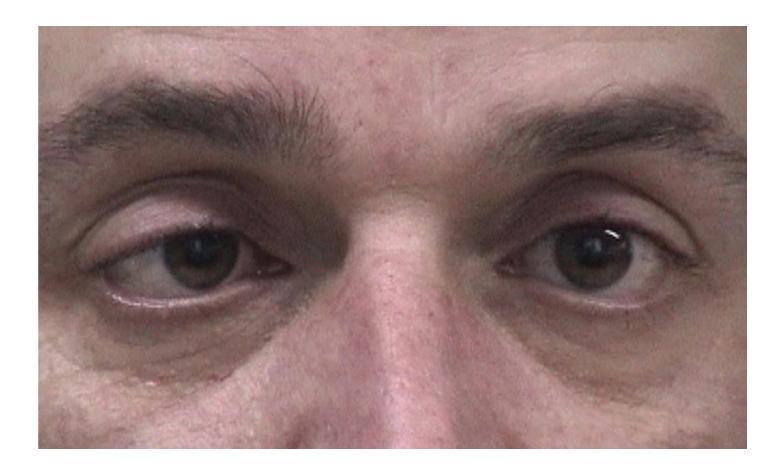
A close-up clinical photograph of the periorbital region in an adult male, demonstrating ocular misalignment consistent with right esotropia. While the left eye maintains a primary forward gaze, the right eye shows significant medial deviation toward the nasal bridge. The image illustrates a manifest strabismus, which clinically correlates with a sixth nerve (abducens) palsy, causing an inability to abduct the right eye and resultant convergent misalignment. The pupils appear slightly dilated, and the surrounding skin and eyelids show no acute inflammatory changes. This visual is intended for medical education regarding neuro-ophthalmological examination, cranial nerve palsies, and ocular motility disorders.
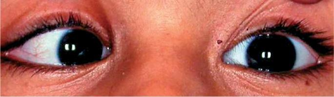
This clinical photograph shows a close-up view of a child's eyes demonstrating ocular misalignment. The patient exhibits right esotropia, characterized by a visible inward deviation of the right eye toward the nose (nasal deviation) while the left eye maintains primary gaze fixation. The pupils are equally round and black with visible light reflections centrally located, suggesting symmetric pupillary size. In the left eye, mild injection of the sclera is noted with visible fine blood vessels. The surrounding periorbital skin is intact, and a small, pigmented nevus is visible near the medial canthus of the right eye. This image serves as a clinical example of strabismus (squint) in a pediatric patient, emphasizing the lack of parallel ocular alignment. It is used in educational contexts to teach the visual identification of ocular deviations and the importance of screening for vision conditions such as amblyopia that may result from untreated strabismus.
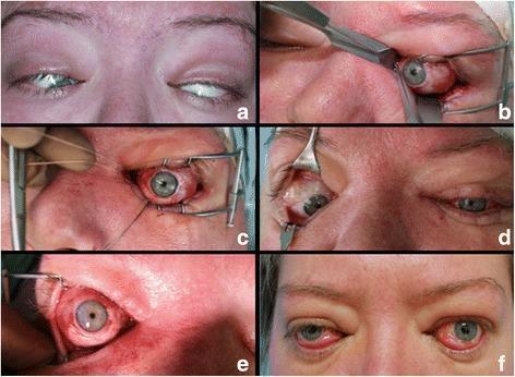
This composite of clinical photographs documents the surgical management of severe restrictive strabismus in a patient with Graves' ophthalmopathy. Panel (a) shows the preoperative presentation of extreme bilateral esotropia and hypotropia, with significant globe deviation and restricted visibility of the pupils. Panels (b), (c), and (e) illustrate intraoperative stages of extraocular muscle release and recession. Specifically, (b) displays the identification and isolation of the inferior rectus muscle using a squint hook through a transconjunctival approach. Panel (c) shows the globe in a corrected primary position after the release of the inferior and medial rectus muscles, with non-absorbable sutures and a speculum visible. Panel (d) demonstrates the asymmetrical interim results one week after surgery on the left eye only. Finally, panel (f) depicts the three-month postoperative outcome, showing restored vertical and horizontal primary alignment in both eyes, despite mild persistent exophthalmos and conjunctival hyperemia. This sequence highlights the effectiveness of extraocular muscle repositioning for correcting fixed globe deviation in advanced autoimmune thyroid eye disease.
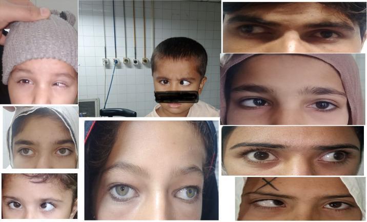
A collage of clinical photographs showcasing various manifestations of strabismus (squint) in pediatric and adult patients. The images demonstrate different types of ocular misalignment, including esotropia (inward deviation of one eye) and exotropia (outward deviation of one eye) relative to the fixating eye. Several frames highlight the asymmetry in pupillary alignment and the visible corneal light reflex displacement, which are key diagnostic signs. The collection illustrates clinical variations in the degree of deviation, ranging from subtle misalignment to more pronounced, large-angle strabismus. These visual findings are frequently associated with amblyopia (lazy eye), where the brain favors the visual input from the correctly aligned eye. The clinical relevance of this visual resource is in ophthalmology and optometry education, specifically for identifying sensory and motor ocular abnormalities and understanding the visual presentation of binocular vision disorders.
Medial Squint (Convergent Squint / Esotropia)
Terminology Clarification
| Term | Meaning |
|---|---|
| Orthophoria | Perfect alignment; rare in the true sense |
| Esophoria | Latent inward tendency, controlled by fusion (no visible squint) |
| Esotropia (medial squint) | Manifest inward deviation - the visual axes do not meet at the fixation point |
| Heterophoria | Latent deviation only apparent when fusion is disrupted |
| Heterotropia | Manifest, constant or intermittent deviation visible at rest |
- Kanski's Clinical Ophthalmology, 10th ed.
Anatomy Relevant to Medial Squint
- Kanski's Clinical Ophthalmology, 10th ed.
Clinical Features
- One eye turns inward while the other fixes on the target
- The corneal light reflex (Hirschberg test) is displaced laterally (outward) on the deviating eye
- In adults: homonymous (uncrossed) diplopia - the false image appears on the same side as the deviating eye
- In children: the brain suppresses the image from the deviating eye to avoid diplopia, risking amblyopia (lazy eye)
- May be constant or intermittent
Types of Esotropia
1. Infantile (Congenital) Esotropia
- Present from birth or within the first 6 months of life
- Accounts for ~25% of all strabismus cases
- Deviation is usually large (>40 prism diopters / >20°) and constant
- Children do NOT "grow out of" large-angle deviations
- Not typically caused by hyperopia
- Treatment: Surgical correction at 6-12 months of age; early correction (before 11 months) improves visual cortical development
- Bradley and Daroff's Neurology in Clinical Practice; Textbook of Family Medicine 9e
2. Accommodative Esotropia (Most Common Type - ~50%)
- Onset between 6 months and 7 years (average ~2.5 years)
- Caused by hyperopia (far-sightedness): the child over-accommodates to see clearly, triggering excessive convergence through the accommodation-convergence reflex
- Initially intermittent, then becomes constant
- Treatment: Corrective spectacles for hyperopia; bifocals if near deviation exceeds distance deviation. This relieves the need to accommodate and allows the eyes to straighten
- High-level stereopsis can be restored if treatment starts within 3 months of constant esotropia onset
- Bradley and Daroff's Neurology in Clinical Practice; Textbook of Family Medicine 9e
3. Non-Accommodative (Acquired) Esotropia
- Not caused by excessive accommodation
- Causes include: anisometropia (unequal refractive errors), cataracts, corneal scars
- Treatment: Address the underlying condition; amblyopia treatment; possible surgery
- Textbook of Family Medicine 9e
4. Microtropia
- Very small-angle esotropia (usually <10 prism diopters)
- Often missed on routine examination
- Associated with amblyopia despite small deviation
5. Cyclic Esotropia
- Very rare; esotropia alternates with straight eyes, each phase lasting ~24 hours
- May persist for months to years
- Kanski's Clinical Ophthalmology
6. Secondary Esotropia Causes (Adults)
- VI nerve (abducens) palsy - inability to abduct the eye causes the medial rectus to go unopposed
- Divergence insufficiency/paralysis
- Spasm of the near reflex
- Thalamic esotropia (central cause)
- Sagging eye syndrome (elderly)
- Thyroid eye disease (restrictive - medial rectus fibrosis)
- Acute acquired comitant esotropia - warrants neurological evaluation (can indicate Chiari malformation or posterior fossa tumor)
- Localization in Clinical Neurology 8e; Bradley and Daroff's Neurology
Sensory Consequences
- Confusion - simultaneous appreciation of two superimposed dissimilar images (both foveae stimulated by different objects)
- Diplopia - in esotropia, diplopia is homonymous (uncrossed): the false image appears on the nasal side (same side as the deviating eye)
- Suppression - the child's brain actively inhibits the image from the deviating eye (occurs due to plasticity of the developing visual cortex, up to age 6-8 years)
- Amblyopia - chronic suppression leads to reduced central vision in the deviating eye ("functional blindness")
- Abnormal retinal correspondence (ARC) - a sensory adaptation unique to childhood strabismus
- Kanski's Clinical Ophthalmology, 10th ed.
Diagnosis
| Test | How It Works |
|---|---|
| Hirschberg (corneal light reflex) test | Penlight shone at both eyes - in esotropia, the reflex is displaced laterally (outward) in the squinting eye |
| Cover test | Covering the fixing eye causes the deviant eye to move outward to take up fixation |
| Cover-uncover test | Reveals latent phoria vs. manifest tropia |
| Prism cover test | Quantifies the angle of deviation in prism diopters |
| Red reflex | Screens for underlying causes (e.g., cataract, retinoblastoma) |
- Textbook of Family Medicine 9e
- Onset after age 6
- Complaints of diplopia or headache
- Incomitance (deviation varies with gaze direction)
- Esotropia greater at distance than near
- Any neurological signs
Management Summary
| Type | Primary Treatment |
|---|---|
| Infantile esotropia | Surgery (6-12 months) |
| Accommodative esotropia | Spectacle correction (full hyperopic prescription) |
| Partially accommodative | Spectacles + surgery for residual deviation |
| Amblyopia (complication) | Occlusion (patching) of the better eye |
| CN VI palsy | Treat underlying cause; prisms; Botulinum toxin; surgery |
| Thyroid restrictive | Orbital decompression; then muscle recession surgery |
- Kanski's Clinical Ophthalmology; Textbook of Family Medicine 9e; Bradley and Daroff's Neurology