

Tell me the diagnosis and treatment
Wolff-Parkinson-White syndrome ECG delta wave pre-excitation
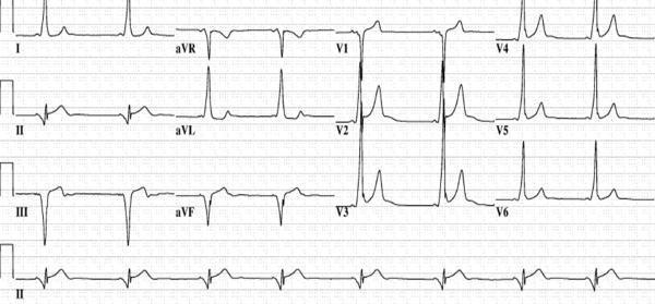
This diagnostic image is a 12-lead electrocardiogram (ECG) demonstrating the classic pre-excitation pattern of Wolff-Parkinson-White (WPW) syndrome associated with a posteroseptal accessory pathway. Key features include a shortened PR interval and the presence of delta waves—slurred upstrokes at the start of the QRS complexes. In the precordial leads, the delta wave is positive in V1, becomes less prominent in V2, and disappears by V3. Crucially, the delta waves are negative in the inferior leads (II, III, and aVF), mimicking a pseudo-infarct Q-wave pattern. This negative polarity in the inferior leads is highly suggestive of an accessory pathway originating in the posteroseptal region, potentially localized near the coronary sinus or middle cardiac vein. The tracing serves as an educational tool for medical students and electrophysiologists to practice electrocardiographic localization of bypass tracts prior to catheter ablation.
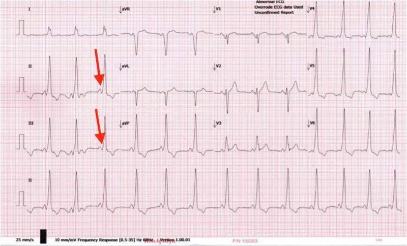
This diagnostic image is a 12-lead electrocardiogram (ECG) tracing demonstrating classic features of Wolff-Parkinson-White (WPW) syndrome. The primary finding is ventricular pre-excitation, characterized by a shortened PR interval (typically less than 120 ms) and a slurred upstroke of the QRS complex, known as a delta wave. Red arrows in leads II and III highlight these delta waves, which indicate early ventricular activation via an accessory pathway (Bundle of Kent). The QRS complexes are widened across multiple leads, particularly evident in the precordial leads V1-V3, and the rhythm is regular but appears tachycardic. The morphology of the QRS complexes in the limb leads and the prominent R waves in V1 are suggestive of an anteroseptal accessory pathway. This visual serves as a key educational resource for identifying pre-excitation patterns in cardiology and understanding the electrophysiological manifestations of accessory conduction pathways in pediatric and adult patients presenting with palpitations or syncope.
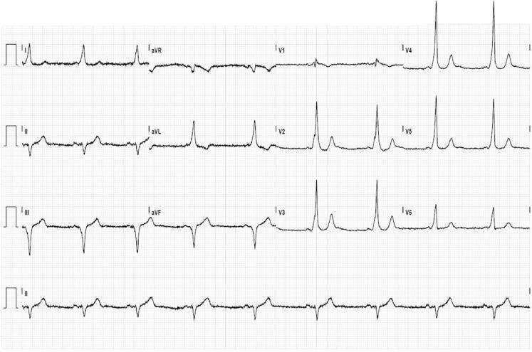
A 12-lead baseline electrocardiogram (ECG) demonstrating classic features of ventricular pre-excitation, characteristic of Wolff-Parkinson-White (WPW) syndrome. The tracing shows a short PR interval and widened QRS complexes across multiple leads. A prominent delta wave (slurring of the initial QRS portion) is visible, most notably in leads I, aVL, and the precordial leads V2-V6. The QRS morphology, specifically the negative delta waves in the inferior leads (II, III, and aVF), suggests a posteroseptal location for the accessory pathway. Secondary repolarization abnormalities are present, including ST-segment depression in the lateral precordial leads (V3-V6), which can occur as a result of altered ventricular activation rather than primary ischemia. The rhythm is sinus, and the overall tracing serves as an educational example of how accessory pathways modify cardiac conduction and QRS morphology.
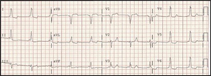
A standard 12-lead electrocardiogram (ECG) tracing demonstrating classic features of manifest ventricular pre-excitation, characteristic of Wolff-Parkinson-White (WPW) syndrome. The primary diagnostic findings include a shortened PR interval (<120 ms) and a widened QRS complex with a distinctive 'delta wave,' visible as initial slurring or notching of the QRS upstroke. These features are most prominent in the precordial leads (V1-V6) and lateral limb leads. The polarity of the delta waves—specifically the negative delta wave in lead III and a predominantly positive QRS in leads V4-V6—is consistent with a mid-septal accessory pathway location. The rhythm is a regular sinus rhythm, and secondary ST-T wave changes are visible, which are common in pre-excitation due to altered ventricular depolarization. This diagnostic image is essential for medical students and clinicians studying cardiac electrophysiology and the interpretation of tachyarrhythmia substrates.
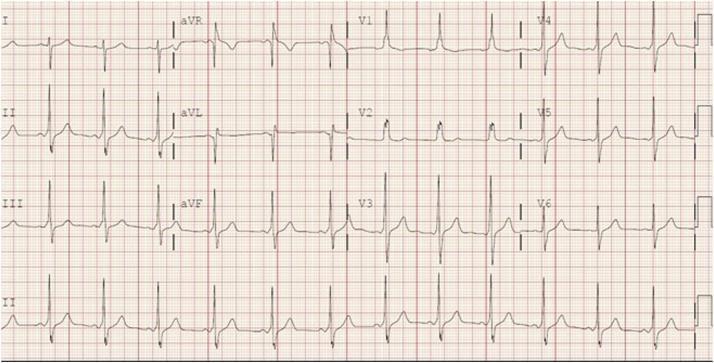
A standard 12-lead electrocardiogram (ECG) tracing printed on red grid paper, displaying a baseline rhythm consistent with Wolff-Parkinson-White (WPW) syndrome. The tracing demonstrates classic signs of ventricular pre-excitation. Key diagnostic features include a shortened PR interval and widened QRS complexes (prolonged duration) across multiple leads. Distinctive 'delta waves'—characterized by a slurred initial upstroke of the QRS complex—are clearly visible, particularly in the lateral leads (I, aVL, V5, and V6). The axis and delta wave morphology suggest an accessory pathway located in the left anterolateral region. Precordial leads show tall R waves in V1 and V2, further supporting the localization of the bypass tract. There are associated secondary ST-segment and T-wave changes opposite to the main QRS deflection, which are common in pre-excitation patterns. This ECG serves as an educational example of cardiac electrophysiology and the identification of accessory pathways in pediatric and adolescent patients presenting with supraventricular tachycardia.
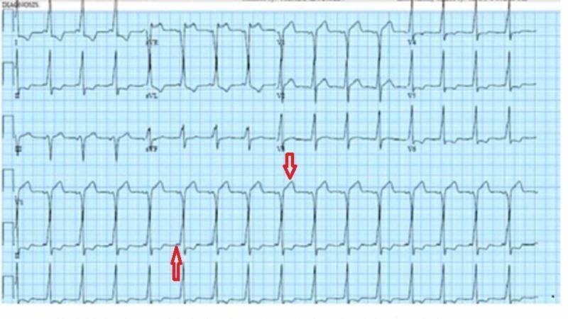
A 12-lead diagnostic electrocardiogram (ECG) demonstrating classic features of ventricular pre-excitation, characteristic of Wolff-Parkinson-White (WPW) syndrome. The tracing shows a notably shortened PR interval (less than 120 ms) followed by widened QRS complexes. A prominent finding is the presence of delta waves, which appear as slurred upstrokes at the beginning of the QRS complexes, most visible in the precordial leads. Red arrows highlight the specific morphology of the slurred QRS complexes and the associated ST-segment changes. Secondary repolarization abnormalities are present, manifesting as ST-segment and T-wave changes discordant with the QRS polarity. This ECG serves as a critical educational example of an accessory pathway (Bundle of Kent) bypassing the atrioventricular node, leading to early ventricular depolarization. The image is categorized under cardiovascular medicine and electrophysiology, suitable for medical students and residents studying cardiac arrhythmias and syncope evaluation.
ECG Diagnosis: Wolff-Parkinson-White (WPW) Syndrome
ECG Findings
| Feature | Finding |
|---|---|
| PR interval | Short (<120 ms) |
| QRS duration | Widened (>120 ms) |
| Delta waves | Present - slurred initial upstroke on QRS complexes |
| Rhythm | Sinus rhythm |
| ST/T changes | Secondary repolarization abnormalities (discordant to QRS) |
Diagnosis: Wolff-Parkinson-White (WPW) Syndrome
- Orthodromic AVRT (most common) - antegrade via AV node, retrograde via accessory pathway - produces narrow QRS tachycardia
- Antidromic AVRT - antegrade via accessory pathway, retrograde via AV node - produces wide QRS tachycardia
- Pre-excited AF - the most dangerous form; rapid conduction through the accessory pathway (not limited by AV nodal delay) can drive ventricular rates >250-300 bpm and degenerate into ventricular fibrillation
Treatment
Acute Management
- Vagal maneuvers first
- IV adenosine (drug of choice) - terminates by blocking AV node
- IV procainamide - if adenosine fails; also blocks accessory pathway
- DC synchronized cardioversion immediately
- Hemodynamically unstable: immediate electrical cardioversion
- Hemodynamically stable: IV procainamide or ibutilide (procainamide preferred - blocks the accessory pathway and slows ventricular rate before restoring sinus rhythm)
CRITICAL WARNING: AV-nodal blocking drugs are contraindicated in pre-excited AF:
- Adenosine, digoxin, beta-blockers, verapamil/diltiazem must NOT be used
- These agents block the AV node and can accelerate conduction through the accessory pathway, paradoxically increasing the ventricular rate and precipitating VF - Braunwald's Heart Disease
Long-Term / Definitive Management
| Option | Details |
|---|---|
| Catheter ablation | First-line (Class I, ACC/AHA/HRS) - curative in >95% of cases, very low complication rate, eliminates need for long-term antiarrhythmics |
| Pharmacologic | Class Ic agents (flecainide, propafenone) or amiodarone for those who decline ablation or are poor candidates |
| Risk stratification | Electrophysiology study (EPS) recommended for high-risk occupations (pilots), competitive athletes, or asymptomatic pre-excitation |
| Observation | Reasonable for asymptomatic pre-excitation with demonstrated intermittent/abrupt loss of pathway conduction (indicating low-risk pathway) |
Reference ECG Image (WPW Pattern)