
Scrub Typhus its clinical features, its complications and treatment guidelines
"scrub typhus" AND treatment
scrub typhus eschar skin lesion chigger bite
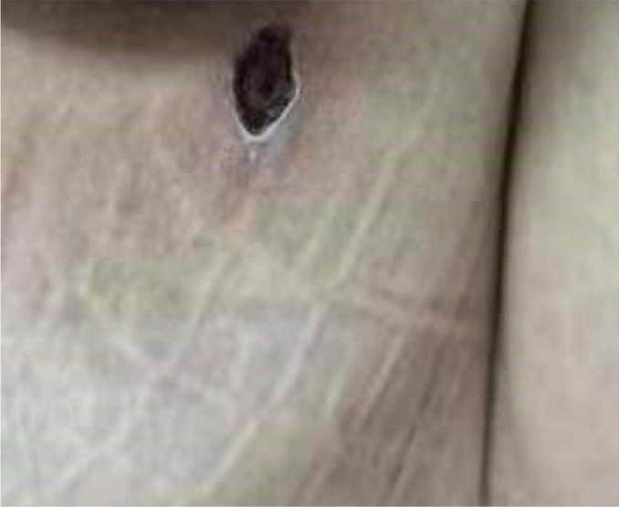
Clinical photograph of a pathognomonic eschar on the left buttock, a hallmark sign of scrub typhus (Orientia tsutsugamushi infection). The lesion is approximately 1 cm in diameter, presenting as a dark brown to black necrotic center (crust) with an oval-to-circular shape and irregular borders. A distinct pale, slightly raised inflammatory halo surrounds the central eschar, creating the characteristic 'cigarette burn' appearance. The surrounding skin exhibits faint erythema and prominent striae distensae (stretch marks). This visual finding is a critical diagnostic indicator in rickettsial diseases, typically representing the site of a larval mite (chigger) bite. Clinically, it aids in differentiating scrub typhus from other febrile illnesses in endemic regions.
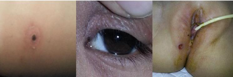
This composite of three clinical photographs displays the characteristic eschar of scrub typhus (Orientia tsutsugamushi) across different anatomical sites. The leftmost image shows a classic, well-circumscribed, necrotic eschar with a dark black central crust surrounded by an erythematous halo and slight edema. The middle image illustrates an eschar located on the inner canthus/eyelid region, appearing as a smaller dark-pigmented lesion. The rightmost image depicts a similar necrotic lesion in the perianal/groin region, accompanied by adjacent skin erythema and a medical catheter in situ. These lesions represent the site of a larval mite (chigger) bite and are pathognomonic clinical markers for rickettsial diseases like scrub typhus. The images emphasize the importance of thorough skin examination in febrile patients, particularly in skin folds and obscured areas where these diagnostic lesions are frequently found.
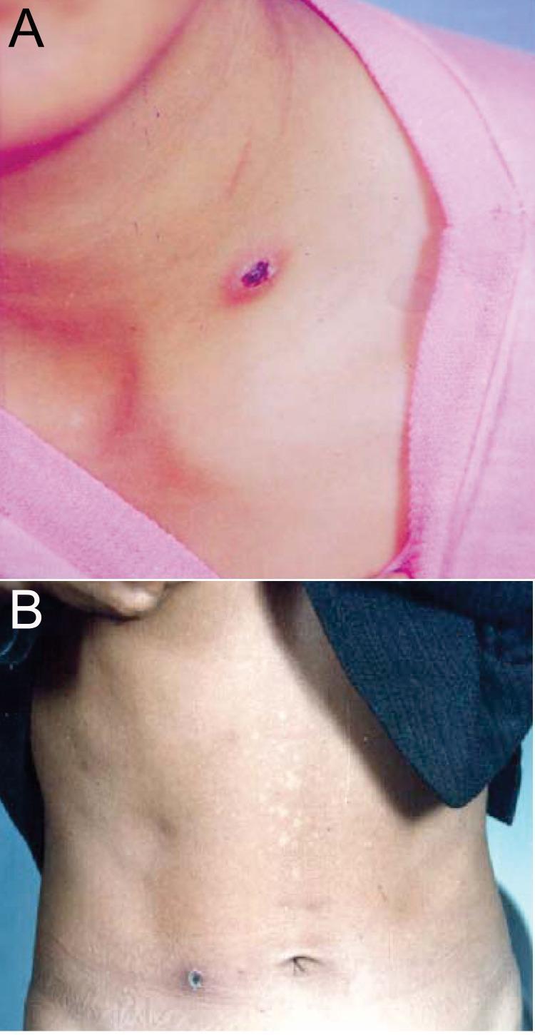
This clinical photograph consists of two panels (A and B) demonstrating the characteristic skin eschars of scrub typhus, caused by Orientia tsutsugamushi. Panel A shows an anatomical view of the neck and upper chest of a patient. A single, small, necrotized lesion is visible, characterized by a dark purple to black central crust (eschar) with an oval-to-irregular shape. The lesion is surrounded by a faint erythematous halo, suggesting localized inflammation at the chigger bite site. Panel B shows a second patient with an eschar located on the waist/lower abdominal region. This lesion is similarly dark and crusty but appears slightly more circular. The surrounding abdominal skin shows subtle mottling or hypopigmentation. These images serve as classic diagnostic examples of the primary lesion found in rickettsial diseases, particularly scrub typhus, which often appears in areas where clothing is tight or skin is thin. The clinical significance of these visual findings is their role as a hallmark diagnostic sign during the acute phase of infection.
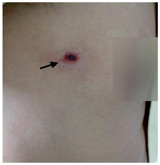
A clinical photograph of a human chest area demonstrating a pathognomonic eschar, a classic skin lesion associated with scrub typhus. The lesion, indicated by a black arrow, features a small, dark, necrotic central crust (eschar) that is circular to oval in shape. This necrotic center is surrounded by a prominent, well-demarcated erythematous halo and a slightly raised, indurated rim, characteristic of an inflammatory reaction at the site of a mite bite (chigger). The surrounding fair skin appears otherwise unremarkable without diffuse rash or additional lesions. This image serves as a key diagnostic educational tool for infectious disease and dermatology, illustrating the hallmark physical finding used in the clinical diagnosis of Orientia tsutsugamushi infection.
Scrub Typhus (Orientia tsutsugamushi infection)
Clinical Features
- Incubation period: 6-21 days (typically ~10 days) after the chigger bite.
- Onset: Usually sudden - fever, headache, myalgia, and often a nonproductive cough and GI symptoms.
- Eschar: The classic finding is a painless, black, necrotic scab ("cigarette-burn" lesion) at the mite bite site, surrounded by an erythematous halo, often with regional tender/draining lymphadenopathy. It is detected in a variable proportion of patients depending on skin pigmentation and how carefully skin folds/axillae/groin are examined.
- Rash: A maculopapular rash may appear on the trunk (less often extremities), usually a few days into illness; it is rarely petechial and rarely involves the face, palms, or soles.
- Relative bradycardia is common despite fever.
- Generalized lymphadenopathy and hepatosplenomegaly can occur.
- Neurologic: neuromeningeal symptoms are relatively common - confusion, meningoencephalitis, ataxia, seizures.
- Pulmonary: cough, interstitial pneumonitis, pulmonary edema/ARDS in severe cases.
- Laboratory: leukopenia (early) with later leukocytosis, thrombocytopenia, elevated hepatic transaminases, hyponatremia, hypoalbuminemia.
- Severe disease can present as septic shock with multiorgan dysfunction.
Complications
- Acute respiratory distress syndrome / respiratory failure, interstitial pneumonia, pleural effusion
- Acute kidney injury (associated with higher ICU admission and mortality)
- Meningoencephalitis, seizures, coma
- Septic shock, multiorgan failure
- Myocarditis
- Hepatic dysfunction/jaundice
- Hemorrhagic manifestations (rarely, hematemesis, cerebral hemorrhage in related typhus group illness)
- Disseminated intravascular coagulation in severe cases
- In pregnancy, scrub typhus is associated with abortion
Treatment Guidelines
- Doxycycline 100 mg orally twice daily for 7-15 days is the treatment of choice for uncomplicated disease.
- In severe scrub typhus, IV doxycycline 200 mg/day is recommended, and combination therapy with doxycycline plus azithromycin has been shown to be more beneficial than monotherapy with either agent alone.
- Azithromycin 500 mg for 3 days is an effective alternative, particularly favored in pregnancy and children, and in areas with doxycycline-poor-responder strains (e.g., parts of Thailand).
- Chloramphenicol 500 mg four times daily (or 2 g/day) for 7-15 days
- Rifampin 600 mg/day - useful for cases poorly responsive to doxycycline or chloramphenicol
- Quinolones should be avoided - they are inferior/ineffective and have been associated with treatment failure.
- Single-day doxycycline courses are followed by relapse; a full 7-day (or longer, up to 15 days in some regimens) course is needed.
- Doxycycline-resistant/poorly-responsive cases have been reported, notably in northern Thailand, where azithromycin and rifampin remain effective alternatives.