
IgA nephropathy
Reading File
IgA nephropathy (Berger's disease) is the most common primary glomerulonephritis worldwide. It is characterized by mesangial IgA deposits, typically presenting with recurrent macroscopic hematuria (often following mucosal infections), or asymptomatic microscopic hematuria with/without proteinuria. It ranges from benign to progressive, with ~20-40% reaching ESRD over 20 years.
Searching sources...
Finding Sources
Searching PubMed
"IgA nephropathy" AND treatment
Good. Now let me read the primary sources in parallel - Comprehensive Clinical Nephrology (most detailed nephrology text) and Robbins (pathology/mechanism), plus Brenner & Rector.
Reading File
Reading File
Reading File
Excellent - I have very rich content from multiple textbooks. Let me fetch the key histology images and also read the treatment/management section from Comprehensive Clinical Nephrology.
Loading Image
Reading File
I now have comprehensive material. Let me compile the full answer.
IgA Nephropathy (Berger's Disease)
Overview
IgA nephropathy (IgAN) is the most common primary glomerulonephritis worldwide, particularly prevalent in Asia (accounting for >45% of primary glomerular diseases in Chinese biopsy registries). It predominantly affects males over females (2:1 to 3:1 in White populations; near 1:1 in Asian populations), most commonly in the second to third decade of life. - Comprehensive Clinical Nephrology, 7th Ed.
Pathogenesis - The "Four-Hit" Model
The pathogenesis follows a well-established sequence: - Brenner & Rector's The Kidney
-
Hit 1 - Aberrant IgA1 glycosylation: B cells (especially mucosal) produce galactose-deficient IgA1 (Gd-IgA1) - the hinge-region O-linked glycans lack galactose and instead carry N-acetylgalactosamine, making the molecule immunogenic.
-
Hit 2 - Autoantibody formation: IgG and IgA autoantibodies recognize the exposed N-acetylgalactosamine on Gd-IgA1 as "foreign" and form immune complexes.
-
Hit 3 - Immune complex deposition: These large polymeric complexes deposit in the glomerular mesangium (they cannot be cleared efficiently by the liver in patients with liver disease, worsening the condition).
-
Hit 4 - Mesangial injury: Deposited complexes activate complement (predominantly via the alternative and lectin pathways - classical pathway components C1q/C4 are typically absent) and trigger mesangial cell proliferation, cytokine/chemokine overproduction, and progressive fibrosis.
Genetic factors: GWAS studies identify susceptibility loci in MHC, the DEFA gene cluster, and the alternative complement pathway. Familial IgAN variants have been found in DEFA4, MYCT1, CARD8, and ZNF543. More than 25% of blood relatives have elevated serum Gd-IgA1 levels.
Why mucosal infections trigger attacks: Infections increase mucosal IgA production, driving surges in Gd-IgA1 and immune complex formation - hence the "synpharyngitic hematuria" pattern.
Clinical Presentations
No single presentation is pathognomonic. The three main patterns are: - Comprehensive Clinical Nephrology, 7th Ed., p. 333
| Presentation | Frequency | Features |
|---|---|---|
| Episodic macroscopic hematuria | 40-50% | Brown urine (rarely red), loin pain, onset within 24 hours of mucosal infection (vs. 2-3 week delay in poststreptococcal GN) |
| Asymptomatic micro-hematuria ± proteinuria | 30-40% | Detected on screening; proteinuria usually <2 g/24 h |
| Nephrotic/nephritic syndrome, hypertension, CKD | Less common | Older patients; worse prognosis |
Macroscopic hematuria episodes typically resolve spontaneously over days; microscopic hematuria persists between attacks. Episodes may be accompanied by acute kidney injury from tubular injury, which is usually reversible.
Histology (Renal Biopsy - Required for Diagnosis)
Light microscopy: Ranges from normal to mesangial widening/hypercellularity, focal proliferative GN, diffuse mesangioproliferative GN, or (rarely) crescentic GN.
Immunofluorescence: Dominant mesangial IgA deposits (often with C3, properdin, and smaller amounts of IgG or IgM). C1q is absent. This is the diagnostic hallmark.
Electron microscopy: Electron-dense deposits in the mesangium.
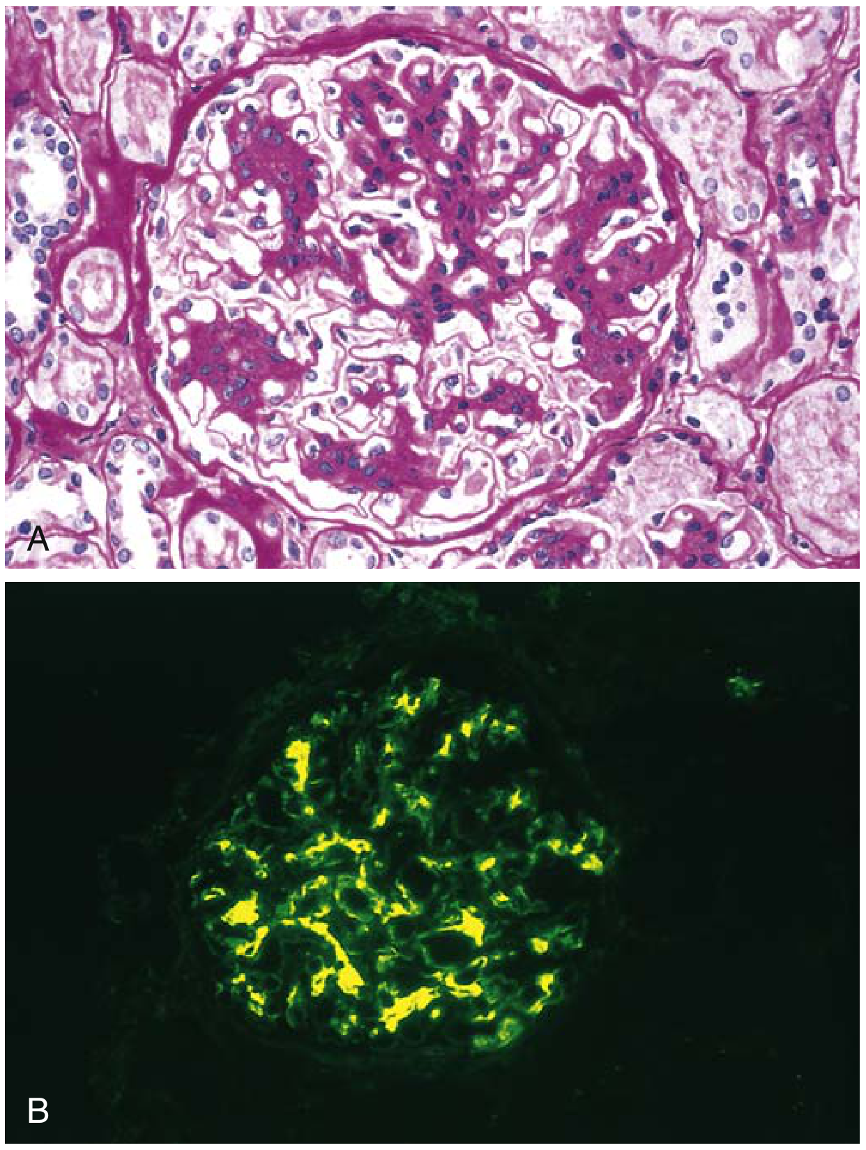
Fig. 12.12 - Robbins Basic Pathology: (A) mesangial proliferation on H&E; (B) characteristic IgA deposition (yellow/green) in mesangium on immunofluorescence.
Oxford (MEST-C) Classification (2009, updated 2016)
Biopsy scored on five features to predict progression risk: - Comprehensive Clinical Nephrology, 7th Ed.
| Score | Feature | Categories |
|---|---|---|
| M | Mesangial hypercellularity | M0 / M1 |
| E | Endocapillary hypercellularity | E0 / E1 |
| S | Segmental glomerulosclerosis | S0 / S1 |
| T | Tubular atrophy / interstitial fibrosis | T0 / T1 / T2 |
| C | Crescents | C0 / C1 / C2 |
Higher T and C scores correlate strongly with worse renal outcomes.
Prognosis
- ~30-40% of patients reach ESRD within 20-30 years
- If diagnosed very early: only ~5% develop CKD stage 4-5 at 20 years
- In typical presentations: ~25% ESRD + further 20% significant GFR decline at 20 years
- A Swedish study found patients with IgAN die ~6 years earlier than the general population, mostly from cardiovascular causes
Adverse prognostic markers (Box 24.2, CCN): hypertension, proteinuria >1 g/day, reduced eGFR at biopsy, older age, high MEST-C scores, male sex.
Associated Conditions
IgAN can be secondary to: - Comprehensive Clinical Nephrology, 7th Ed., p. 333
- Rheumatic/autoimmune: Ankylosing spondylitis, rheumatoid arthritis, Reiter syndrome, uveitis
- GI: Celiac disease, ulcerative colitis, Crohn's disease, Whipple disease
- Hepatic: Alcoholic liver disease, nonalcoholic cirrhosis (impaired hepatic clearance of IgA complexes)
- Skin: Dermatitis herpetiformis
- IgA vasculitis (Henoch-Schonlein Purpura): Systemic counterpart with identical renal lesion plus rash, arthritis, GI involvement
Treatment
Supportive (All Patients)
- RAS blockade (ACEi or ARB): First-line for proteinuria >0.5 g/day and/or hypertension; target BP <130/80 mmHg; aim to reduce proteinuria to <1 g/day (or <0.5 g/day if possible)
- Optimized BP control
- Fish oil (omega-3): Modest evidence; used in some centers
- Tonsillectomy: Practiced widely in Japan; evidence remains limited
Immunosuppression (High-Risk Patients)
- Corticosteroids: Used in patients with proteinuria >1 g/day despite optimized RAS blockade and eGFR >30 mL/min/1.73m². The TESTING trial showed benefits but also serious infection risk.
- Targeted-release budesonide (TRF-budesonide / Nefecon): Delivers budesonide to the ileum (the primary site of aberrant IgA production). A 2025 meta-analysis (PMID 40721561) confirms efficacy and safety in IgAN treatment, with reduced systemic side effects versus systemic steroids.
- Sparsentan: Dual endothelin/angiotensin receptor antagonist. A 2024 systematic review and meta-analysis (PMID 39333921) confirmed safety and efficacy versus irbesartan in IgAN.
- A 2025 network meta-analysis (PMID 40606442) comparing multiple agents for IgAN found significant antiproteinuric effects from several newer targeted therapies.
Emerging/Novel Agents
- Complement inhibitors (anti-C5, anti-factor B, anti-MASP-2): Targeting the alternative/lectin complement pathway
- Atacicept, blisibimod: Anti-BAFF/APRIL agents targeting B-cell IgA production
- Iptacopan: Factor B inhibitor
ESRD
- Renal replacement therapy; IgAN recurs in ~30% of transplants (usually mild)
Key Differentials
- Poststreptococcal GN: Hematuria 2-3 weeks after infection (vs. concurrent in IgAN); low C3; resolves completely
- Thin basement membrane disease: Persistent micro-hematuria, family history, normal IgA on IF
- IgA vasculitis: Systemic features (rash, joint, GI); identical renal pathology
- Alport syndrome: X-linked, thinning/splitting of GBM on EM, family history of renal failure/deafness
This is a shared conversation. Sign in to Orris to start your own chat.