
Pancreas histology
histology of normal pancreas showing exocrine acini islets of Langerhans and ducts
pancreas histology
pancreas histology exocrine acini islets centroacinar cells site:nih.gov OR site:ncbi.nlm.nih.gov
Pancreas histology
- Exocrine portion - produces digestive enzymes and bicarbonate-rich fluid
- Endocrine portion - the islets of Langerhans, which release hormones into blood
1. Exocrine pancreas
- Acinar cells are pyramidal.
- Their basal cytoplasm is basophilic because it contains abundant rough endoplasmic reticulum.
- Their apical cytoplasm is eosinophilic/granular, due to enzyme-containing zymogen granules.
- The round nucleus is usually basal.
- Digestive enzyme precursors include trypsinogen, chymotrypsinogen, lipase, amylase, and nucleases.
2. Centroacinar cells
- They are pale, flattened or cuboidal cells seen within the lumen of an acinus.
- They represent the beginning of the intercalated duct.
- Together with duct cells, they contribute bicarbonate-rich secretion, helping neutralize acidic chyme in the duodenum.
3. Pancreatic duct system
- Intercalated ducts are lined by low cuboidal cells.
- Larger ducts have cuboidal to columnar epithelium.
- Striated ducts are absent, unlike in major salivary glands.
4. Endocrine pancreas: Islets of Langerhans
- They are more numerous in the tail of the pancreas.
- Cells are arranged in cords around many fenestrated capillaries.
- They have no ducts.
| Cell type | Main hormone | Major action |
|---|---|---|
| Beta cells | Insulin | Lowers blood glucose |
| Alpha cells | Glucagon | Raises blood glucose |
| Delta cells | Somatostatin | Inhibits insulin, glucagon, and GI secretion |
| PP/F cells | Pancreatic polypeptide | Modulates pancreatic and GI activity |
How to identify pancreas on an H&E slide
- Dark serous acini
- Pale islets of Langerhans
- Centroacinar cells inside acini
- Absence of striated ducts
- No mucous acini or serous demilunes
Ureter histology
normal ureter histology cross section urothelium lamina propria muscularis layers
ureter histology
ureter histology urothelium muscularis externa layers
Ureter histology
- Mucosa
- Muscularis
- Adventitia
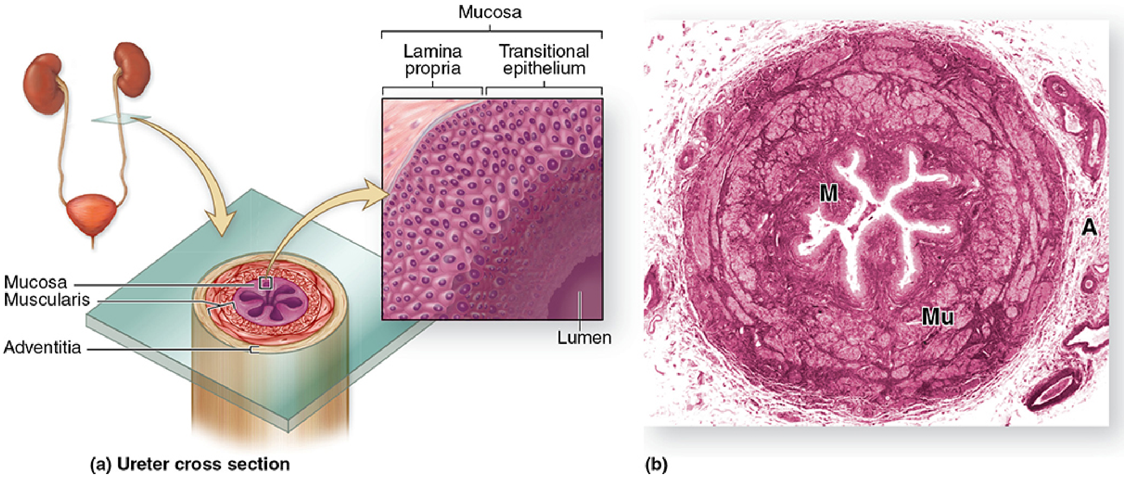
1. Mucosa
- The lumen is usually stellate (star-shaped) in cross-section because the mucosa is thrown into prominent longitudinal folds.
- It is lined by transitional epithelium (urothelium), specialized to withstand and distend with urine.
- Urothelium has:
- Basal cells: small cells resting on the basement membrane
- Intermediate cells: cuboidal to low columnar cells
- Superficial umbrella cells: large, dome-shaped, sometimes binucleate cells that form the urine barrier
- Beneath it is a dense connective-tissue lamina propria.
- There is no muscularis mucosae and no submucosa.
2. Muscularis
- In most of the ureter:
- Inner longitudinal smooth-muscle layer
- Outer circular smooth-muscle layer
- In the lower/distal one-third, an additional outer longitudinal layer develops.
3. Adventitia
- Outer layer of fibroelastic connective tissue
- Contains blood vessels, lymphatics, nerves, and surrounding adipose tissue
- The ureter is retroperitoneal, so it normally has an adventitia, not a serosa.
Identification points on a slide
- Star-shaped lumen
- Longitudinal mucosal folds
- Urothelium with umbrella cells
- Thick smooth muscle wall
- Adventitia
- No glands and no muscularis mucosae
Important distinction
Uterus histology
normal uterus histology endometrium myometrium perimetrium proliferative secretory phase
endometrium histology
uterus histology endometrium myometrium proliferative secretory phase site:ncbi.nlm.nih.gov
Uterus histology
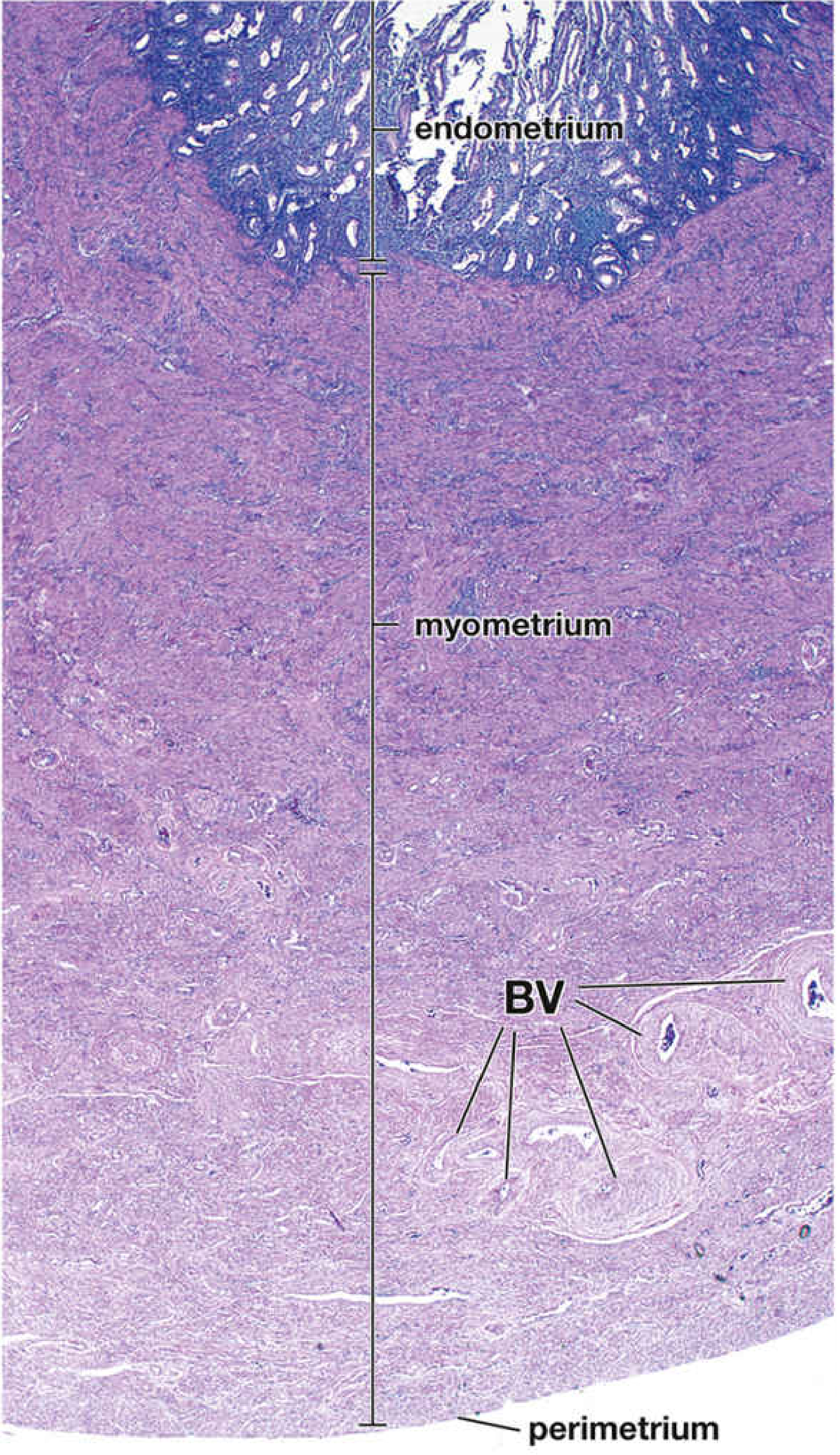
1. Endometrium
- Simple columnar surface epithelium, with ciliated and secretory cells
- Endometrial stroma, a cellular connective tissue
- Simple tubular uterine glands that extend from the surface toward the myometrium
| Layer | Features and fate |
|---|---|
| Stratum functionalis | Superficial two-thirds. Contains most of the glands and spiral arteries. It undergoes cyclic change and is shed during menstruation. |
| Stratum basalis | Deep one-third, adjacent to myometrium. Supplied by straight arteries. It is retained during menstruation and regenerates the functionalis. |
Menstrual-cycle appearances
| Phase | Hormonal influence | Histological features |
|---|---|---|
| Proliferative phase | Estrogen | Thin-to-thickening endometrium; straight, narrow tubular glands; frequent mitoses; compact cellular stroma. |
| Secretory phase | Progesterone | Thick endometrium; tortuous, coiled “saw-toothed” glands with secretion in their lumina; edematous stroma; prominent spiral arteries. |
| Menstrual phase | Fall in estrogen and progesterone | Spiral-artery constriction, ischemia, breakdown, and shedding of the stratum functionalis. |
2. Myometrium
- Made of interlacing bundles of smooth muscle with connective tissue.
- Its layers are not sharply demarcated.
- The middle vascular layer, the stratum vasculare, contains large blood vessels.
- During pregnancy, smooth-muscle cells undergo hypertrophy and hyperplasia.
- At delivery, myometrial contraction expels the fetus and compresses blood vessels to reduce bleeding.
3. Perimetrium
- Where covered by peritoneum, it is a thin serosa consisting of mesothelium and loose connective tissue.
- Parts of the uterus not covered by peritoneum have an adventitia instead.
Identification points on a histology slide
- A lumen lined by simple columnar epithelium
- Numerous uterine glands in endometrial stroma
- A very thick layer of irregularly arranged smooth muscle forming the myometrium
- An outer thin serosal layer when present
- Endometrium that changes substantially with the menstrual phase
Fallopian tube histology
normal fallopian uterine tube histology cross section mucosal folds ciliated cells secretory cells ampulla
fallopian tube histology
fallopian uterine tube histology ciliated secretory cells mucosal folds site:ncbi.nlm.nih.gov
Fallopian tube histology
- Mucosa
- Muscularis
- Serosa
1. Mucosa
- Lined by simple columnar epithelium
- Lamina propria: loose, vascular connective tissue
- No submucosa and no muscularis mucosae
| Cell | Histological feature | Function |
|---|---|---|
| Ciliated cells | Apical cilia | Beat toward the uterus, moving the oocyte or zygote uterineward |
| Nonciliated secretory cells, or peg cells | Narrow apex, lack cilia | Produce nutritive tubal fluid for sperm, oocyte, and early embryo |
2. Muscularis
- Inner circular layer - thicker
- Outer longitudinal layer - thinner
3. Serosa
- Mesothelium, or simple squamous epithelium
- Thin underlying connective tissue
Regional differences
| Segment | Histological appearance |
|---|---|
| Infundibulum | Numerous mucosal folds and many ciliated cells |
| Ampulla | Widest lumen; most elaborate, branched mucosal folds; usual site of fertilization |
| Isthmus | Narrower lumen, fewer and lower folds; relatively thicker muscular wall |
| Intramural part | Very narrow lumen; minimal folding; passes through uterine wall |
How to identify it on a slide
- A very irregular, folded or labyrinth-like lumen
- Simple columnar epithelium with cilia
- No glands in the mucosa
- Smooth-muscle wall beneath the mucosa
- Outer serosa
Histology of testis