Positive findings & PG level assessment for Parkinson's disease & explain anatomy ,types & pathology of disease & difference between parkinson's & parkinsonism
"Parkinson disease"[MeSH Terms] AND diagnosis
Parkinson's disease positive findings clinical signs PG level assessment pathology 2025
Parkinson's Disease - Complete Clinical Review
1. Anatomy Relevant to Parkinson's Disease
The Basal Ganglia Circuit
- Pars compacta (SNpc): contains melanin-pigmented dopaminergic neurons that project to the striatum via the nigrostriatal pathway - this is the primary site of neurodegeneration in PD
- Pars reticulata (SNpr): a GABAergic output nucleus of the basal ganglia
- Direct pathway: striatum → GPi/SNpr → thalamus → cortex (facilitatory; uses D1 receptors, activated by dopamine)
- Indirect pathway: striatum → GPe → subthalamic nucleus (STN) → GPi/SNpr → thalamus → cortex (inhibitory; uses D2 receptors, inhibited by dopamine)
- Olfactory bulb and enteric nervous system (early; stages 1-2)
- Locus coeruleus, dorsal raphe, dorsal motor nucleus of vagus (stages 1-2)
- Substantia nigra and basal forebrain (stages 3-4 - symptomatic phase)
- Limbic cortex, neocortex (late stages 5-6)
2. Types of Parkinson's Disease
A. Idiopathic (Primary) Parkinson's Disease
| Subtype | Features | Prognosis |
|---|---|---|
| Tremor-dominant | Prominent rest tremor, slow progression | Slower disease course, longer levodopa benefit |
| Akinetic-rigid | Bradykinesia + rigidity dominate, minimal tremor | Faster progression |
| Mixed/Postural instability & gait difficulty (PIGD) | Prominent gait freeze, falls | More rapid decline, worse prognosis |
B. Genetic Forms of PD
| Gene | Locus | Inheritance | Notes |
|---|---|---|---|
| SNCA (α-synuclein) | 4q21 | Autosomal dominant | First PD gene identified; protein aggregates as Lewy bodies |
| LRRK2 (leucine-rich repeat kinase 2) | 12q12 | Autosomal dominant | Most common cause of late-onset familial PD |
| Parkin (PARK2) | 6q26 | Autosomal recessive | Most common early-onset (<40 yrs) |
| PINK1 | 1p36 | Autosomal recessive | Mitochondrial dysfunction |
| DJ-1 (PARK7) | 1p36 | Autosomal recessive | Rare; oxidative stress mechanism |
| GBA | 1q22 | Risk factor | Glucocerebrosidase; increased PD susceptibility |
3. Positive Findings in Parkinson's Disease
Cardinal Motor Signs (TRAP)
- Characteristic 4-6 Hz "pill-rolling" rest tremor
- Appears when limb is fully at rest; disappears with voluntary movement
- Asymmetric onset (typically one hand)
- Re-emergent postural tremor (appears after a latency of a few seconds when arms are outstretched)
- Velocity-independent lead-pipe resistance to passive movement
- Cogwheel rigidity: tremor superimposed on rigidity, gives a ratchet-like quality
- Present at wrist, elbow, neck; tested by passive joint movement
- Froment's maneuver (contralateral voluntary movement amplifies rigidity on the tested side - positive finding)
- Slowness of movement (bradykinesia) with progressive decrement in speed and amplitude on repetitive movements
- Assessed by finger tapping, hand opening/closing, foot tapping
- Hypomimia: masked/poker face, reduced blinking (2-10/min vs normal 12-20/min)
- Hypophonia: soft, monotone voice
- Micrographia: progressively smaller handwriting
- Hypodiadochokinesia: impaired rapid alternating movements
- Pull test (Retropulsion test): examiner pulls patient backward by shoulders; patient fails to take a corrective step or falls - strongly positive in PD
- Usually a late feature (if early, suggests atypical parkinsonism)
Other Positive Clinical Findings
- Festination: shuffling gait with short steps, accelerating to keep up with shifted centre of gravity
- Reduced arm swing (often unilateral early)
- Freezing of gait (FOG): sudden inability to initiate or continue walking
- Camptocormia: abnormal forward flexion of thoracolumbar spine
- Procerus sign (furrowing between brows): more typical of PSP; PD shows hypomimia instead
- Seborrhoeic dermatitis over face
- Orthostatic hypotension
- Constipation (often precedes motor symptoms by years)
- Urinary dysfunction
- Hyperhidrosis
- Sialorrhoea (drooling)
- Hyposmia/anosmia (often precedes motor symptoms; one of earliest markers)
- REM Sleep Behavior Disorder (RBD): acting out dreams; a major prodromal feature
- Depression and anxiety
- Cognitive impairment / dementia (Parkinson's disease dementia, PDD)
- Visual hallucinations (later stage)
- Pain and sensory symptoms
4. PG Level Assessment (Prognostic Grading / Clinical Scales)
A. Hoehn and Yahr Scale (H&Y)
| Stage | Description |
|---|---|
| Stage I | Unilateral involvement only; minimal or no functional impairment |
| Stage II | Bilateral or midline involvement; no impairment of balance |
| Stage III | First sign of impaired righting reflex (positive pull test); mild-moderate disability; physically independent |
| Stage IV | Fully developed, severely disabling disease; patient still able to walk/stand unassisted |
| Stage V | Confinement to bed or wheelchair unless aided |
B. MDS-UPDRS (Movement Disorder Society - Unified Parkinson's Disease Rating Scale)
| Part | What it Measures |
|---|---|
| Part I | Non-motor aspects of daily living (mentation, behavior, mood) |
| Part II | Motor aspects of daily living (ADL impairment) |
| Part III | Motor examination (clinician-scored; tremor, rigidity, bradykinesia, gait, postural stability) |
| Part IV | Motor complications (dyskinesia, fluctuations) |
C. Mini-Mental State / MoCA
D. Schwab & England ADL Scale
5. Pathology of Parkinson's Disease
Macroscopic Pathology
- Depigmentation of the substantia nigra: loss of melanin-containing neurons causes visible pallor of the SN in cross-sections of the midbrain (compared to the normal dark appearance)
- Pallor also seen in the locus coeruleus
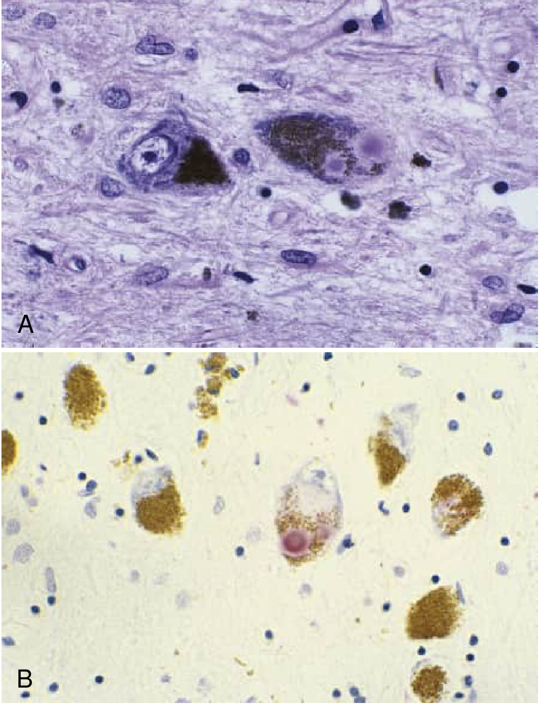
Microscopic Pathology
- Selective loss of dopaminergic neurons in the SNpc (60-80% lost before symptoms appear)
- Remaining neurons show degenerative changes
- Intraneuronal, round, eosinophilic cytoplasmic inclusions with a pale halo
- On H&E: dense core (eosinophilic, 5-25 µm) surrounded by a clear halo, displacing neuromelanin
- Composed of: α-synuclein (major protein), ubiquitin, neurofilaments, proteasome components
- On α-synuclein immunostaining: stain intensely red/brown
- Found in SNpc, locus coeruleus, dorsal vagal nucleus, cortex (in PDD)
- Abnormal, dystrophic neuritic processes containing α-synuclein aggregates
- Found in the CA2/3 region of the hippocampus and elsewhere
- Astrocytic gliosis in areas of neuronal loss
Braak Staging of PD Pathology
| Stage | Location of Lewy Pathology | Clinical Correlate |
|---|---|---|
| 1-2 | Olfactory bulb, dorsal motor nucleus of vagus, enteric NS | Anosmia, constipation, RBD (prodrome) |
| 3-4 | Substantia nigra, basal forebrain, amygdala | Motor symptoms begin |
| 5-6 | Limbic cortex → neocortex | Dementia, psychosis |
Neurochemical Pathology
| Neurotransmitter | Change | Circuit Effect |
|---|---|---|
| Dopamine | Severely reduced in striatum (>80% at symptom onset) | Bradykinesia, rigidity, tremor |
| Norepinephrine | Reduced (locus coeruleus loss) | Autonomic dysfunction, depression |
| Serotonin | Reduced (raphe nuclei) | Depression, mood changes |
| Acetylcholine | Reduced (nucleus basalis of Meynert) | Dementia, cognitive decline |
6. Parkinson's Disease vs. Parkinsonism - Key Differences
| Feature | Parkinson's Disease (PD) | Parkinsonism |
|---|---|---|
| Definition | A specific neurodegenerative disease caused by idiopathic dopaminergic loss with Lewy body pathology | A clinical syndrome of bradykinesia + rigidity ± tremor ± postural instability, from ANY cause |
| Relationship | PD IS a form of parkinsonism (most common cause; ~80%) | Parkinsonism is the umbrella term; PD is a subtype |
| Pathology | Lewy bodies (α-synuclein) + SNpc degeneration | Varies by cause; no Lewy bodies in most secondary forms |
| Onset | Asymmetric; unilateral at onset | Often bilateral/symmetric at onset (atypical or secondary forms) |
| Tremor | Prominent 4-6 Hz resting tremor | Minimal or absent in most atypical and secondary forms |
| Levodopa response | Excellent (70-100%) initially | Poor or absent (atypical); good (drug-induced, after withdrawal) |
| Progression | Slow (years to decades) | Faster in atypical forms |
| Postural instability | Late feature | Early prominent feature in PSP, MSA |
| Dementia | Late feature (PDD) or with DLB spectrum | Early in DLB, PSP |
| Brain imaging (MRI) | Normal on standard imaging | Abnormal in many: midbrain atrophy (PSP), putaminal atrophy (MSA), white matter lesions (vascular) |
| DAT scan | Reduced (asymmetric) | Reduced in neurodegenerative forms; normal in drug-induced/functional |
Causes of Parkinsonism (Non-PD)
| Condition | Key Distinguishing Features |
|---|---|
| Progressive Supranuclear Palsy (PSP) | Vertical supranuclear gaze palsy, axial (nuchal) rigidity > limb, early falls backward, Procerus sign ("astonished look"), midbrain atrophy ("hummingbird sign" on MRI) |
| Multiple System Atrophy (MSA) | Profound early dysautonomia, cerebellar signs (MSA-C), anterocollis, no/poor levodopa response, "hot cross bun sign" on MRI |
| Corticobasal Degeneration (CBD) | Alien limb, cortical sensory loss, apraxia, limb dystonia, asymmetric fronto-parietal atrophy on MRI |
| Dementia with Lewy Bodies (DLB) | Cognitive decline preceding/co-emerging with parkinsonism, early visual hallucinations, neuroleptic sensitivity, RBD, fluctuating cognition |
| Cause | Key Feature |
|---|---|
| Drug-induced | Onset during offending drug (antipsychotics, metoclopramide, reserpine); improves after withdrawal |
| Vascular (VP) | Lower body predominance, pyramidal signs, white matter lesions/lacunar infarcts on MRI, stepwise progression |
| Post-infectious/Encephalitis lethargica | History of encephalitis, oculogyric crises |
| Toxic | MPTP (meperidine analog), manganese, carbon monoxide exposure |
| Normal Pressure Hydrocephalus (NPH) | Gait apraxia + dementia + urinary incontinence triad; communicating hydrocephalus on CT/MRI |
| Wilson's disease | Young age, Kayser-Fleischer rings, liver disease, low ceruloplasmin |
7. Diagnostic Criteria Summary (MDS Clinical Criteria)
- Absence of absolute exclusion criteria
- At least two supportive criteria
- No red flags
- Unilateral onset
- Clear and dramatic response to levodopa (70-100%)
- Levodopa-induced dyskinesia
- Rest tremor
- Hyposmia or loss of smell
- Cardiac sympathetic denervation on MIBG scintigraphy
- Presence of Lewy bodies on biopsy (definitive)
- Unequivocal cerebellar features
- Downward vertical supranuclear gaze palsy
- Frontotemporal dementia within first 5 years
- Purely lower-limb parkinsonism for >3 years
- No response to levodopa despite adequate dose
- Cortical sensory loss, alien limb
Recent Evidence Note
- Bradley and Daroff's Neurology in Clinical Practice (9780323642613)
- Adams and Victor's Principles of Neurology, 12th Edition (9781264264520)
- Harrison's Principles of Internal Medicine 22E (9781265977061)
- MDS Clinical Diagnostic Criteria for Parkinson's Disease (Postuma et al., 2015)
Explain to me types in detail & how to diagnose those types clinically & then give me management for same or clinical feature wise (make a short term & long term goals & then give me management)
"Parkinson disease"[MeSH Terms] AND management AND treatment
PSP MSA CBD DLB clinical diagnosis features management guidelines 2024 2025
PSP MSA CBD DLB clinical diagnosis management treatment
What Was Covered
Part 1 - Types in Detail (7 types)
- Idiopathic PD - with 4 motor subtypes (Tremor-dominant, Akinetic-Rigid, PIGD, Young-onset)
- PSP - 5 phenotypes, MDS OPAC diagnostic framework, hummingbird sign
- MSA - MSA-P vs MSA-C, autonomic criteria, hot cross bun sign
- CBD/CBS - alien limb, asymmetric rigidity, apraxia, cortical sensory loss
- DLB - McKeith criteria, fluctuating cognition, RBD, neuroleptic sensitivity
- Drug-Induced Parkinsonism - symmetric, reversible, normal DAT scan
- Vascular Parkinsonism - lower body predominance, pyramidal signs, MRI white matter changes
Part 2 - Goals
- Short-term: motor control, fall prevention, ADL independence, dysphagia screening, education
- Long-term: delay motor complications, manage dementia/autonomic/psychiatric features, DBS candidacy, advance care planning
Part 3 - Management
- Drug-wise: Levodopa, DAs, MAO-B inhibitors, COMT inhibitors, Amantadine, Anticholinergics - each with mechanism, indication, and when to use
- Feature-wise: tremor, rigidity, gait/freezing, speech, dysphagia, depression, psychosis, RBD, constipation, orthostatic hypotension
- Atypical types: individual management tables for PSP, MSA, CBD, DLB, DIP, Vascular
- Non-pharmacological: MDT roles, exercise evidence (2024 Cochrane), LSVT, PT/OT/SLT
- Palliative: late-stage goals including aspiration management and advance care planning
Typical physiotherapy management
"Parkinson disease"[MeSH Terms] AND "physical therapy"[MeSH Terms]
physiotherapy management Parkinson's disease evidence based guidelines 2024 2025 NICE European
"Parkinson disease" AND "exercise" AND "balance" AND rehabilitation
physiotherapy interventions Parkinson balance gait exercise cueing
"Parkinson disease" AND "treadmill" OR "tai chi" OR "dance" AND "gait" rehabilitation
Physiotherapy Management of Parkinson's Disease
Overview and Rationale
Physiotherapy Assessment Framework
| Domain | Assessment Tool |
|---|---|
| Motor severity | MDS-UPDRS Part III, Hoehn and Yahr stage |
| Balance | Berg Balance Scale (BBS), Mini-BESTest, Functional Reach Test |
| Gait | Timed Up and Go (TUG), 10-metre walk test, 6-minute walk test |
| Falls risk | Falls history, Dynamic Gait Index, ABC Scale (Activities-specific Balance Confidence) |
| Freezing of gait | Freezing of Gait Questionnaire (FOG-Q) |
| Functional mobility | Functional Independence Measure (FIM), Barthel Index |
| Posture | Visual postural analysis; kyphosis/camptocormia measurement |
| Muscle strength | Manual muscle testing; grip dynamometry |
| Flexibility | ROM at trunk, hip, shoulder |
| Respiratory | Peak flow, chest expansion (relevant in late stage) |
| Cognitive | MoCA (influences therapy delivery) |
Physiotherapy Goals
Short-Term Goals (4-12 weeks)
| Goal | Target Outcome |
|---|---|
| Improve gait pattern | Increase step length, walking speed, arm swing |
| Reduce fall risk | Improve reactive and proactive balance responses |
| Improve transfers | Sit-to-stand, bed mobility, rolling |
| Reduce rigidity | Improve range of motion through stretching and active exercise |
| Improve posture | Reduce kyphosis; improve axial extension |
| Patient education | Teach home exercise program; cueing strategies; fall prevention |
| Improve confidence | Reduce fear of falling; improve self-efficacy |
Long-Term Goals (Months to Years)
| Goal | Target Outcome |
|---|---|
| Slow functional decline | Maintain independence in ambulation and ADLs as long as possible |
| Prevent complications | Reduce falls, contractures, pressure injuries, aspiration |
| Maintain cardiovascular fitness | Reduce deconditioning; promote neuroprotective exercise |
| Manage disease progression | Adapt program as disease advances through stages |
| Caregiver training | Teach safe handling, transfers, cueing techniques |
| Advance to assistive devices | Timely introduction of walking aids when indicated |
Core Physiotherapy Interventions
1. Gait Training
A. Treadmill Training
- One of the most well-studied interventions
- Body-Weight Supported Treadmill Training (BWS-TT): rated highest for improving overall balance scores (BBS p-score 0.97) and dynamic steady-state balance in a 2023 network meta-analysis of 24 exercise types (PMID 37641007)
- Provides a rhythmic, forced pace that bypasses the basal ganglia deficit
- Improves walking speed, stride length, endurance
- Protocol: typically 3x/week, 20-30 min sessions, moderate intensity
B. Overground Gait Training
- Focus on attentional strategies: "big steps," "high steps," "swing arms"
- Lee Silverman Voice Treatment BIG (LSVT BIG): translates the amplitude-focused LSVT principle to whole-body movements - patients trained to take LARGE movements; improves gait and motor function
C. Cueing Strategies
| Cue Type | Method | Evidence |
|---|---|---|
| Auditory cueing | Metronome beat; music with rhythmic beat; verbal rhythm ("1-2-3-step") | BEST EVIDENCE: auditory cueing + walking training improves walking speed by +0.09 m/s more than walking training alone (PMID 38897907) |
| Visual cueing | Floor stripes (3-5 cm bands at step-length intervals); laser pointer on walking frame; transverse lines on floor | Helps initiate stepping; improves step length; useful for FOG |
| Attentional strategies | Conscious focus on step size ("think big step") | Overrides defective automatic movement generation |
| Somatosensory cueing | Rhythmic vibration on wrist; tactile metronome | Emerging evidence; adjunct |
D. Freezing of Gait (FOG) Strategies
- Mental imagery before stepping: visualise a large stride before moving
- Step-over obstacle: laser pointer on walking frame; stepping over imaginary line
- Counting or marching in place before initiating
- Shifting weight: rock side to side to initiate stepping
- Backward walking: can paradoxically unfreeze some patients
- Environmental modifications: remove rugs, clutter; widen doorways; avoid narrow spaces
2. Balance Training
A. Static Balance Exercises
- Narrow base of support standing (feet together)
- Single-leg stance (with support as needed)
- Tandem standing
- Standing on foam/unstable surface (proprioceptive challenge)
B. Dynamic Balance Exercises
- Weight shifting: side-to-side and forward-backward
- Stepping in all directions
- Reaching tasks (moving centre of gravity outside base of support)
- Functional reach training
C. Reactive Balance / Perturbation Training
- Therapist applies unexpected perturbations to the patient while standing
- Patient practices rapid stepping responses
- Evidence suggests reactive balance training specifically reduces fall frequency
- BGT-ECA (Balance and Gait Training with External Cue or Attention) - top ranked for reactive balance (PMID 37641007)
D. Dual-Task Training
- PD patients are highly susceptible to dual-task interference (walking + talking = falls)
- Training: practice balance/walking while performing cognitive tasks (counting backwards, carrying a tray)
- Gradually progress cognitive challenge
- Motor-cognitive dual-task training improves both gait and cognition
E. Technology-Assisted Balance
- Wii Fit / Balance Board: visual biofeedback of centre of pressure; engaging and motivating
- Robotic-assisted gait and balance (RA-GT): top-ranked for reactive balance (PMID 37641007)
- Virtual reality (VR): immersive environments for obstacle avoidance, balance challenges
3. Strengthening Exercises
- Lower limb strengthening: squats, sit-to-stand, step-ups, leg press, calf raises
- Trunk stabilisation: core exercises (bridges, dead bug, bird-dog), particularly important for camptocormia/Pisa syndrome
- Upper limb: shoulder external rotation, rowing exercises (counteract the flexed stooped posture)
- Resistance training: 2024 Cochrane (PMID 38588457) - small but meaningful improvement in UPDRS motor scores (MD -4.96 vs passive control)
- Frequency: 2-3x/week; 2-3 sets of 8-15 repetitions; progressive overload
4. Flexibility and Stretching
- Active and passive ROM exercises for all major joints
- Trunk rotation exercises: rotational movements in lying, sitting, standing
- Chest wall stretches: doorway stretches, thoracic extension over foam roller
- Hip flexor stretches: lunge position
- Postural correction: shoulder retraction, scapular squeezes, chin tucks
- Yoga: systematic review evidence supports yoga for rigidity, flexibility, and quality of life in PD
- Timing: best performed during medication "on" phase when rigidity is reduced
5. Postural Training
- Thoracic extension exercises over foam roller
- Wall standing: standing with back against wall; practice maintaining upright posture
- Mirror feedback: patient observes and corrects posture in mirror
- Taping techniques: kinesiotaping to facilitate erect posture
- McKenzie extension exercises (adapted)
- Scapular stabilisation program
- Body awareness training: Feldenkrais, Alexander Technique
- For severe camptocormia: thoracolumbar orthosis (TLSO) as adjunct
6. Transfers and Functional Mobility
Sit-to-Stand
- Move to edge of seat first
- Feet hip-width apart, tuck feet under chair
- Lean forward ("nose over toes")
- Push through arms and legs simultaneously
- Verbal counting cue to initiate ("1-2-3-up")
- Chair height modifications (raised seat cushion)
Bed Mobility
- Log roll technique: bend knees, arms crossed, roll to side
- Push up to sitting from side-lying
- Satin/silk sheets to reduce friction during turning in bed
- Bed rails or rope ladder if needed
Floor Rise
- Essential life skill to address early
- Sequence: roll to side → hands and knees → step one foot up → use chair to push to standing
- Practise regularly; family to be taught also
Car Transfers
- Swivel cushion for car seat
- Grab handles; avoiding low sports car seats
7. Aerobic / Cardiovascular Exercise
- Cycling: stationary cycling (including forced-rate cycling at higher cadence than voluntary); improvements in motor symptoms
- Walking programs: regular brisk walking; Nordic walking (poles improve posture and arm swing)
- Swimming: aquatic exercise - ranked highly for static balance (sSSB, p-score 0.85; PMID 37641007); water buoyancy reduces fall risk, allows larger movements
- Dance: strongest evidence for motor signs in 2024 Cochrane NMA - UPDRS-M MD -10.18 (dance ranked FIRST; moderate confidence); tango has most evidence among dance forms; Irish set dancing, ballroom also studied
- Tai Chi: high-quality evidence for balance and fall reduction; improves reactive balance; accessible and enjoyable
- Target: ≥150 minutes/week moderate-intensity aerobic activity (WHO/NICE recommendation)
8. Respiratory Physiotherapy
- Diaphragmatic breathing exercises: deep belly breathing; pursed lip breathing
- Inspiratory muscle training (IMT): threshold device (eg Threshold IMT); improves inspiratory muscle strength
- Active cycle of breathing technique (ACBT): for secretion clearance
- Thoracic mobility exercises: rotational and lateral flexion
- Postural drainage (if secretions present)
- Huffing and assisted cough: important in late-stage disease
9. Aquatic Physiotherapy (Hydrotherapy)
- Warm water (34-36°C) reduces rigidity and facilitates movement
- Buoyancy support allows balance training with reduced fall risk
- Hydrostatic pressure assists proprioception
- Exercises: walking in water, stepping, balance challenges, rotation, swimming
- Aquatic exercise ranked top for improving static steady-state balance (PMID 37641007)
- Particularly useful for patients with severe postural instability or fear of falling
10. Mind-Body Therapies
| Approach | Evidence | Notes |
|---|---|---|
| Tai Chi | Strong - reduces falls, improves balance (Li et al., 2012) | 2x/week recommended; improves reactive balance particularly |
| Yoga | Moderate - improves flexibility, rigidity, quality of life, balance | Evidence supports adapted yoga; requires modification for PD posture |
| Dance (Tango) | Strongest motor evidence in Cochrane 2024 NMA | Social engagement benefit; improves rhythm, balance, dual-tasking |
| Pilates | Top-ranked for proactive balance (p-score 0.95; PMID 37641007) | Core strengthening; postural control |
| Qi Gong | Moderate - balance and well-being | Related to Tai Chi |
| Feldenkrais | Small studies - body awareness, posture | |
| Alexander Technique | Small evidence - postural re-education |
11. Home Exercise Programme (HEP)
- 2023 meta-analysis (PMID 38114897): home-based exercise improves motor symptoms, quality of life, and functional performance significantly vs control
- Components: stretching, strengthening, balance, walking, breathing
- Frequency: daily practice preferred; minimum 5 days/week
- Technology: smartphone apps (Parkinson's Exercise: PD Warrior), YouTube programs, Nintendo Wii/games
- Caregiver involvement in monitoring and assistance
- Review and progress every 4-8 weeks
Stage-Based Physiotherapy Approach
| H&Y Stage | Main Problems | Physiotherapy Focus |
|---|---|---|
| Stage 1-2 (Mild, unilateral) | Reduced arm swing, mild rigidity, early posture changes | Preventive exercise; aerobic fitness; postural correction; education; HEP |
| Stage 2.5-3 (Moderate) | Balance impairment, bilateral symptoms, early gait changes | Balance training; cueing for gait; transfer training; fall prevention; strengthening |
| Stage 3-4 (Moderate-Severe) | Frequent falls, FOG, postural instability, transfer difficulty | Reactive balance; FOG strategies; assistive device prescription; carer training; respiratory PT |
| Stage 5 (Severe, wheelchair/bedbound) | Immobility, contractures, pressure injuries, aspiration | Passive ROM; positioning; chest PT; pressure area care; carer education; palliative focus |
Assistive Devices and Equipment
| Device | Indication |
|---|---|
| Walking poles / Nordic walking poles | Mild-moderate gait disorder; improves posture and arm swing |
| Rollator (wheeled walking frame with brakes) | Moderate gait disorder; FOG - some models have laser line projector attached |
| Weighted walker | PSP (where retrocollis/backward falling is the problem) |
| Laser cane / walking frame with laser | Provides a visual cue line to step over; reduces FOG |
| Raised toilet seat | Difficulty with sit-to-stand from toilet |
| Grab rails | Bathroom, stairs, bedside |
| Recliner / high-seat chair | Easier sit-to-stand |
| Satin sheets | Night-time turning difficulty |
| Cushioned insoles | Foot dystonia / striatal deformity |
Physiotherapy in Atypical Parkinsonism
| Condition | Key PT Focus | Special Considerations |
|---|---|---|
| PSP | Prevent backward falls (retrocollis); use weighted walker (counteracts backward pull); transfer training; early introduction of seating adaptations | Do NOT use rollator (backward fall risk); patient tends to fall BACKWARD |
| MSA | Manage orthostatic hypotension before exercise (gradual position changes); balance and gait; respiratory PT for stridor/sleep apnea | Compression garments before standing; shorter sessions due to autonomic intolerance |
| CBD/CBS | Alien limb management (mirror therapy, splinting); apraxia retraining; asymmetric balance training; ADL retraining with unaffected limb | Functional goal-setting critical as motor function is very asymmetric |
| DLB | Cognitive engagement during PT; avoid complex dual-task (cognitive fluctuations); fall prevention; gentle exercise | Adapt to cognitive fluctuations; avoid high-stimulation environments |
Multidisciplinary Integration
- Physiotherapy + Speech-Language Therapy (LSVT LOUD for voice; LSVT BIG for movement - these are linked programmes)
- Physiotherapy + Occupational Therapy (ADL integration of movement gains)
- Physiotherapy + Neurologist (timing PT sessions in medication "on" phase for maximum benefit)
- Physiotherapy + Dietitian (nutritional support for exercise; weight management)
- ParkinsonNet model (Netherlands): specialist PT network trained specifically in PD - associated with better outcomes and lower healthcare costs
Evidence Summary Table
| Intervention | Best Outcome | Evidence Level | Key Reference |
|---|---|---|---|
| Dance | Motor signs (UPDRS-M MD -10.18) | Moderate confidence | PMID 38588457, Cochrane 2024 |
| Gait/Balance/Functional training | Motor signs (UPDRS-M MD -7.50) | Moderate confidence | PMID 38588457, Cochrane 2024 |
| BWS Treadmill | Overall balance (BBS p-score 0.97) | Low-moderate | PMID 37641007 |
| Aquatic exercise | Static steady-state balance | Low-moderate | PMID 37641007 |
| Pilates | Proactive balance (p-score 0.95) | Low | PMID 37641007 |
| Auditory cueing + walking | Walking speed (+0.09 m/s) | Low | PMID 38897907 |
| Balance-specific training | Mini-BESTest, BBS, TUG | Moderate | npj Parkinson's 2026 |
| Home exercise | Motor + QoL + functional performance | Moderate | PMID 38114897 |
| Tai Chi | Balance, fall reduction | Moderate | Li et al. 2012 |
| Multi-domain training | Motor signs (UPDRS-M MD -5.90) | Moderate | PMID 38588457 |
Key Clinical Tips for Physiotherapists
- Time sessions during medication "on" phase - typically 45-90 minutes after levodopa dose; motor performance is significantly better during "on"
- Start early - physiotherapy in early PD has better long-term outcomes than starting late; neuroprotective exercise principles apply
- High intensity when possible - forced-rate exercise (cycling at cadence above voluntary maximum) shows particular benefit; intensity appears important
- Progress the programme - static → dynamic → dual-task → perturbation; continue adding challenge
- Group sessions - peer motivation; social engagement; cost-effective; evidence supports group exercise formats
- Avoid over-fatigue - PD patients tire easily; balance effort with rest; monitor for medication wearing-off during sessions
- Educate family and carers - cueing strategies, transfer assistance, fall response should be taught to carers
- Fear of falling is common and disabling - address psychological component alongside physical; graded exposure to challenging situations
- Adams and Victor's Principles of Neurology, 12th Edition
- European Physiotherapy Guideline for Parkinson's Disease (ParkinsonNet, 2014 - still primary reference)
- ANPT Clinical Practice Guideline for Physical Therapist Management of Parkinson Disease (2022)
- Cochrane NMA on Exercise in PD (PMID 38588457, 2024)
- Network meta-analysis of 24 exercise types (PMID 37641007, 2023)
- Auditory cueing systematic review (PMID 38897907, 2024)
- npj Parkinson's Disease balance physiotherapy dose-response meta-analysis (2026)