Write a short note on radiohumeral joint for Ms orthopedics theory examination

Reading File
Finding Sources
Finding Sources
Reading File
Reading File
Finding Sources
Reading File
Finding Sources
Searching Images

radiohumeral joint elbow anatomy diagram
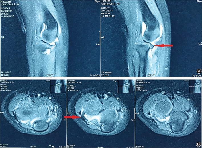
This diagnostic image consists of multi-planar T2-weighted MRI scans of a human elbow joint, presented in sagittal (A) and axial (B) views. The scans demonstrate the complex anatomy of the radiohumeral (humeroradial) joint, showing the distal humerus, proximal radius, and ulna. A prominent clinical finding is highlighted by red arrows in both views: a localized area of low signal intensity representing the annular ligament, which has become incarcerated or 'stuck' within the radiohumeral joint space. In the sagittal view, this interposed soft tissue is seen disrupting the normal articulation between the radial head and the humeral capitellum. In the axial slices, the displaced ligament is visualized as a dark, hypointense structure contrasting against the brighter (hyperintense) joint fluid. This finding is characteristic of a complex radial head dislocation where soft tissue entrapment prevents successful closed reduction. The image is a critical educational resource for understanding mechanical blocks in pediatric elbow trauma and the utility of MRI in identifying ligamentous interposition.
This diagnostic image consists of multi-planar T2-weighted MRI scans of a human elbow joint, presented in sagittal (A) and axial (B) views. The scans demonstrate the complex anatomy of the radiohumeral (humeroradial) joint, showing the distal humerus, proximal radius, and ulna. A prominent clinical finding is highlighted by red arrows in both views: a localized area of low signal intensity representing the annular ligament, which has become incarcerated or 'stuck' within the radiohumeral joint space. In the sagittal view, this interposed soft tissue is seen disrupting the normal articulation between the radial head and the humeral capitellum. In the axial slices, the displaced ligament is visualized as a dark, hypointense structure contrasting against the brighter (hyperintense) joint fluid. This finding is characteristic of a complex radial head dislocation where soft tissue entrapment prevents successful closed reduction. The image is a critical educational resource for understanding mechanical blocks in pediatric elbow trauma and the utility of MRI in identifying ligamentous interposition.

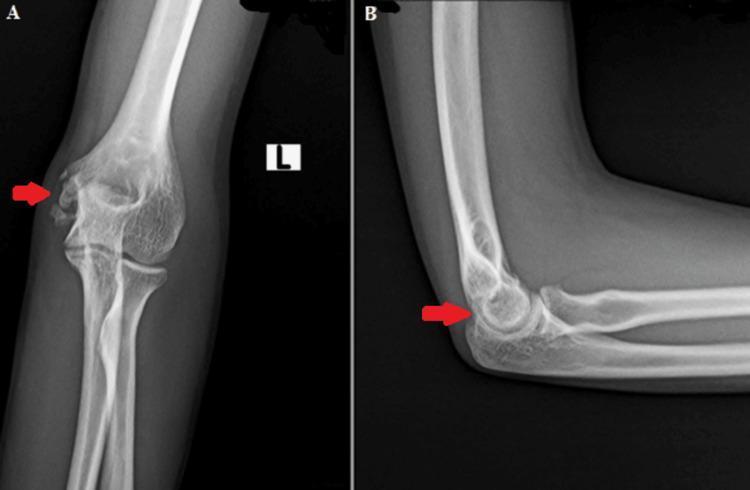
This diagnostic image consists of two radiographic views of the left elbow joint: an anteroposterior (AP) view (A) and a lateral view (B). The X-rays demonstrate the skeletal anatomy of the distal humerus, proximal radius, and proximal ulna. In the AP view, a red arrow points to the medial epicondyle of the humerus, where the bony architecture appears preserved with normal cortical thickness and trabecular patterns. The radiohumeral and ulnohumeral joint spaces are well-maintained, indicating a lack of significant degenerative changes or acute inflammatory destruction. The lateral view shows congruent alignment of the humeroulnar and radiocapitellar joints, with the red arrow again highlighting the medial epicondylar region from a side profile. These follow-up images show no evidence of active lytic lesions, cortical erosions, or recurrence of previously treated disease such as musculoskeletal tuberculosis. The surrounding soft tissues appear normal without significant swelling or calcification, confirming clinical stability at one year post-treatment.
This diagnostic image consists of two radiographic views of the left elbow joint: an anteroposterior (AP) view (A) and a lateral view (B). The X-rays demonstrate the skeletal anatomy of the distal humerus, proximal radius, and proximal ulna. In the AP view, a red arrow points to the medial epicondyle of the humerus, where the bony architecture appears preserved with normal cortical thickness and trabecular patterns. The radiohumeral and ulnohumeral joint spaces are well-maintained, indicating a lack of significant degenerative changes or acute inflammatory destruction. The lateral view shows congruent alignment of the humeroulnar and radiocapitellar joints, with the red arrow again highlighting the medial epicondylar region from a side profile. These follow-up images show no evidence of active lytic lesions, cortical erosions, or recurrence of previously treated disease such as musculoskeletal tuberculosis. The surrounding soft tissues appear normal without significant swelling or calcification, confirming clinical stability at one year post-treatment.

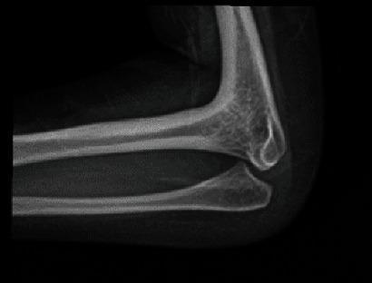
This diagnostic X-ray (radiography) captures a lateral view of a human elbow joint, illustrating a congenital or acquired radiohumeral synostosis. The image demonstrates a clear bony fusion between the distal humerus and the proximal head of the radius. Specifically, there is a total lack of a visible joint space at the radiohumeral articulation, with continuous cortical and trabecular bone spanning the two structures. In contrast, the ulnohumeral articulation appears relatively spared, with a discernible joint space maintained between the trochlea of the humerus and the trochlear notch of the ulna. The olecranon process of the ulna is visible posteriorly. This radiographic finding is clinically significant as a manifestation of rare skeletal dysplasias or as a solitary orthopedic anomaly, leading to a complete loss of pronation and supination at the forearm. The image serves as a textbook example for orthopedic and radiological training in identifying joint fusion and anomalous skeletal developments.
This diagnostic X-ray (radiography) captures a lateral view of a human elbow joint, illustrating a congenital or acquired radiohumeral synostosis. The image demonstrates a clear bony fusion between the distal humerus and the proximal head of the radius. Specifically, there is a total lack of a visible joint space at the radiohumeral articulation, with continuous cortical and trabecular bone spanning the two structures. In contrast, the ulnohumeral articulation appears relatively spared, with a discernible joint space maintained between the trochlea of the humerus and the trochlear notch of the ulna. The olecranon process of the ulna is visible posteriorly. This radiographic finding is clinically significant as a manifestation of rare skeletal dysplasias or as a solitary orthopedic anomaly, leading to a complete loss of pronation and supination at the forearm. The image serves as a textbook example for orthopedic and radiological training in identifying joint fusion and anomalous skeletal developments.

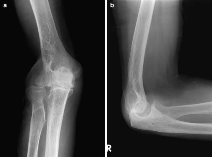
Two-view conventional radiographs of a human right elbow joint, including an anteroposterior (AP) view (a) and a lateral view (b), demonstrating post-surgical sequelae and secondary osteoarthritis. The AP view shows a widening or separation at the proximal radioulnar joint (PRUJ) and marked articular surface irregularity of the radiohumeral joint. The lateral view, taken in 90 degrees of flexion, reveals joint space narrowing and prominent anterior osteophyte formation on the distal humerus near the radiohumeral compartment. There is evidence of previous surgical intervention in the lateral epicondyle region of the humerus, leading to altered joint morphology. Findings are consistent with advanced degenerative changes following ligamentous instability and wide resection for an orthopedic tumor. Key educational features include the radiographic signs of secondary elbow osteoarthritis: subchondral sclerosis, osteophytosis, and joint space loss, particularly affecting the radiohumeral and ulnohumeral articulations.

Two-view conventional radiographs of a human right elbow joint, including an anteroposterior (AP) view (a) and a lateral view (b), demonstrating post-surgical sequelae and secondary osteoarthritis. The AP view shows a widening or separation at the proximal radioulnar joint (PRUJ) and marked articular surface irregularity of the radiohumeral joint. The lateral view, taken in 90 degrees of flexion, reveals joint space narrowing and prominent anterior osteophyte formation on the distal humerus near the radiohumeral compartment. There is evidence of previous surgical intervention in the lateral epicondyle region of the humerus, leading to altered joint morphology. Findings are consistent with advanced degenerative changes following ligamentous instability and wide resection for an orthopedic tumor. Key educational features include the radiographic signs of secondary elbow osteoarthritis: subchondral sclerosis, osteophytosis, and joint space loss, particularly affecting the radiohumeral and ulnohumeral articulations.

Loading Image
Loading Image
I now have all the content needed. Here is the complete short note for the MS Orthopedics theory examination:

SHORT NOTE: Radiohumeral Joint

Introduction
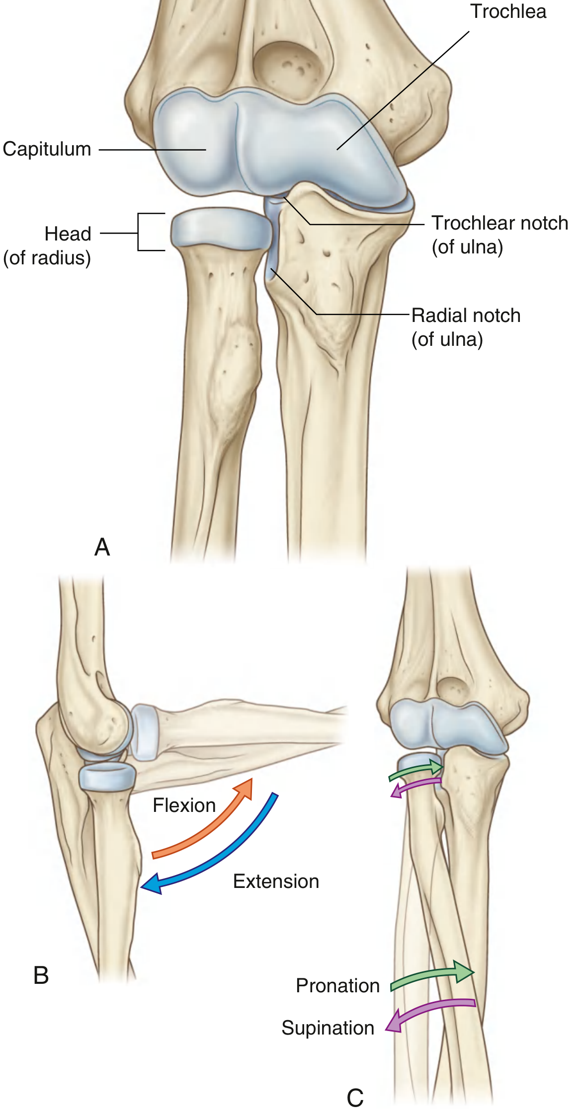
The radiohumeral joint (also called the humeroradial or radiocapitellar joint) is one of three articulations that together constitute the elbow joint. All three share a single synovial cavity. The other two are the humeroulnar joint (between the trochlear notch of the ulna and the trochlea of the humerus) and the proximal radioulnar joint (between the radial head and the radial notch of the ulna).
(Gray's Anatomy for Students, Fig. 7.71)
Articulating surfaces of the elbow: capitulum with head of radius, trochlea with trochlear notch, showing flexion/extension and pronation/supination movements

Type of Joint

The radiohumeral joint is a ball-and-socket (condyloid) type of synovial joint in isolation, but functionally it participates in two distinct movements as part of the overall elbow complex:
  • Flexion and extension - the concave fovea of the radial head articulates with the convex capitulum (a hemispheric projection on the anteroinferior lateral aspect of the distal humerus), acting as a hinge at the elbow.
  • Pronation and supination - the radial head spins on the capitulum while simultaneously sliding against the radial notch of the ulna.

Articular Surfaces

StructureDescription
Capitulum of humerusRounded, hemispherical projection on the anteroinferior lateral distal humerus
Head of radiusDisc-shaped; the superior surface bears a central concavity (fovea articularis radii) that articulates with the capitulum
Both surfaces are covered with hyaline articular cartilage.

Joint Capsule

The fibrous membrane of the joint capsule encloses the entire elbow joint complex. On the lateral side, the free inferior margin of the capsule passes around the neck of the radius from the anterior coronoid process attachment to the posterior olecranon base, allowing rotatory movement.
The capsule is reinforced laterally by the radial collateral ligament and is further cuffed around the radial head by the anular ligament of the radius.

Anular Ligament

  • A strong ring of fibrous tissue that encircles the head and neck of the radius
  • Attached at both ends to the anterior and posterior margins of the radial notch of the ulna
  • Blends with the radial collateral ligament and the fibrous capsule (though it remains separate from the capsule posteriorly)
  • Its deep surface (where it contacts the sides of the radial head) is lined with articular cartilage
  • Permits rotation of the radial head during pronation/supination
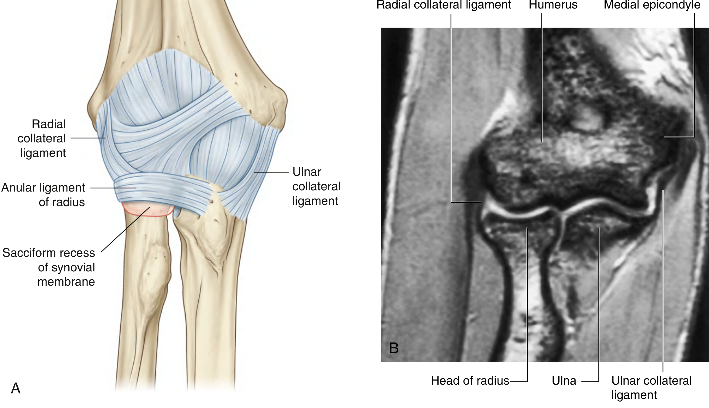
Elbow joint - capsule, radial collateral ligament, anular ligament, sacciform recess, and coronal MRI showing radiohumeral articulation

Synovial Membrane

  • Lines the radial fossa, coronoid fossa, olecranon fossa, deep surface of the capsule, and the medial surface of the trochlea
  • Separated from the fibrous capsule by fat pads (anterior, posterior, and radial fossae fat pads)
  • A sacciform recess protrudes inferiorly from the free margin of the capsule - this accommodates rotation of the radial head during forearm rotation

Blood Supply

A rich anastomotic network derived from:
  • Collateral and recurrent branches of the brachial artery
  • Profunda brachii artery
  • Radial artery (radial recurrent branch)
  • Ulnar artery (anterior and posterior ulnar recurrent branches)

Nerve Supply

Predominantly by branches of the radial and musculocutaneous nerves; additional contributions from the ulnar and median nerves.

Movements

MovementArticulation Involved
Flexion / ExtensionRadiohumeral + humeroulnar (hinge)
Pronation / SupinationRadiohumeral (spin) + proximal and distal radioulnar joints

Clinical Relevance

1. Radial Head Fractures

The radial head is commonly fractured in adults following a fall on an outstretched hand - valgus stress forces the radial head against the capitulum. This is the most common cause of elbow hemarthrosis after trauma when radiographs show no visible fracture (nondisplaced fracture). Anterior fat pad displacement ("sail sign") or a posterior fat pad sign on lateral radiograph indicates joint effusion.

2. Pulled Elbow (Nursemaid's Elbow) - Subluxation of the Radial Head

Common in children under 5 years. Sudden longitudinal traction on the forearm causes the radial head to slip partially through the anular ligament, leading to pain and refusal to use the arm. Treated by supination and flexion maneuver.

3. Radiohumeral Bursitis / Lateral Epicondylitis ("Tennis Elbow")

Epicondylitis was first described as an inflammatory process involving the radiohumeral joint or lateral epicondyle. Radiohumeral bursitis occurs at the juncture of the radial head and lateral epicondyle. Managed conservatively with corticosteroid injection into the radiohumeral bursa.

4. Radiohumeral Synostosis

Congenital or acquired bony fusion between the distal humerus and proximal radial head - results in loss of pronation/supination and reduced elbow flexion. May be seen in rare skeletal dysplasias (e.g., Pfeiffer syndrome).

5. Arthrocentesis of the Radiohumeral Joint

Landmarks: lateral epicondyle of humerus and head of radius. Technique: flex elbow to 90 degrees, pronate forearm, place palm flat on a table, and insert a 20-gauge needle just distal to the lateral epicondyle directed medially. Gout and septic arthritis commonly affect this joint.

Summary

The radiohumeral joint is the lateral articulation of the elbow complex - the convex capitulum of the humerus articulates with the concave fovea of the radial head within a shared synovial cavity. It participates in both flexion/extension (as a hinge) and forearm rotation (as a pivot point for the spinning radial head). Its key supporting structure is the anular ligament. Clinically, it is relevant in radial head fractures, pulled elbow, lateral epicondylitis, and as a site for arthrocentesis.

Sources: Gray's Anatomy for Students (9780323934237); Roberts and Hedges' Clinical Procedures in Emergency Medicine (9780323354783); Rosen's Emergency Medicine (9780323757898); Imaging Anatomy Vol. 3 (9781626239845)
This is a shared conversation. Sign in to Orris to start your own chat.