
Short note on varicose veins with definition, epidemiology, symptoms, signs, investigation and treatment
Reading File
Now let me search the medical library for varicose veins content across multiple angles simultaneously.
Finding Sources
Finding Sources
Searching PubMed
"varicose veins"[MeSH Terms] AND treatment
Excellent results. Now let me read the key textbook sections in parallel - Bailey & Love (surgical perspective), Harrison's (internal medicine), and Schwartz's (surgery).
Reading File
Reading File
Reading File
Let me now read the rest of the Bailey & Love signs/investigations/treatment section and also fetch the Harrison's treatment section, plus a clinical image.
Reading File
Reading File
Loading Image
Let me get the rest of the Bailey & Love treatment section:
Reading File
Reading File
Reading File
I now have comprehensive information to write the full short note. Let me compile it.
Varicose Veins
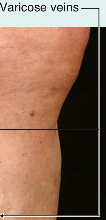
Clinical photograph demonstrating varicose veins - Gray's Anatomy for Students
Definition
Varicose veins are dilated, bulging, tortuous superficial veins measuring at least 3 mm in diameter, typically occurring in the lower limbs. They are distinct from:
- Reticular veins: dilated intradermal veins, 1-3 mm diameter, blue-green, do not protrude from the skin surface
- Telangiectasias (spider veins): <1 mm diameter, near the skin surface, forming blue/purple/red linear or web patterns
Varicose veins are classified as:
- Primary: originate in the superficial system; result from defective valve structure and function in the saphenous veins, intrinsic wall weakness, or high intraluminal pressure
- Secondary: result from venous hypertension due to deep-venous insufficiency, deep-venous obstruction, incompetent perforating veins, or arteriovenous fistulas
(Harrison's Principles of Internal Medicine 22E, p.2223; Bailey and Love's 28th Ed.)
Epidemiology
- Adult prevalence of visible varicose veins: 30-50% (Bailey and Love's 28th Ed.)
- At least 10% of the general population is affected (Schwartz's 11th Ed.)
Risk factors:
| Factor | Detail |
|---|---|
| Gender | Higher prevalence in women in most studies |
| Age | Prevalence rises sharply with age; Edinburgh Vein Study: 11.5% (18-24 yrs) rising to 55.7% (55-64 yrs) |
| Pregnancy | Increases risk due to hormonal changes and elevated intra-abdominal pressure |
| Family history | ~50% have a family history; genetic susceptibility is well-documented |
| Obesity/BMI | Increasing BMI associated with higher prevalence |
| Prolonged standing | Inconclusive but commonly cited occupational factor |
| Ethnicity | Influences prevalence; Western populations more affected |
| Hormonal therapy | Exogenous estrogens associated with increased risk |
Symptoms
Symptoms may be absent (asymptomatic) or significant. Typical complaints include:
- Aching, heaviness, throbbing, burning, or bursting sensation over affected areas or the whole limb
- Pruritus (itching) - more frequent in the presence of complications
- Ankle swelling (edema) - mild in early disease
- Early fatigue of the affected leg
- Cosmetic concern - unsightly dilated veins
Key feature: Symptoms typically worsen throughout the day or with prolonged standing and improve with leg elevation or compression hosiery. Symptoms are independent of the degree of venous incompetence and can significantly impair health-related quality of life.
Telangiectasias and reticular veins without significant reflux rarely cause physical symptoms but patients often seek cosmetic treatment.
(Bailey and Love's 28th Ed., p.1052; Schwartz's 11th Ed.)
Signs
On examination (standing position):
- Tortuous, dilated subcutaneous veins - usually clinically obvious
- Distribution helps identify the affected system:
- Medial thigh and calf varicosities → Great Saphenous Vein (GSV) incompetence (~60% of cases)
- Posterolateral calf varicosities → Small Saphenous Vein (SSV) incompetence (~20% of cases)
- Anterolateral thigh and calf → Anterior accessory GSV (AAGSV) incompetence
- Saphena varix: large dilated vein at the saphenofemoral junction (SFJ) presenting as a lump in the groin - disappears when recumbent, may transmit a cough impulse (can mimic a groin hernia)
- Pitting edema - may extend from ankle to knee or thigh in severe disease
- Signs of chronic venous insufficiency (CVI):
- Stasis dermatitis / eczema (corona phlebectatica)
- Hyperpigmentation (hemosiderin deposition)
- Lipodermatosclerosis: induration + hemosiderin deposition + inflammation, just above the ankle
- Atrophie blanche: white scar patches with focal telangiectasias, near medial malleolus
- Phlebectasia corona: fan-shaped intradermal veins near the ankle
- Venous ulceration: shallow, irregular border, medial/lateral malleolus
Bedside clinical tests (now largely replaced by duplex ultrasound):
- Brodie-Trendelenburg test: leg elevated to empty veins, tourniquet placed on proximal thigh, patient stands - rapid filling after tourniquet release = superficial venous insufficiency (primary); filling before release = deep-venous incompetence (secondary)
- Perthes test: tourniquet on midthigh while standing, patient walks 5 min - collapse of superficial veins = patent deep system; distension = deep venous obstruction
(Bailey and Love's 28th Ed.; Harrison's 22E, p.2224)
Investigations
Duplex Ultrasound Scanning (gold standard)
Tourniquet tests and hand-held Doppler have now been abandoned. Duplex ultrasound is recommended for all patients prior to any intervention. It should be performed with the patient standing, using a 7.5-13 MHz high-frequency linear array transducer.
Duplex scanning establishes:
- Presence of reflux in deep and superficial venous systems
- Exact distribution and extent of reflux in the superficial system (affected junctions, perforators)
- Presence of obstruction in the deep venous system
- Suitability of incompetent veins for various treatment options (based on diameter, tortuosity, presence of saphena varix)
- Presence of thrombus within superficial veins
- Indication of a pelvic source of reflux
Reflux definitions on duplex:
- Superficial/crural vein reflux: retrograde flow lasting ≥0.5 seconds
- Proximal deep vein reflux: retrograde flow lasting ≥1 second
The "Mickey Mouse sign" on transverse view shows the GSV and CFV lying medial to the common femoral artery.
Other investigations (occasionally required):
- MR venography (non-invasive) - for pelvic or iliac vein assessment
- Contrast venography / IVUS - invasive, reserved for complex cases
- Varicography is now historical
(Bailey and Love's 28th Ed., p.1053-1054)
Treatment / Management
1. Conservative Management
Compression hosiery - the cornerstone of conservative treatment:
- Graduated external compression (20-30, 30-40, or 40-50 mmHg)
- Stockings range from knee-high to waist-high
- Provides sufficient symptomatic relief in many patients
- First-line trial before interventional management
Other conservative measures: weight loss, leg elevation, regular exercise, avoiding prolonged standing.
2. Sclerotherapy
- Destroys the venous endothelium to obliterate the vein
- Sclerosants used:
- Hypertonic saline (11.7-23.4%)
- Sodium tetradecyl sulfate (STS) (0.125-1%)
- Polidocanol (0.5-1.0%)
- Foam sclerotherapy: ultrasound-guided foam injection - randomized trials show significant symptom relief and improved cosmetic appearance
- Elastic bandaging after injection worn for 3-5 days (to appose inflamed walls), then compression stockings for ≥2 weeks
- Complications: allergic reaction, hyperpigmentation, thrombophlebitis, DVT, skin necrosis
3. Endovenous Thermal Ablation
Catheter-based minimally invasive techniques, performed under tumescent local anaesthesia under ultrasound guidance. Both have similar outcomes and have largely replaced open surgery.
a) Endovenous Laser Ablation (EVLA):
- A laser fibre is introduced via catheter; energy delivery of ~60-80 J/cm causes thermal occlusion
- Compression applied post-procedure
b) Radiofrequency Ablation (RFA):
- Electromagnetic current heats the vein wall to 120°C in 20-second treatment cycles
- Most popular device: ClosureFast™ (Medtronic)
- Multiple RCTs show EVLA and RFA have comparable efficacy and safety; recent meta-analysis (Jiang et al., 2024) confirmed this
4. Non-Thermal, Non-Sclerosant Ablation (Newer Techniques)
- Mechanochemical ablation (MOCA): combines catheter-based mechanical endoluminal injury with liquid sclerosant - no tumescent anaesthesia required
- Cyanoacrylate adhesive (VenaSeal): proprietary glue injected under ultrasound guidance - promising early results, avoids heat and sclerosants
5. Surgical Treatment
- High ligation and stripping (Trendelenburg operation): ligation of the SFJ or SPJ + stripping of the GSV or SSV - now less commonly performed, reserved for cases not amenable to endovenous techniques
- Phlebectomy (avulsions): removal of tributary varicosities through small stab incisions; often performed alongside thermal ablation as a combined procedure
Indications for intervention (beyond conservative therapy):
- Symptoms not relieved by compression
- Lipodermatosclerosis
- Venous ulceration
- Bleeding from varicosities
- Superficial vein thrombosis (thrombophlebitis)
(Bailey and Love's 28th Ed., p.1054-1055; Schwartz's Principles of Surgery 11th Ed.; Harrison's 22E)
Complications
| Acute | Chronic |
|---|---|
| Superficial vein thrombosis (thrombophlebitis) | Lipodermatosclerosis |
| Haemorrhage (spontaneous or traumatic) | Venous eczema/stasis dermatitis |
| Venous ulceration | |
| Hyperpigmentation | |
| Atrophie blanche |
Recent evidence note: A 2024 meta-analysis (PMID 38316290) comparing endovenous RFA vs. EVLA confirmed comparable efficacy, safety, and quality-of-life outcomes - supporting either technique as first-line interventional treatment for GSV/SSV reflux over open surgery.
Classification of varicose vein in detail
Varicose veins are classified by etiology (primary/secondary/congenital), by the affected anatomical system (GSV/SSV), and by the CEAP classification system which is the internationally accepted standard grading from C0 (no signs) to C6 (active venous ulcer). Let me pull the detailed classification from the textbooks.
Searching sources...
Finding Sources
Finding Sources
Excellent results. Now let me read the most comprehensive sources in parallel - Bailey & Love's classification section, Sabiston's CEAP table, Current Surgical Therapy, and Harrison's CEAP table.
Reading File
Reading File
Reading File
Finding Sources
Let me now read the full CEAP table from Sabiston and the Bailey & Love classification section, plus the Venous Clinical Severity Score and anatomic classification details.
Reading File
Reading File
Finding Sources
Finding Sources
Now let me read the Fischer's VCSS section and Bailey & Love's classification content to get complete detail:
Reading File
Reading File
Finding Sources
Let me also fetch the C6 active venous ulcer image and get Bailey & Love's classification of varicose veins by type:
Reading File
Loading Image
I now have all the material needed to write a comprehensive, detailed classification of varicose veins. Let me compile the full answer.
Classification of Varicose Veins
Varicose veins are classified across several overlapping frameworks: by etiology, by morphology/type, by anatomical system, and most comprehensively by the internationally accepted CEAP classification system (updated 2020). Two additional scoring tools - the Venous Clinical Severity Score (VCSS) and the Venous Segmental Disease Score (VSDS) - supplement CEAP for outcomes assessment.
I. Classification by Etiology
This is the most fundamental and clinically practical division.
1. Primary Varicose Veins (Ep)
- Arise from intrinsic abnormalities of the venous wall (structural weakness) or venous valve leaflets
- Result in valvular incompetence and reflux within the superficial venous system
- No identifiable underlying deep venous disease
- Approximately 50% have a positive family history, suggesting genetic susceptibility
- Other contributing factors: aging, female sex, pregnancy, obesity, hormonal therapy, prolonged standing
- The most common type encountered in clinical practice
2. Secondary Varicose Veins (Es)
Result from an identifiable acquired cause that elevates venous pressure and drives dilation of superficial veins.
The 2020 CEAP revision further subclassifies secondary causes as:
| Subtype | Meaning | Examples |
|---|---|---|
| E | Secondary - intravenous | Post-thrombotic syndrome (previous DVT leading to valve destruction and reflux), intraluminal obstruction |
| E | Secondary - extravenous | External compression of deep veins (May-Thurner syndrome, Nutcracker syndrome, pelvic tumours, pregnancy), arteriovenous fistula |
Arteriovenous fistulas cause varicose veins in the affected limb by raising venous pressure directly.
3. Congenital Varicose Veins (Ec)
- Present from birth; result from a developmental defect in the venous system
- Associated syndromes include:
- Klippel-Trenaunay syndrome: port wine stain + varicose veins + bony/soft tissue hypertrophy of a limb
- Parkes-Weber syndrome: similar but with arteriovenous fistulas
- Ehlers-Danlos syndrome: connective tissue disorder with venous fragility
- Clonal trisomies and other chromosomal anomalies
4. No Cause Identified (En)
- Venous disease present but no etiology can be established after workup
II. Classification by Morphology / Type of Vessel
| Type | Diameter | Appearance | Protrudes? |
|---|---|---|---|
| Telangiectasias (spider veins) | < 1 mm | Blue, purple, or red; linear, branching, or spider-web patterns | No |
| Reticular veins | 1-3 mm | Dilated intradermal/subdermal veins; blue-green | No |
| Varicose veins (trunk varicosities) | ≥ 3 mm | Dilated, tortuous, bulging subcutaneous veins | Yes |
| Saphena varix | Variable | Dilated vein at the saphenofemoral junction; disappears supine; cough impulse present | Yes (groin lump) |
(Harrison's Principles of Internal Medicine 22E, p.2223)
III. Classification by Anatomical System
Based on which superficial venous axis is incompetent (confirmed by duplex ultrasound):
| System | Distribution of Varicosities | Approximate Frequency |
|---|---|---|
| Great Saphenous Vein (GSV) incompetence | Medial thigh and calf varicosities | ~60% of cases |
| Small Saphenous Vein (SSV) incompetence | Posterolateral calf varicosities | ~20% of cases |
| Anterior Accessory GSV (AAGSV) incompetence | Anterolateral thigh and calf varicosities | Less common |
| Perforator vein incompetence | Localised clusters of varicosities at specific perforator sites | May coexist with above |
| Pelvic/gonadal vein reflux | Perineal, posterior thigh, or bilateral varicosities | Especially in parous women |
(Bailey and Love's Short Practice of Surgery 28th Ed., p.1152)
IV. Classification by Presentation / Complication Status
From clinical practice (Bailey and Love's):
Varicose Veins
├── Uncomplicated
│   ├── Asymptomatic
│   └── Symptomatic (aching, heaviness, itching, etc.)
└── Complicated
    ├── Chronic (skin changes, venous eczema, lipodermatosclerosis, ulceration)
    └── Acute (superficial vein thrombosis / thrombophlebitis, haemorrhage)
V. CEAP Classification (International Standard, Updated 2020)
Devised in 1994 by the American Venous Forum (AVF); revised in 2004 and again in 2020 (Lurie et al., J Vasc Surg Venous Lymphat Disord. 2020;8:342-352). Modelled after the TNM cancer staging system. Classifies chronic venous disease (CVD) across four domains: Clinical (C), Etiologic (E), Anatomic (A), Pathophysiologic (P).
The 2020 update added:
- C4c (corona phlebectatica) as a new subclass
- The "r" modifier for recurrent varicose veins and recurrent venous ulcers
- Replacement of numeric venous segment descriptions with standard abbreviations
C - Clinical Classification
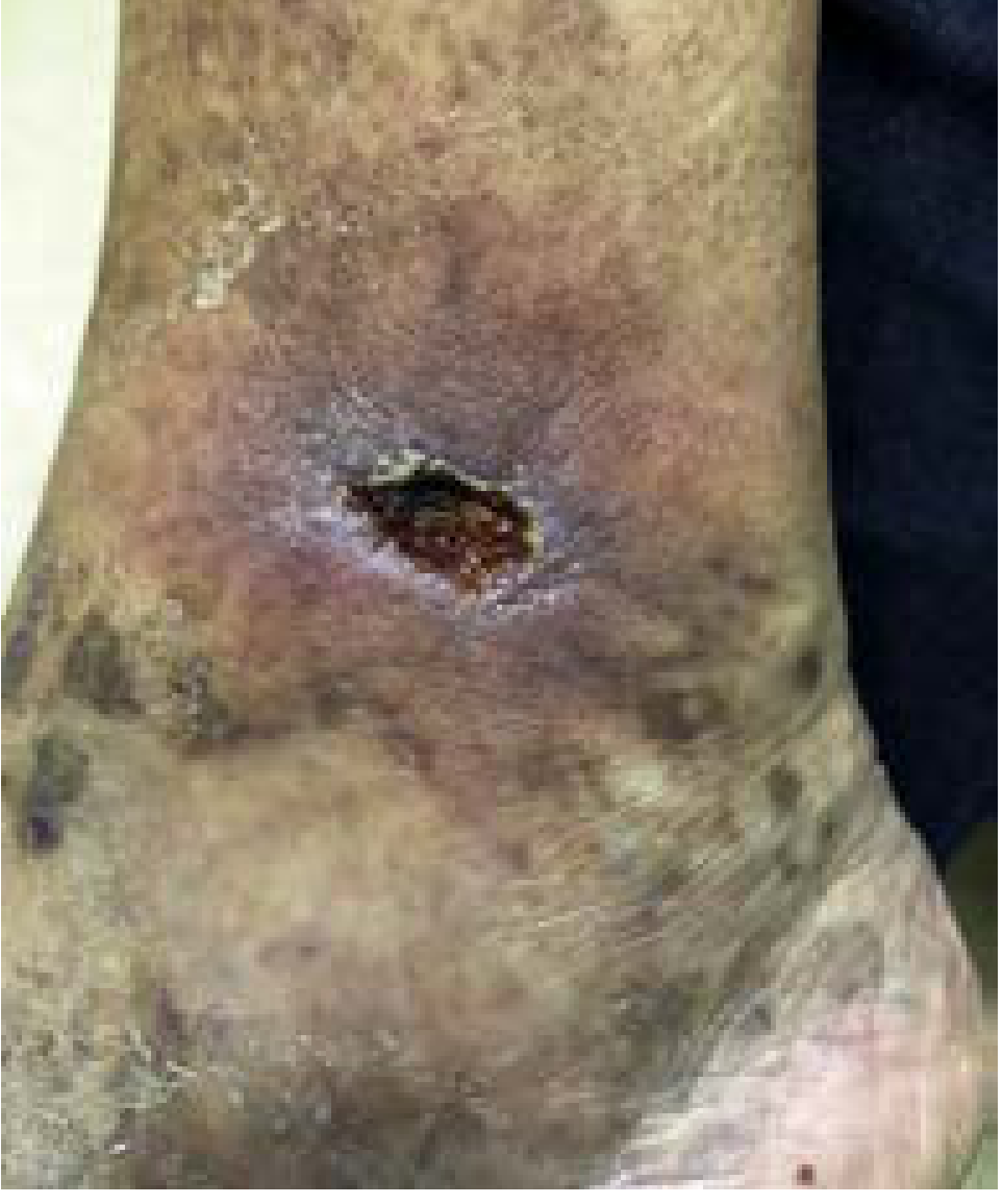
| C Class | Description | Clinical Features |
|---|---|---|
| C0 | No visible or palpable signs of venous disease | Normal appearance; patient may have symptoms |
| C1 | Telangiectasias or reticular veins | Spider veins (<1 mm) or reticular veins (1-3 mm) |
| C2 | Varicose veins | Dilated, tortuous, subcutaneous veins ≥3 mm |
| C2r | Recurrent varicose veins | Varicosities recurrent after previous treatment |
| C3 | Edema | Pitting or non-pitting edema; no skin changes yet |
| C4 | Skin and subcutaneous tissue changes secondary to CVD | (subdivided below) |
| C4a | Pigmentation or eczema | Hemosiderin staining; stasis dermatitis/eczema |
| C4b | Lipodermatosclerosis or atrophie blanche | Induration + inflammation above ankle; white scars |
| C4c | Corona phlebectatica | Fan-shaped pattern of intradermal veins near ankle/foot (new in 2020) |
| C5 | Healed venous ulcer | Ulcer previously present but now healed; residual scar |
| C6 | Active venous ulcer | Open ulcer; shallow, irregular edges, granulation tissue |
| C6r | Recurrent active venous ulcer | Active ulcer in a patient with previously healed venous ulcer |
Each limb is further designated:
- "s" = symptomatic (e.g., C2s)
- "a" = asymptomatic (e.g., C2a)
Important: C3-C6 reflects progressive chronic venous insufficiency (CVI). Skin changes (C4b) and ulceration carry significant morbidity - venous disease accounts for ~85% of all chronic lower limb ulcers.
E - Etiologic Classification
| Code | Etiology |
|---|---|
| Ep | Primary (intrinsic valve/wall defect) |
| Es | Secondary (acquired cause) |
| Esi | Secondary - intravenous (e.g., post-DVT) |
| Ese | Secondary - extravenous (e.g., compression syndromes) |
| Ec | Congenital (present since birth) |
| En | No cause identified |
A - Anatomic Classification
The 2020 revision uses standard venous abbreviations rather than numbered segments.
| Code | Location | Key Veins Included |
|---|---|---|
| As | Superficial | Great saphenous vein (GSV), small saphenous vein (SSV), anterior accessory GSV (AAGSV), non-saphenous superficial veins, telangiectasias, reticular veins |
| Ap | Perforator | Thigh perforators, calf perforators (e.g., Cockett's perforators above medial malleolus) |
| Ad | Deep | Inferior vena cava (IVC), iliac veins (common, internal, external), femoral veins, popliteal vein, tibial veins (anterior, posterior, peroneal), gastrocnemius/soleal veins |
| An | No venous anatomic location identified |
Multiple anatomic locations can be listed together (e.g., As,p,d).
P - Pathophysiologic Classification
| Code | Pathophysiology | Mechanism |
|---|---|---|
| Pr | Reflux | Incompetent valves → retrograde flow → venous hypertension |
| Po | Obstruction | Thrombosis or extrinsic compression blocking outflow |
| Pr,o | Reflux and obstruction | Combined (e.g., post-thrombotic syndrome) |
| Pn | No pathophysiology identified |
How to Write a Full CEAP Code
A complete CEAP classification is written as: C_E_A_P
Examples:
C2s,Ep,As,Pr= Symptomatic varicose veins; primary etiology; superficial system; reflux only - classic uncomplicated primary varicose veinsC6,Es,Ad,Pr,o= Active venous ulcer; secondary etiology (post-DVT); deep system affected; both reflux and obstruction - severe post-thrombotic syndromeC4a,Ep,As,p,Pr= Pigmentation/eczema; primary; superficial + perforators; reflux - CVI with skin changesC2r,Es,As,Pr= Recurrent varicose veins after treatment; secondary; superficial; reflux
VI. Supplementary Scoring Systems
CEAP classifies but does not quantify severity for outcomes tracking. Two validated tools complement it:
Venous Clinical Severity Score (VCSS / r-VCSS)
- Assigns a quantitative score to 10 clinical attributes (pain, varicose veins, edema, skin pigmentation, inflammation, induration, ulcer count, duration, size, compression use)
- Each attribute scored 0-3 (absent/mild/moderate/severe)
- Total score 0-30; used to track treatment outcomes before and after intervention
- Current standard for validated patient-reported outcomes following endovascular and open surgical interventions
Venous Segmental Disease Score (VSDS)
- Maps the anatomic extent and severity of reflux and obstruction across individual venous segments
- Complements CEAP and VCSS for research and complex cases
Aberdeen Varicose Vein Score (AVVS)
- Patient-reported outcome measure (PROM) specific to varicose veins
- Helps identify patients most likely to benefit from intervention and assess treatment expectations
(Sabiston Textbook of Surgery; Fischer's Mastery of Surgery 8th Ed.; Current Surgical Therapy 14th Ed.)
Summary Diagram
VARICOSE VEIN CLASSIFICATION
│
├── 1. By ETIOLOGY
│   ├── Primary (intrinsic valve/wall defect)
│   ├── Secondary
│   │   ├── Intravenous (post-DVT, intraluminal)
│   │   └── Extravenous (compression syndromes, AV fistula)
│   └── Congenital (Klippel-Trenaunay, Parkes-Weber, etc.)
│
├── 2. By MORPHOLOGY
│   ├── Telangiectasias (<1 mm)
│   ├── Reticular veins (1-3 mm)
│   ├── Varicose veins (≥3 mm)
│   └── Saphena varix
│
├── 3. By ANATOMICAL SYSTEM
│   ├── GSV system (60%)
│   ├── SSV system (20%)
│   ├── AAGSV
│   └── Perforator/Pelvic source
│
├── 4. By COMPLICATION STATUS
│   ├── Uncomplicated (asymptomatic / symptomatic)
│   └── Complicated (acute: thrombophlebitis, bleed | chronic: ulcer, LDS)
│
└── 5. CEAP CLASSIFICATION (International Standard 2020)
    ├── C0 → C6r  (Clinical severity)
    ├── Ep/Es/Ec/En  (Etiology)
    ├── As/Ap/Ad  (Anatomic location)
    └── Pr/Po/Pr,o  (Pathophysiology)
(Sources: Bailey and Love's 28th Ed.; Harrison's Principles 22E; Sabiston Textbook of Surgery; Current Surgical Therapy 14e; Fischer's Mastery of Surgery 8th Ed.; Lurie et al. 2020 CEAP Update)
This is a shared conversation. Sign in to Orris to start your own chat.