
Explain 10cimmon heart diseases with ECG changes and ECG images of those diseases
ECG changes common heart diseases STEMI atrial fibrillation heart block images
https://mesimedical.com/insights/frequently-diagnosed-condit…
ECG images STEMI LVH heart block bundle branch block atrial fibrillation Wolff-Parkinson-White pericarditis ventricular fibrillation
ECG examples STEMI inferior anterior lateral LVH first degree heart block complete heart block ventricular tachycardia pericarditis images
site:litfl.com ECG library images atrial fibrillation ventricular tachycardia Brugada syndrome LVH complete heart block pericarditis
https://litfl.com/av-block-3rd-degree-complete-heart-block
ECG atrial fibrillation complete heart block ventricular fibrillation Brugada syndrome LVH pericarditis images litfl ecgwaves
10 Common Heart Diseases: ECG Changes & Images
1. ST-Elevation Myocardial Infarction (STEMI)
- Hyperacute phase: Tall, peaked (hyperacute) T waves - the earliest change
- Acute phase: ST segment elevation ≥1 mm in 2+ contiguous leads (convex/tombstone morphology)
- Pathologic Q waves develop (>40 ms wide, >25% of R wave amplitude) - indicate irreversible necrosis
- Reciprocal ST depression in leads facing opposite wall
- T wave inversion in evolving phase
| Territory | Elevated leads | Reciprocal changes |
|---|---|---|
| Anterior (LAD) | V1-V4 | II, III, aVF |
| Inferior (RCA/LCx) | II, III, aVF | I, aVL |
| Lateral (LCx/diagonal) | I, aVL, V5-V6 | II, III, aVF |


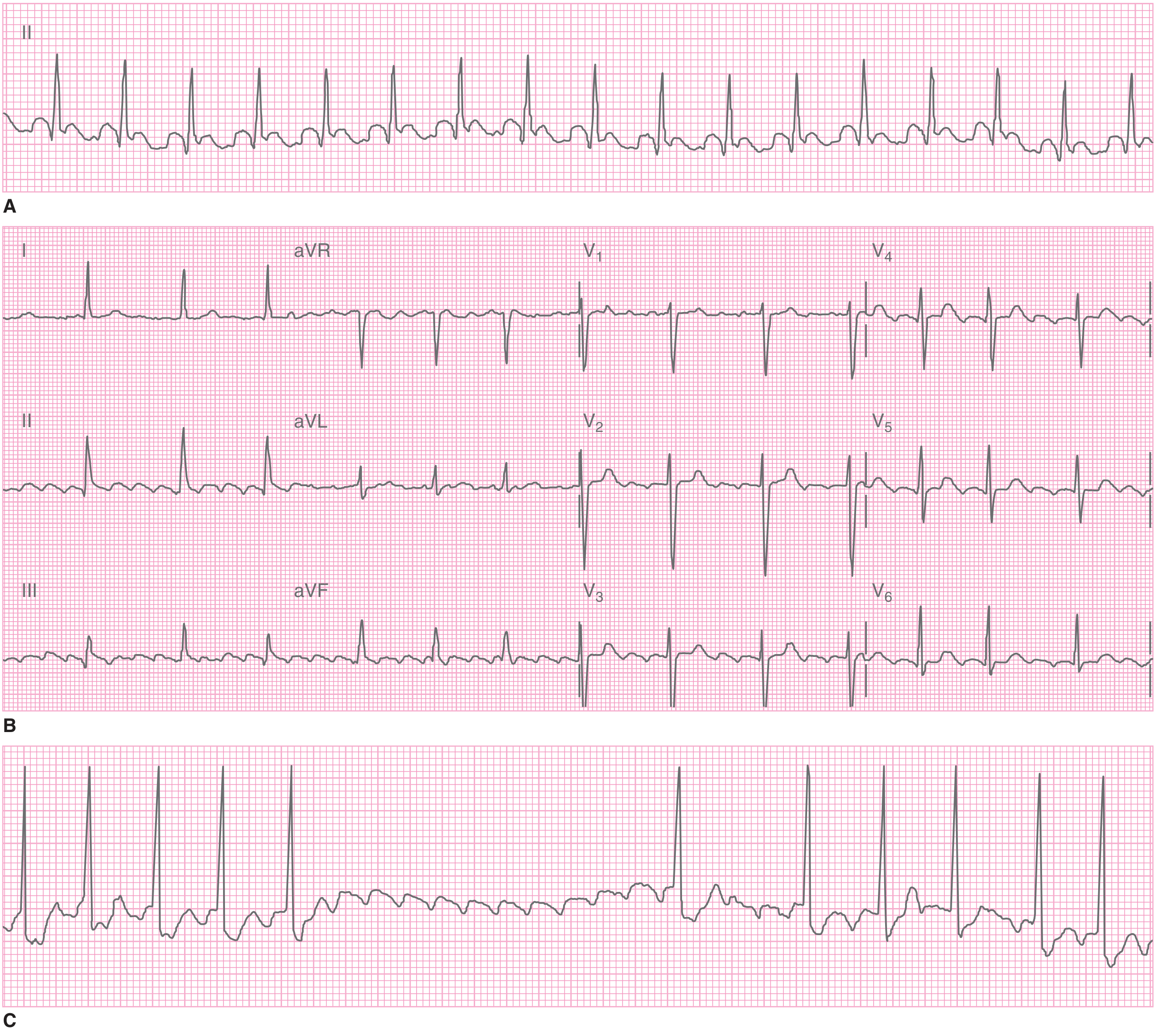
2. Atrial Fibrillation (AF)
- Absent P waves - replaced by chaotic fibrillatory baseline (f waves)
- Irregular irregularity of QRS complexes (no two R-R intervals are the same)
- Narrow QRS unless bundle branch block or pre-excitation coexists
- Ventricular rate typically 100-180 bpm in uncontrolled AF
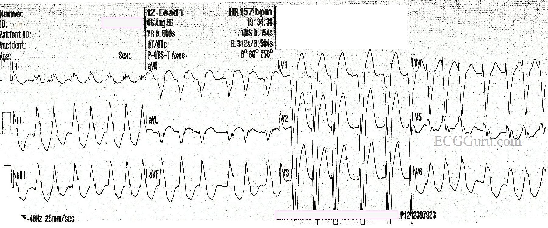
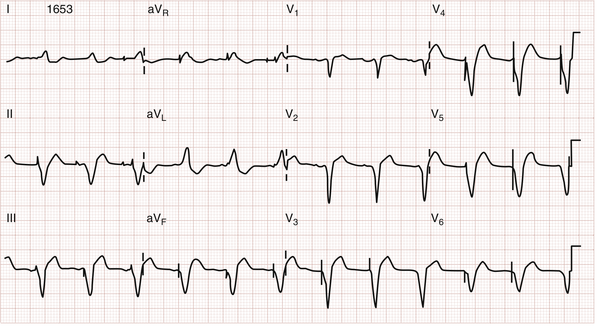
3. Left Bundle Branch Block (LBBB)
- QRS duration ≥120 ms (wide complex)
- Broad, notched ("M-shaped") R waves in I, aVL, V5-V6
- Deep S wave or QS pattern in V1 (broad, downwardly deflected QRS in V1)
- No septal Q waves in I, V5, V6
- Discordant ST-T changes - ST and T wave opposite to main QRS deflection
- Left axis deviation common
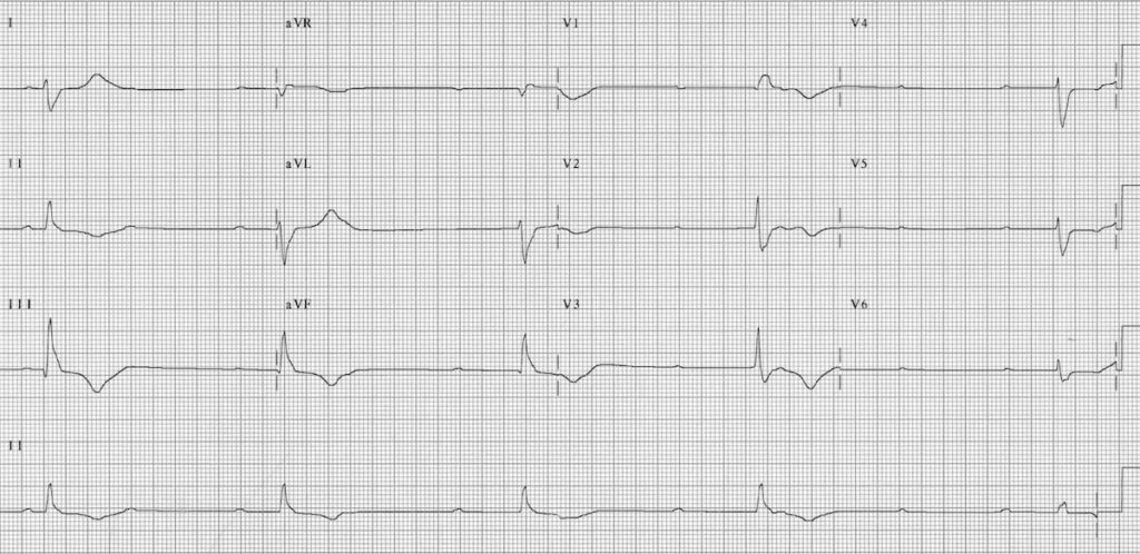
4. Complete (3rd Degree) Heart Block
- Complete AV dissociation - P waves and QRS complexes march independently
- Regular P-P intervals (atrial rate ~60-100)
- Regular R-R intervals but at a slower rate (escape rhythm ~30-50 bpm)
- Wide QRS if ventricular escape, narrow if junctional escape
- No relationship between P waves and QRS complexes (more P waves than QRS)
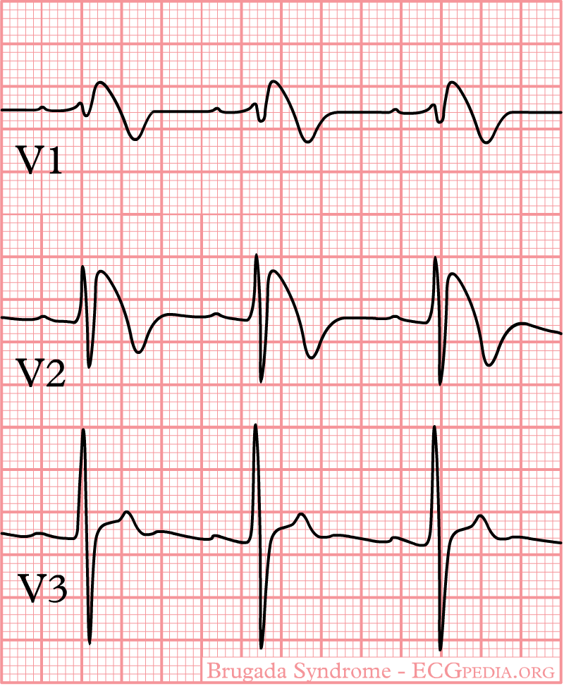
5. Brugada Syndrome
- Type 1 (diagnostic): Coved ST elevation ≥2 mm with a negative T wave in V1-V2 (sometimes V3) - "shark fin" or coved morphology
- Type 2: Saddle-back ST elevation ≥0.5 mm in V1-V2 (not diagnostic alone)
- Incomplete right bundle branch block pattern
- Changes may be dynamic and only appear with sodium channel blockers (ajmaline/flecainide provocation)
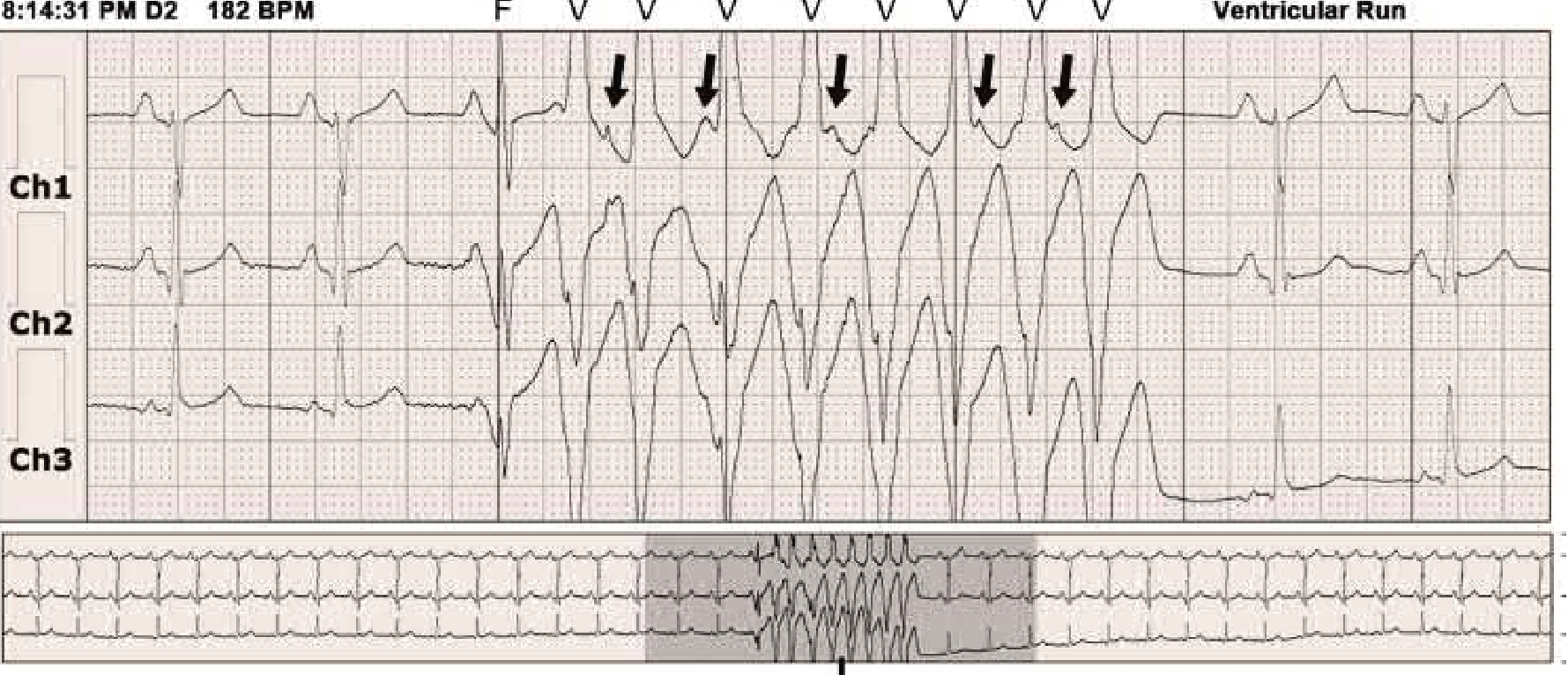
- Risk of VF and sudden cardiac death, especially during fever or sleep
6. Left Ventricular Hypertrophy (LVH)
- Increased QRS voltage (Sokolow-Lyon: SV1 + RV5 or RV6 >35 mm; Cornell: RaVL >28 mm in men, >20 mm in women)
- Left axis deviation
- "Strain" pattern: ST depression and T wave inversion in leads with tall R waves (I, aVL, V5-V6) - indicates pressure overload
- Prolonged QRS duration (may approach LBBB)
- Left atrial enlargement often coexists (broad bifid P wave in II, deep negative P in V1)
- Prominent voltages are a common normal variant in young/athletic individuals - context matters

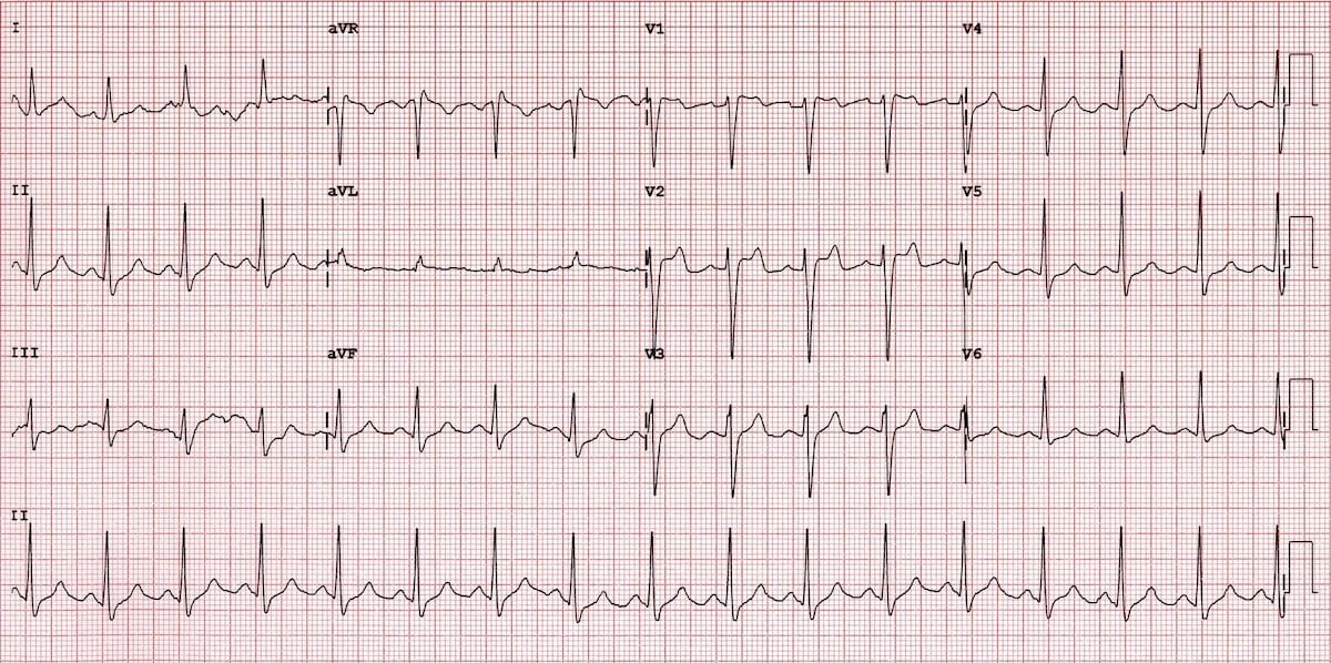
7. Acute Pericarditis
- Stage 1 (acute): Diffuse, concave ("saddle-shaped") ST elevation in multiple leads (I, II, aVF, V2-V6) with PR depression - the key distinguishing feature from STEMI; no reciprocal ST depression (except aVR and V1)
- Stage 2: ST normalizes, T waves flatten
- Stage 3: Diffuse T wave inversion
- Stage 4: ECG normalizes (may take weeks)
- ST elevation is diffuse (not in a single coronary territory)
- ST morphology is concave (vs. convex in STEMI)
- PR segment depression is the pathognomonic sign
- No reciprocal ST depression (except aVR/V1)
- No Q waves
8. Hypertrophic Cardiomyopathy (HCM)
- Voltage criteria for LVH - often dramatic, deeply negative T waves in lateral leads
- Pathologic Q waves in I, aVL, V5-V6 (due to septal hypertrophy - "pseudo-infarction" pattern)
- ST depression and T wave inversion - may be dramatic ("giant T wave inversions") especially in apical HCM (Yamaguchi variant)
- Left axis deviation
- Left atrial enlargement (broad bifid P wave)
- Nonsustained VT may be seen on Holter
9. Pulmonary Embolism (PE)
- Sinus tachycardia - most common finding (present in ~40%)
- S1Q3T3 pattern: Deep S in lead I, Q wave and T wave inversion in lead III - classic but present in only ~20%
- Right axis deviation
- Right bundle branch block (new RBBB suggests massive PE)
- T wave inversions in V1-V4 (right precordial leads) - anterior T inversion pattern
- Atrial fibrillation/flutter may occur
- Right atrial enlargement (tall peaked P waves in II)
- ECG can be completely normal in ~25% of PE cases
10. Wolff-Parkinson-White (WPW) Syndrome
- Short PR interval (<120 ms) - due to ventricular pre-excitation bypassing AV node delay
- Delta wave - slurred, notched upstroke at the beginning of the QRS complex
- Wide QRS (>120 ms) - superficially resembles bundle branch block
- Secondary ST-T changes - discordant to QRS
- Negative delta waves in some leads can mimic pathologic Q waves ("pseudo-infarction")
Quick Reference Summary Table
| Disease | Key ECG Features | Leads Affected |
|---|---|---|
| STEMI | ST elevation, Q waves, T inversion | Territory-dependent (see above) |
| Atrial Fibrillation | Absent P waves, irregular RR | All leads |
| LBBB | Wide QRS ≥120ms, broad R in I/V6, QS in V1 | V1, I, V5-V6 |
| Complete Heart Block | AV dissociation, slow escape rhythm | All leads |
| Brugada Syndrome | Coved ST elevation, RBBB pattern | V1-V2 (±V3) |
| LVH | Tall voltages, strain pattern (ST↓, T inversion) | V4-V6, I, aVL |
| Acute Pericarditis | Diffuse concave ST↑, PR depression | I, II, V2-V6 |
| HCM | LVH + deep Q waves, giant T inversions | I, aVL, V5-V6 |
| Pulmonary Embolism | S1Q3T3, sinus tachycardia, T inversion V1-V4 | I, III, V1-V4 |
| WPW Syndrome | Short PR, delta wave, wide QRS | All leads (pathway-dependent) |
- Harrison's Principles of Internal Medicine 22E (2025) - ECG in cardiac disorders
- Tintinalli's Emergency Medicine: A Comprehensive Study - ECG features of arrhythmias
- Braunwald's Heart Disease: A Textbook of Cardiovascular Medicine - ECG dynamics
- Goldman-Cecil Medicine - Electrocardiography chapter
- ROSEN's Emergency Medicine - Electrocardiographic adjuncts in ACS
- LITFL ECG Library | ECGPedia | Healio Learn the Heart
Clinical note: ECG interpretation must always be correlated with the clinical presentation, symptoms, and additional investigations. A normal ECG does not exclude serious cardiac disease (e.g., ~25% of PEs and some NSTEMIs have normal initial ECGs).The 10 heart diseases covered above are:
- STEMI - ST elevation by coronary territory, evolving Q waves, reciprocal changes
- Atrial Fibrillation - No P waves, irregularly irregular QRS
- LBBB - Wide QRS, broad R in lateral leads, QS in V1, discordant ST-T
- Complete Heart Block - AV dissociation, independent P and QRS rates
- Brugada Syndrome - Coved ST elevation in V1-V2, RBBB pattern, risk of VF
- LVH - Voltage criteria, strain pattern ST depression/T inversion in lateral leads
- Acute Pericarditis - Diffuse concave ST elevation, PR depression (pathognomonic)
- Hypertrophic Cardiomyopathy - LVH + pseudo-infarction Q waves, giant T inversions
- Pulmonary Embolism - Sinus tachycardia, S1Q3T3, right precordial T inversions, RBBB
- WPW Syndrome - Short PR, delta wave, wide QRS, risk of AF with rapid conduction