
Recurrent shoulder dislocation in details according to orthopaedics text books
recurrent shoulder dislocation Bankart lesion Hill-Sachs
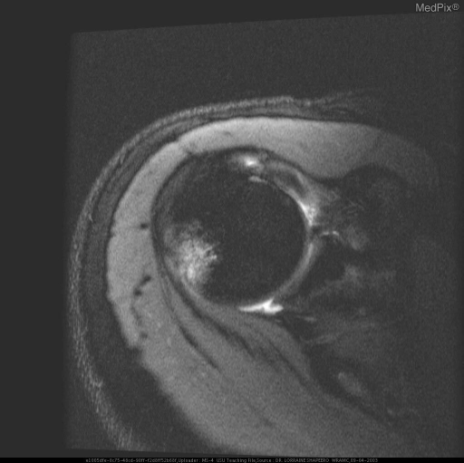
**Imaging Modality:** Magnetic Resonance Imaging (MRI), axial view. **Anatomical Region:** Glenohumeral joint (shoulder). **Observed Pathology:** The image demonstrates findings consistent with sequelae of anterior shoulder dislocation, specifically a bony Bankart lesion and an associated Hill-Sachs deformity. **Characteristic Visual Features:** * **Bankart Lesion:** Evidence of a fracture and fragmentation at the anterior-inferior aspect of the glenoid rim. There is associated disruption of the anterior labrum. * **Hill-Sachs Deformity:** A cortical depression and wedge-shaped defect are visible on the posterolateral aspect of the humeral head. * **Signal Intensity:** High T2-weighted/STIR signal intensity is noted within the humeral head and adjacent glenoid, indicating subchondral bone marrow edema or bruising (bone marrow contusion) at the impact sites. **Clinical Context:** These findings are pathognomonic for recurrent or acute anterior glenohumeral instability. The "kissing lesions" represent the forceful impact between the humeral head and the glenoid during the dislocation event. **Key Diagnostic Features:** Posterolateral humeral head compression fracture (Hill-Sachs) and anterior-inferior glenoid rim osseous avulsion (Bony Bankart).
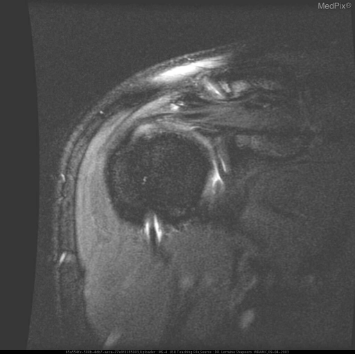
**Imaging Modality:** Magnetic Resonance Imaging (MRI), coronal oblique view, fat-saturated T2-weighted or proton density sequence. **Anatomical Region:** Right shoulder glenohumeral joint. **Observed Pathology:** Post-traumatic changes consistent with a prior anterior glenohumeral dislocation. The image demonstrates a Hill-Sachs lesion, characterized by a cortical depression and marrow signal abnormality (bone bruise) on the posterolateral aspect of the superior humeral head. **Characteristic Visual Features:** * **Hill-Sachs Deformity:** A distinct contour defect and focal hyperintensity in the subchondral bone of the posterolateral humeral head, indicating an impaction fracture. * **Labral Assessment:** There is evidence of a Bankart lesion, visualized as a disruption of the anteroinferior glenoid labrum with associated high signal intensity, suggesting a tear. * **Joint Space:** Mild joint effusion is present. The acromioclavicular joint and subacromial space are visible superiorly. **Clinical Significance:** These findings are pathognomonic for anterior instability of the shoulder. The combination of a Hill-Sachs impaction fracture and a Bankart labral tear is a common sequela of shoulder dislocation, increasing the risk of chronic recurrent instability and secondary osteoarthritis.
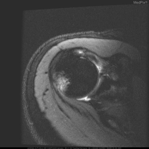
**Imaging Modality:** Magnetic Resonance Imaging (MRI), axial plane. **Anatomical Region:** Shoulder joint (glenohumeral joint). **Observed Pathology:** * **Hill-Sachs Lesion:** A prominent impaction fracture is visible on the posterolateral aspect of the humeral head. This is characterized by a cortical depression and subchondral marrow edema, appearing as an area of increased signal intensity on this fluid-sensitive sequence. * **Bankart Lesion:** Evidence of injury to the anteroinferior glenoid labrum. There is disruption of the labral-ligamentous complex, consistent with an avulsion of the labrum from the glenoid rim. * **Joint Effusion:** Increased intra-articular signal intensity indicates presence of joint fluid/effusion. **Characteristic Visual Features:** The "hatchet" deformity of the posterior humerus and the blunting/detachment of the anterior labrum are classic markers of shoulder instability. **Clinical Context:** These findings are pathognomonic for sequelae of recurrent anterior glenohumeral dislocation. **Diagnostic Differentiating Features:** The co-occurrence of a posterolateral humeral head defect (Hill-Sachs) and an anteroinferior labral tear (Bankart) confirms a mechanism of anterior instability, distinguishing it from posterior dislocation patterns.
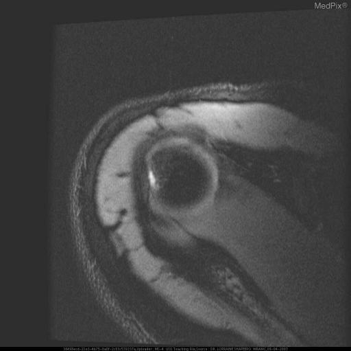
**Imaging Modality:** Magnetic Resonance Imaging (MRI) **Anatomical Region:** Right shoulder (glenohumeral joint) **View:** Axial section **Observed Pathology:** The image demonstrates signs of chronic anterior shoulder instability. There is evidence of a **Bankart lesion**, characterized by a disruption or fracture of the anterior-inferior glenoid labrum/rim. This is accompanied by a **Hill-Sachs deformity**, visible as a cortical depression or flattening of the posterolateral aspect of the humeral head. **Characteristic Visual Features:** - **Bankart Lesion:** Loss of the normal triangular low-signal intensity of the anterior labrum, with associated irregularity of the glenoid margin. - **Hill-Sachs Lesion:** A wedge-shaped defect in the humeral head, resulting from forceful impaction against the glenoid during an anterior dislocation event. - **Joint Space:** The relationship between the humeral head and the glenoid fossa is visualized, showing post-traumatic structural changes. **Clinical Context:** These findings are pathognomonic for recurrent anterior glenohumeral dislocations. The combination of the bony/labral Bankart and the Hill-Sachs lesion represents the "bipolar" nature of many instability-related injuries. **Key Diagnostic Features:** - Anterior-inferior glenoid rim disruption. - Posterolateral humeral head impaction fracture. - Presence of these dual osseous/cartilaginous defects in a patient with a history of shoulder instability.
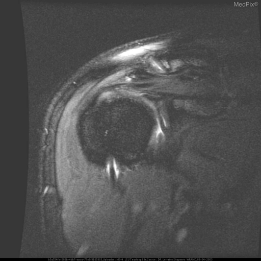
**Imaging Modality:** Magnetic Resonance Imaging (MRI), coronal oblique view, likely T2-weighted with fat suppression or STIR sequence. **Anatomical Region:** Right glenohumeral joint (shoulder). **Observed Pathology:** Evidence of chronic glenohumeral instability and sequelae of recurrent anterior shoulder dislocations. There is a prominent defect in the posterolateral aspect of the humeral head, consistent with a Hill-Sachs lesion (compression fracture). The glenoid margin shows architectural changes suggestive of a Bankart lesion or chronic labral injury. **Characteristic Visual Features:** * **Humeral Head:** Focal loss of bone volume and marrow signal alteration at the posterolateral humeral head superiorly. * **Glenoid:** Blunting or irregularity of the inferior glenoid labrum. * **Soft Tissues:** Increased signal intensity in the subacromial/subdeltoid space, possibly reflecting bursal fluid or reactive changes. The supraspinatus tendon is visible superior to the humeral head. **Clinical Context:** Findings are highly characteristic of recurrent anterior instability commonly seen in contact sports (e.g., rugby). **Key Diagnostic Features:** The "engaging" Hill-Sachs deformity and associated glenoid rim irregularity are the hallmark diagnostic indicators of previous anterior dislocation events.
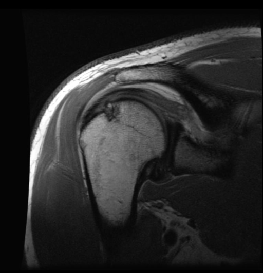
**Imaging Modality:** Magnetic Resonance Imaging (MRI) **Anatomical Region:** Right shoulder (glenohumeral joint) **View:** Coronal oblique T1-weighted sequence **Observed Pathology:** The image demonstrates a classic **Hill-Sachs lesion**, characterized by a cortical depression and marrow signal abnormality on the posterolateral aspect of the superior humeral head. This impaction fracture is a hallmark sign of prior anterior glenohumeral dislocation. **Characteristic Visual Features:** * **Humeral Head:** Focal wedge-shaped defect in the subchondral bone of the superolateral humeral head. * **Glenohumeral Alignment:** The humeral head is currently reduced within the glenoid fossa. * **Surrounding Structures:** Visualization of the acromion, clavicle, and deltoid musculature. There is a suggestion of labral pathology (likely a Bankart lesion) at the inferior glenoid rim, typically associated with this injury pattern. **Clinical Context & Significance:** The presence of a Hill-Sachs lesion is a critical radiologic marker for shoulder instability. It represents the "engagement" of the humeral head against the anterior glenoid rim during a dislocation event. This finding is essential for preoperative planning to assess the risk of recurrent instability and to determine the need for specialized procedures like a Remplissage or bone grafting.
Recurrent Dislocation of the Shoulder
1. Definition and Introduction
2. Pathoanatomy - "The Essential Lesion"
a. Soft Tissue Injuries
- Classic Bankart lesion: Avulsion of the anteroinferior labrum and inferior glenohumeral ligament (IGHL) complex from the glenoid rim
- HAGL (Humeral Avulsion of Glenohumeral Ligament): Less common; IGHL avulses from the humeral side
- Capsular stretching/rupture: Especially the anterior band of the IGHL
- ALPSA (Anterior Labroligamentous Periosteal Sleeve Avulsion): Labrum and periosteum peel medially
b. Bony Injuries ("Bipolar Lesions")
- Hill-Sachs lesion: An impaction fracture of the posterolateral humeral head against the harder glenoid rim during anterior dislocation. Visible as a "hatchet" or wedge-shaped defect
- Bony Bankart lesion: Fracture and fragmentation of the anteroinferior glenoid rim
- Less commonly: coracoid fractures, greater tuberosity fractures, lesser tuberosity fractures
c. Other Associated Injuries
- Rotator cuff tears (especially in patients >40 years)
- Axillary nerve injury
- Brachial plexus injury
- Vascular injury (rare)
3. Static and Dynamic Stabilizers
Static Stabilizers
| Structure | Role |
|---|---|
| Glenoid labrum | Deepens the glenoid socket by ~50%; increases concavity |
| IGHL (anterior band) | Primary restraint to anterior translation in ABER position |
| MGHL | Limits anterior translation at 45° abduction; absent in up to 30% of people |
| SGHL + coracohumeral lig. | Limits inferior translation in adduction |
| Glenohumeral articular geometry | Cartilage arc, version, shape |
| Negative intra-articular pressure | Acts as a suction cup mechanism |
Dynamic Stabilizers
| Structure | Role |
|---|---|
| Rotator cuff | Joint compression, force couple, dynamizes ligaments |
| Biceps tendon | Secondary stabilizer via superior labral attachment |
| Deltoid | Compressive and directional force |
| Proprioception | Mechanoreceptors (Pacinian corpuscles, Ruffini endings, Golgi tendon organs) in capsule/ligaments mediate reflex muscle activation |
4. Classification
Matsen and Thomas Classification (TUBS / AMBRII)
| TUBS | AMBRII |
|---|---|
| Traumatic onset | Atraumatic onset |
| Unidirectional instability | Multidirectional instability |
| Bankart lesion present | Bilateral laxity |
| Surgery required | Rehabilitation first-line |
| Inferior capsule shift if surgery needed | |
| Interval (rotator) closure |
Direction-Based Classification
- Anterior (most common, ~95%)
- Posterior
- Inferior (luxatio erecta - rare)
- Multidirectional
5. Risk Factors for Recurrence
- Age <25 years - single strongest predictor
- Male sex
- Contact/collision sport participation (football, rugby)
- Military service
- First dislocation requiring ER visit with sedation for reduction
- Number and direction of prior dislocations
- Bony lesions (large Hill-Sachs or glenoid bone loss)
- Capsular laxity or collagen disorder (Ehlers-Danlos, Marfan syndrome)
- Prior failed surgical repair
6. Clinical Features
History
- Mechanism: arm abducted and externally rotated (ABER) - classic mechanism for anterior dislocation
- Subsequent episodes occur with less force - eventually spontaneous or with minimal provocation
- Patient may sometimes self-reduce the shoulder
- Painful apprehension with the arm in the "at-risk" position
- Documented prior dislocations, prior reductions (with or without sedation)
Physical Examination
- Apprehension Test: Arm in 90° abduction and maximal external rotation - positive if patient feels apprehension (not just pain)
- Relocation Test: Posterior pressure on the proximal humerus in the apprehension position relieves symptoms
- Anterior Release (Surprise) Test: Sudden removal of the posterior force - the most accurate single test (sensitivity 63.9%, specificity 98.9%)
- Bony Apprehension Test: Apprehension at ≤45° abduction and ≤45° ER - sensitivity 100%, specificity 86% for significant bony lesions
- Load and Shift Test: Humeral head translation assessed anteriorly and posteriorly
- Sulcus Sign: For inferior/multidirectional instability
- Jerk Test: Axial load with arm at 90° abduction and maximal internal rotation, then horizontally adducted - positive if a clunk is felt
- Posterior Drawer Test
7. Imaging
Plain Radiographs
- AP view: Assess glenohumeral alignment, glenoid wear, Hill-Sachs deformity
- True AP (Grashey view): Tangential view of the joint space
- Axillary lateral: Assesses anterior-posterior position of humeral head (essential - cannot assess direction without it)
- Modified axillary view: Patient tolerates this better in acute injury
- Stryker notch view: Best for detecting Hill-Sachs lesion (arm raised vertically, beam 10° cephalad)
- West Point view: Best for detecting bony Bankart lesion (beam 25° downward and 25° medial)
- Bernageau profile view: Measures anterior glenoid bone loss; comparable accuracy to CT
CT and CT Arthrography
- Best for quantifying glenoid bone loss and assessing Hill-Sachs size
- 3D reconstruction helps calculate glenoid surface area deficit
- Critical for surgical planning (Latarjet vs. Bankart vs. bone grafting)
MRI and MR Arthrography
- Gold standard for soft tissue pathology: labral tears, capsular injury, HAGL, rotator cuff
- MR arthrogram with ABER view improves detection of anteroinferior labral tears
- Shows bone marrow edema in Hill-Sachs and Bankart lesions
8. Management
A. Nonoperative Treatment
- Reduction of acute dislocation (with analgesia/sedation)
- Brief immobilization in a sling (2-4 weeks)
- Supervised physiotherapy - rotator cuff strengthening (especially infraspinatus, teres minor, subscapularis), periscapular muscle strengthening, proprioception training
- Activity modification
B. Operative Treatment
- More than one shoulder subluxation or dislocation with anterior labral detachment (Bankart lesion)
- Recurrent anterior instability despite adequate conservative treatment
- Locked dislocation with failed closed reduction under anesthesia
- High-risk athletes (contact/overhead sports) with first-time dislocation and documented labral pathology on MRI
- Primary collagen disorders (Ehlers-Danlos, Marfan)
- Voluntary dislocators or atraumatic MDI (without prior rehabilitation)
- Active infection or severe post-traumatic arthritis
- Neurologic paralysis (axillary/suprascapular nerve)
- Capsular deficiency or prior thermal capsulorrhaphy
Surgical Procedures
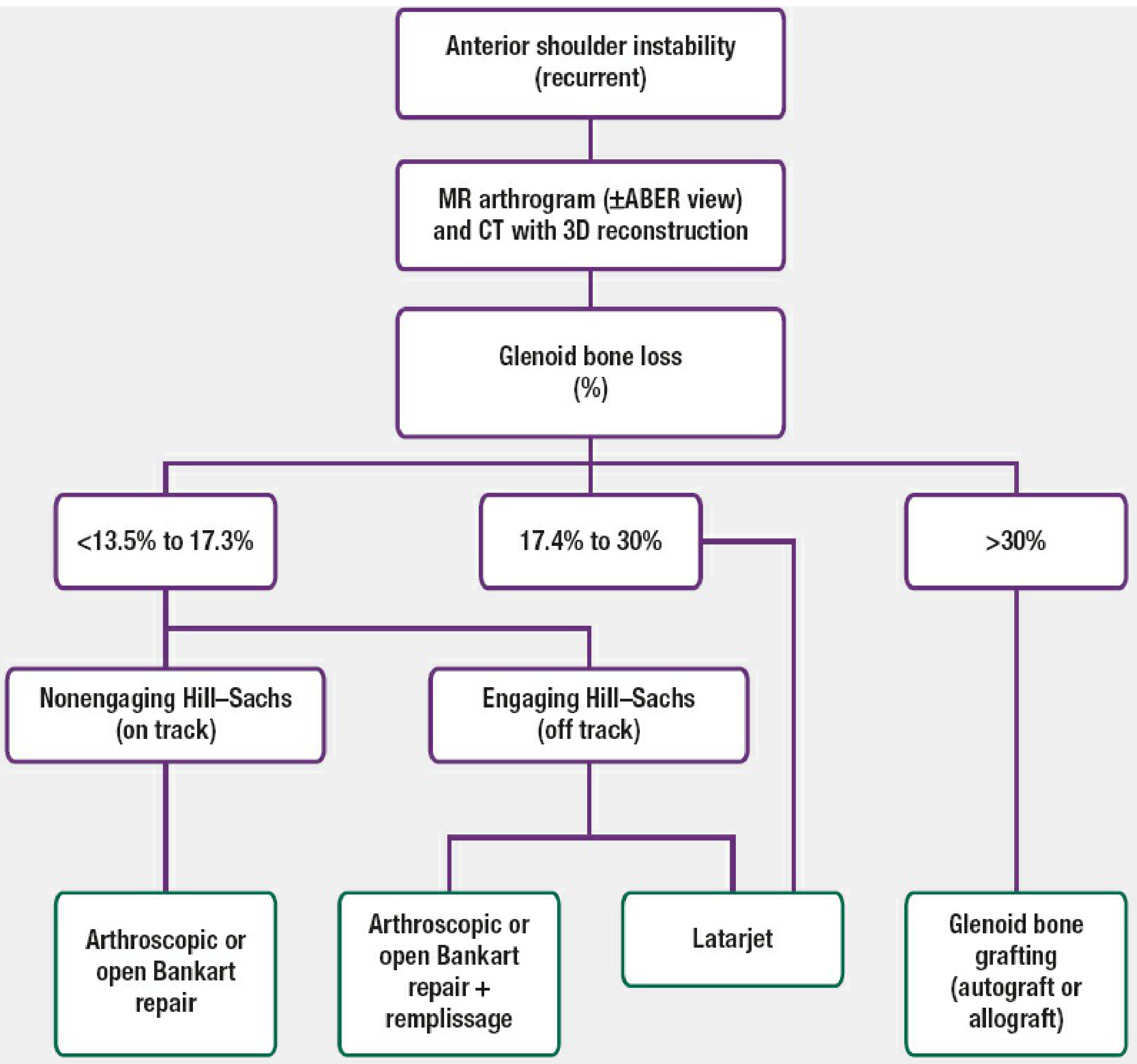
1. Arthroscopic Bankart Repair (ABR)
- ABR now accounts for 90.5% of all shoulder stabilization surgeries
- Success rates comparable to open repair when patient selection is appropriate
- Advantages: minimizes subscapularis morbidity, reduces arthrofibrosis risk
- Recurrence rates increase with: glenoid bone loss >20%, engaging Hill-Sachs, contact sports, age <20 years
2. Open Bankart Repair
3. Putti-Platt Procedure
- Overlapping subscapularis and anterior capsule repair, shortening both
- Tightens anterior structures
- Produces significant restriction of external rotation
- Now largely of historical interest; replaced by anatomic Bankart-type repairs
4. Remplissage Procedure
- Arthroscopic infraspinatus tenodesis into the Hill-Sachs defect (fills the lesion)
- Performed in combination with Bankart repair when an "engaging" (off-track) Hill-Sachs lesion is present with glenoid bone loss 17.4-30%
- Prevents the Hill-Sachs defect from engaging the glenoid rim
5. Latarjet Procedure (Coracoid Transfer)
- Extend the glenoid arc (bone effect)
- Create a sling effect via the conjoined tendon against the subscapularis (dynamic sling)
- Repair of CA ligament to capsule
6. Glenoid Bone Grafting (Autograft or Allograft)
9. Posterior Glenohumeral Instability
- Subscapularis, anterior deltoid, and pectoralis major overpower the posterior cuff during tonic-clonic seizures, creating a locked posterior dislocation
- Treatment: reverse Bankart repair (posterior labral reattachment), posterior capsular plication, or posterior glenoid bone grafting
10. Multidirectional Instability (MDI)
- Characterized by instability in more than one direction, usually with generalized ligamentous laxity
- AMBRII pattern
- First-line: Intense physiotherapy (rotator cuff, deltoid, periscapular muscles) for 6-12 months
- If surgery needed: Inferior capsular shift (open or arthroscopic capsulorrhaphy) with closure of the rotator interval
11. Complications
| Complication | Notes |
|---|---|
| Recurrent instability | Most common; related to bony deficiency, patient factors |
| Glenohumeral arthritis | Occurs in ~2/3 of patients at 25-year follow-up |
| Axillary nerve injury | With dislocation or during surgery |
| Rotator cuff tear | Especially in >40 years age group |
| Loss of external rotation | After Putti-Platt or over-tightened capsulorrhaphy |
| Hardware failure (Latarjet) | Screw breakage, coracoid nonunion |
| Subscapularis rupture | Open anterior surgery complication |
| Infection | Rare |
12. Summary Table
| Feature | Detail |
|---|---|
| Most common direction | Anterior (~95%) |
| Essential lesion | Bankart (anteroinferior labral avulsion) |
| Impaction fracture | Hill-Sachs (posterolateral humeral head) |
| Key classification | TUBS vs. AMBRII (Matsen) |
| Strongest recurrence predictor | Age <25 years |
| Best imaging for bone loss | CT with 3D reconstruction |
| Best imaging for soft tissue | MR arthrogram |
| Most common surgery (2025) | Arthroscopic Bankart repair (90.5%) |
| Bone graft procedure | Latarjet (for >17-30% glenoid loss) |
| MDI initial treatment | Physiotherapy (not surgery) |
- Rockwood and Green's Fractures in Adults, 10th ed. (2025), Chapter 35: Glenohumeral Instability, pp. 1248-1325
- Campbell's Operative Orthopaedics, 15th ed. (2026), Shoulder Chapter
- Pye's Surgical Handicraft, 22nd ed. - Recurrent dislocation of the shoulder