
Functional endoscopy for swallowing disorder short bote
FEES - Short Clinical Note
Definition
- Murray & Nadel's Textbook of Respiratory Medicine, p. 948
Indications
-
Dysphagia with suspected neurological cause (stroke, motor neurone disease, Parkinson's, myasthenia gravis)
-
Suspected aspiration or silent aspiration
-
Post-surgical pharyngeal/laryngeal defects
-
Assessment of secretion management (pooling of saliva in piriform fossa)
-
Evaluating compensatory swallowing strategies
-
Bedside assessment when radiology is unavailable or contraindicated
-
Scott-Brown's Otorhinolaryngology Head & Neck Surgery, p. 819
Procedure
- A flexible endoscope is passed transnasally into the nasopharynx/oropharynx
- The larynx and hypopharynx are visualized at rest - looking for:
- Structural lesions, vocal fold mobility
- Pooling of secretions in piriform fossa or vallecula
- Patient then swallows food/liquid of various consistencies, typically:
- Thin liquids
- Thickened liquids (nectar or honey consistency)
- Pureed food (e.g., applesauce)
- Mixed consistency (solid + liquid, e.g., fruit cocktail)
- Dry solid (e.g., cracker)
- Residue, penetration, and aspiration are noted pre- and post-swallow
- Murray & Nadel's Textbook of Respiratory Medicine, p. 948
What FEES Can Assess
| Finding | Visible on FEES? |
|---|---|
| Pre-swallow pooling of secretions | Yes |
| Post-swallow residue in pharynx | Yes |
| Laryngeal penetration | Yes |
| Frank aspiration | Yes (can be missed in whiteout) |
| Vocal fold motion / paralysis | Yes |
| Structural lesions (mass, tumor) | Yes |
| Oral phase of swallowing | No |
| Vocal fold closure during swallow | No (obscured by "whiteout") |
The "Whiteout" Phase - Key Limitation
-
Direct visualization of vocal fold closure is not possible during the actual swallow
-
Frank aspiration occurring during this phase can be missed
-
Murray & Nadel's Textbook of Respiratory Medicine, p. 948
Advantages of FEES
- No radiation exposure (unlike Modified Barium Swallow/MBS)
- Bedside use - does not require a radiology suite
- Well-tolerated by patients (high tolerability)
- Can be performed in ICU/ward settings
- Allows real-time feedback for therapeutic manoeuvres and compensatory strategies
- Can assess laryngeal sensation (with FEESST variant)
- Repeated testing possible without cumulative radiation risk
Disadvantages / Limitations
- Whiteout phase prevents direct visualization during the swallow
- Does not assess the oral phase of swallowing
- Cannot fully evaluate the esophageal phase
- Operator-dependent skill required
- May cause minor patient discomfort on scope insertion
FEES vs. Modified Barium Swallow (MBS) - Comparison
| Feature | FEES | MBS (Videofluoroscopy) |
|---|---|---|
| Radiation | None | Small dose |
| Setting | Bedside / clinic | Radiology suite |
| Oral phase | Not visible | Visible |
| Pharyngeal phase | Visible (except whiteout) | Fully visible |
| Esophageal phase | Not visible | Partially visible |
| Aspiration detection | Good | Excellent |
| Compensatory strategies | Can be tested | Can be tested |
| Portability | High | Low |
- Murray & Nadel's Textbook of Respiratory Medicine, p. 948-949
- Scott-Brown's Otorhinolaryngology Head & Neck Surgery, p. 819
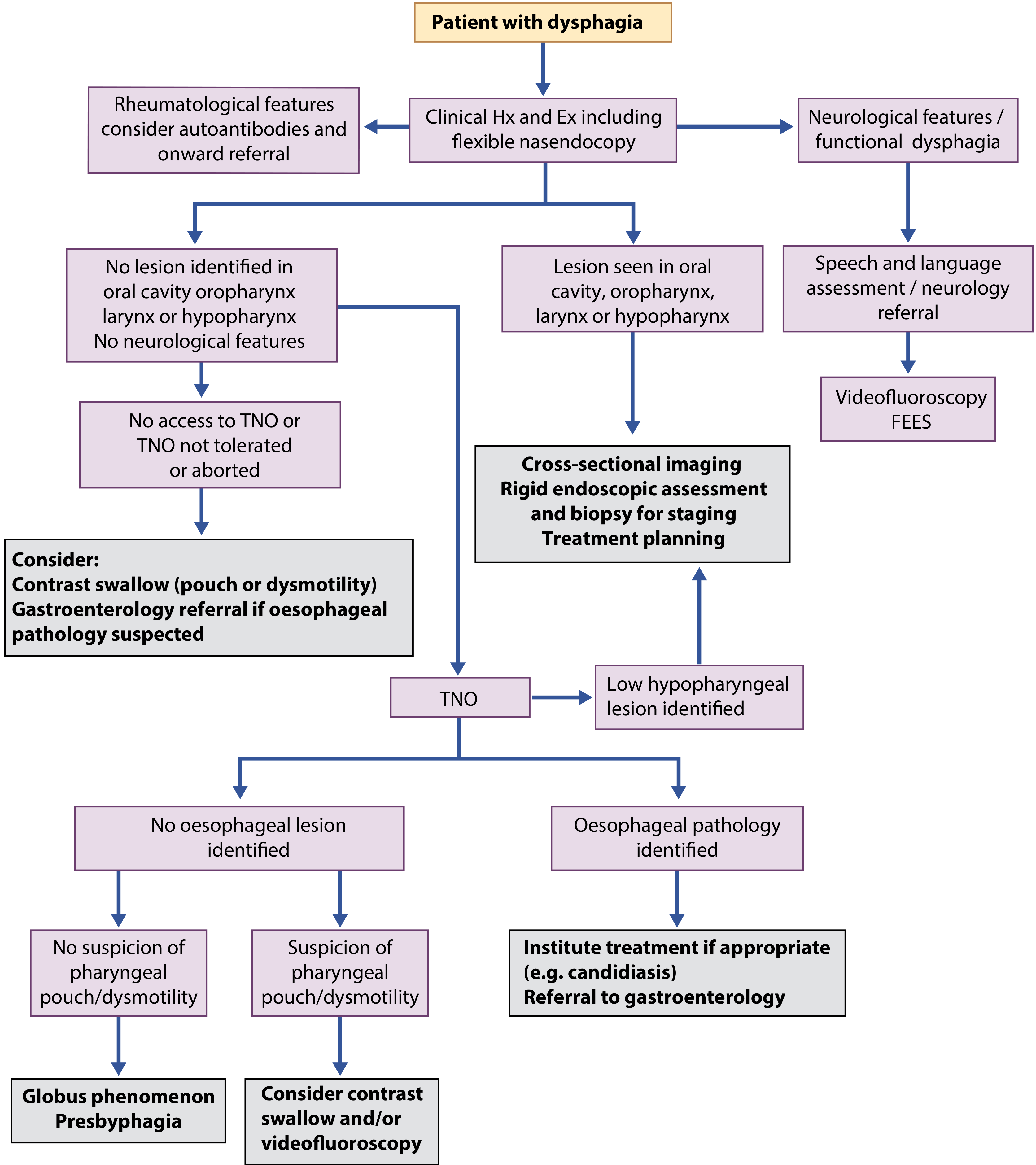
Clinical Algorithm for Dysphagia Assessment
Key Clinical Points
-
Bedside swallow assessment alone by trained SLPs is inadequate to detect aspiration - instrumental testing (FEES or MBS) is required
-
Silent aspiration (aspiration without coughing/choking) can only be reliably detected by instrumental methods like FEES
-
FEES is the first-line instrumental test in neurological dysphagia due to portability and no radiation
-
Results guide diet modification (consistency recommendations) and referral decisions
-
Bradley and Daroff's Neurology in Clinical Practice, p. 232
Intratympanic injection of gentamicin in ENT
Intratympanic Injection of Gentamicin (ITG) - Short Note
Background & History
-
First described by Schuknecht in 1956-57, who injected streptomycin into the middle ear via a microcatheter - vertigo was controlled but severe hearing loss occurred in most patients
-
Gentamicin was subsequently adopted because it has high vestibulotoxicity relative to cochleotoxicity, allowing vestibular ablation while sparing hearing better than earlier aminoglycosides
-
Also called "partial chemolabyrinthectomy" or chemical labyrinthectomy
-
Shambaugh Surgery of the Ear, p. 591; Scott-Brown's Otorhinolaryngology Vol 2, p. 869
Indication
-
Tumarkin crisis / drop attacks - sudden falls without loss of consciousness due to acute otolithic dysfunction
-
End-stage Ménière's disease with disabling vertigo
-
Scott-Brown's Otorhinolaryngology Vol 2, p. 869; Shambaugh Surgery of the Ear, p. 591
Mechanism of Action
- Perilymph concentration reaches 5-10% of the applied solution
- Elimination half-life in perilymph: ~75 minutes
- Selectively concentrated in vestibular hair cells and supporting cells
- Blocks ion currents through stereocilia
- Causes adhesion/fusion of stereocilia
- Ultimately causes hair cell degeneration or extrusion
- Type I hair cells > Type II (more avid gentamicin uptake)
- Also acts via NMDA receptor binding (excitotoxicity) and reactive oxygen species production
- Shambaugh Surgery of the Ear, p. 591; Scott-Brown's Otorhinolaryngology Vol 2, p. 869
Injection Technique
-
Anaesthetize the tympanic membrane (TM) using:
- Topical phenol (Duperstein applicator to pinpoint area), OR
- Lidocaine injection into external auditory canal, OR
- EMLA cream (Lidocaine 2.5% + Prilocaine 2.5%)
-
Two-needle technique using a 25-gauge needle:
- Superior port - to allow air to exit the middle ear
- Inferior port - for injection of gentamicin
-
Volume: Middle ear holds approximately 0.5-0.8 mL of fluid
-
Post-injection positioning: Patient lies in slight Trendelenburg position with the treated ear up for 30 minutes - keeps gentamicin over the round window membrane and prevents escape through the Eustachian tube
-
Warm the solution to body temperature before injection to reduce the brief episode of vertigo that typically follows
-
Injection through an inserted ventilation tube
-
Indwelling catheter into the middle ear
-
Sponge placed through TM
-
Direct injection into round window niche
-
Minipumps (for sustained delivery)
-
Efficacy does not appear to differ between routes
-
Shambaugh Surgery of the Ear, p. 591-592
Dosing Regimens
| Regimen | Description | Notes |
|---|---|---|
| Multiple daily doses | 3x/day via catheter | Higher hearing loss risk (~57%) |
| Weekly doses | Once weekly x 4 weeks | Hearing loss ~19% |
| Single-shot / Titration (current preferred) | One injection; repeat only if vertigo recurs | Lowest hearing risk |
- Shambaugh Surgery of the Ear, p. 591-592
Efficacy
| Source | Vertigo Control (Class A+B) | Duration |
|---|---|---|
| Huon et al. meta-analysis (2012) | 87.5% | - |
| Scott-Brown RCT (ITG vs IT dexamethasone) | 93.5% (ITG) vs 61% (dex) | 2 years |
| Shambaugh retrospective (large) | 91% | ≥2 years |
-
Almost one-third of patients will eventually require additional gentamicin injections (unlike surgical labyrinthectomy)
-
Hair cells may have some capacity for self-repair after gentamicin
-
Scott-Brown's Otorhinolaryngology Vol 2, p. 868-869; Shambaugh Surgery of the Ear, p. 591
Complications & Risks
| Complication | Notes |
|---|---|
| Sensorineural hearing loss (SNHL) | Most important risk; 0-38.7% reported (wide range depending on regimen) |
| Imbalance / disequilibrium | Expected side effect from vestibular ablation; usually compensated |
| Visual-vestibular mismatch | Oscillopsia during head movement |
| TM perforation | Uncommon with careful technique |
| Middle ear infection | Rare |
-
SNHL risk is lowest with titration (single-dose) therapy and highest with multiple daily dose regimens
-
Hearing loss must be weighed against the benefit of vertigo control when selecting candidates - ITG is less preferred in patients with useful residual hearing in the only hearing ear
-
Scott-Brown's Otorhinolaryngology Vol 2, p. 869; Shambaugh Surgery of the Ear, p. 591
ITG vs. Vestibular Neurectomy - Comparison
| Feature | ITG | Vestibular Neurectomy |
|---|---|---|
| Invasiveness | Office procedure | Major surgery (craniotomy) |
| Hearing risk | 0-38% SNHL | Low (hearing-preserving) |
| Vertigo control | ~90-94% | ~90% |
| Reversibility | Partial (hair cells may recover) | Permanent |
| Repeat treatment | May be needed (1/3 of patients) | Not needed |
- Scott-Brown's Otorhinolaryngology Vol 2, p. 869
Treatment Algorithm Position
- Medical therapy - low-sodium diet, diuretics, vestibular suppressants
- IT dexamethasone - if medical failure (Grade A recommendation; 80% vertigo control, no hearing risk)
- IT gentamicin (titration) - if IT dexamethasone fails (Grade A recommendation)
- Surgery - endolymphatic sac decompression, vestibular neurectomy, or labyrinthectomy for extreme refractory cases
- Scott-Brown's Otorhinolaryngology Vol 2, p. 868-869
Botox in ENT
Botulinum Toxin in ENT - Short Note
Pharmacology Basics
-
Produced by Clostridium botulinum - eight serotypes (A-H); Type A (BoNT-A) has the widest clinical use
-
Mechanism: Prevents presynaptic release of acetylcholine by cleaving SNAP-25 protein, blocking vesicle fusion at the nerve terminal
-
Acts on both neuromuscular junctions (motor paralysis) and parasympathetic nerve terminals (glandular secretion reduction)
-
Onset: 2-3 days after injection
-
Duration: 3-9 months (varies by product and tissue type)
-
Repeated injections possible; effect is temporary and reversible
-
Cummings Otolaryngology Head and Neck Surgery
ENT Applications - Overview
| Application | Target | Route |
|---|---|---|
| Spasmodic dysphonia | Thyroarytenoid / posterior cricoarytenoid muscle | Transcricothyroid / transcutaneous |
| Sialorrhea | Parotid + submandibular glands | Intraglandular (US/EMG guided) |
| Frey syndrome | Skin overlying parotid | Intracutaneous |
| Cricopharyngeal spasm | Cricopharyngeus muscle | Endoscopic or percutaneous |
| Tracheoesophageal voice failure | Pharyngeal constrictor / neoglottis | Percutaneous (3 sites) |
| Hyperfunctional voice disorders | Laryngeal adductors | Transcricothyroid injection |
1. Spasmodic Dysphonia (SD)
-
Adductor SD (most common): involuntary adduction of vocal folds causing strained, strangled voice
- Inject thyroarytenoid (TA) muscle - bilateral or unilateral
- Route: Transcricothyroid membrane approach (most common); needle angled superolaterally into adductor muscle group
- Produces controlled, temporary paresis restoring voice fluency
-
Abductor SD: involuntary abduction causing breathy, whispering voice
- Inject posterior cricoarytenoid (PCA) muscle
-
Injection guidance: EMG guidance or direct laryngoscopy via nasendoscope
-
Effect lasts ~3-6 months; repeat injections required
-
Scott-Brown's Otorhinolaryngology, p. 1033
2. Sialorrhea (Drooling)
- BoNT-A injected into parotid and/or submandibular glands
- Causes chemical parasympathetic denervation of glands - reduces saliva production
- Delivery techniques:
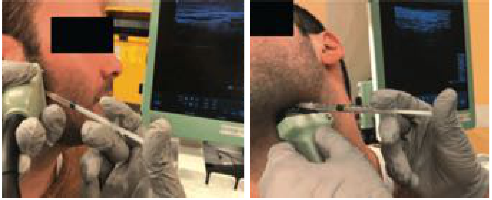
- Anatomic landmarks + manual palpation
- Ultrasound guidance (preferred - most accurate)
- EMG guidance
- Can be performed outpatient/clinic without general anaesthesia (GA may be needed for uncooperative patients)
- Duration: 3-9 months; re-injection required for recurrence
- ~10% non-responders regardless of dose
- Important complication: dental caries - salivary pH drops significantly, increasing acid and reducing protective saliva volume; provide special dental care
- Dysphagia
- Aspiration pneumonia
- Muscle weakness distal to injection (from toxin diffusion into surrounding neck muscles)
- Cummings Otolaryngology Head and Neck Surgery
3. Frey Syndrome (Gustatory Sweating)
-
Complication of parotidectomy - aberrant reinnervation of sympathetic sweat glands by parasympathetic salivary nerve fibers
-
Patient sweats and flushes over the face when eating
-
Confirmed by Minor starch-iodine test
-
BoNT-A injected intracutaneously over the affected facial skin
-
Described as effective in severe cases not responding to topical antiperspirants or glycopyrrolate
-
Cummings Otolaryngology, p. 1521
4. Cricopharyngeal Spasm / Dysfunction
-
Cricopharyngeus fails to relax during swallowing - causes dysphagia, especially post-laryngopharyngectomy
-
BoNT injection into cricopharyngeal muscle relaxes the UES (upper esophageal sphincter)
-
Routes: endoscopic or percutaneous
-
Indications: functional stenosis/spasm; used if dilation fails or at second occurrence of spasm
-
Often combined with dilation; may avoid the need for open surgical cricopharyngeal myotomy
-
Cummings Otolaryngology, p. 1950
5. Post-Laryngectomy Tracheoesophageal Voice Failure (Pharyngospasm / Hypertonicity)
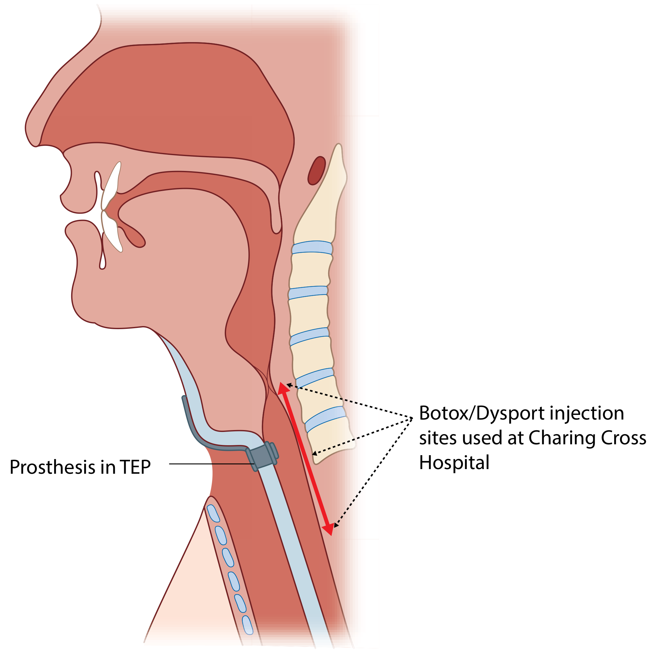
- After total laryngectomy + tracheoesophageal puncture (TEP), some patients develop pharyngeal/neoglottic hypertonicity - the vibratory segment goes into spasm, preventing tracheoesophageal voice
- Confirmed by videofluoroscopy + lidocaine injection test
- BoNT (Botox/Dysport) injected at 3 sites along the myotomy line (from cricopharyngeus level down to below TEP), unilaterally
- Dose adjusted for degree of hypertonicity, neck fibrosis, and type of reconstruction
- A single treatment is often sufficient for continued tonic voice; can be repeated
- Scott-Brown's Otorhinolaryngology Head & Neck Surgery, p. 1033
Injection Approaches to the Larynx (for BoNT)
| Approach | Description |
|---|---|
| Transcricothyroid membrane | Most common; needle through CTM, angled superolaterally into TA muscle |
| Transcutaneous via thyroid cartilage | Through inferior half of thyroid ala (may be blocked by ossified cartilage) |
| Transthyrohyoid | Needle at thyroid notch, angled inferiorly under direct endoscopic visualization |
| Transoral (Abraham cannula) | Curved rigid cannula transorally to inject superior surface of lateral vocal fold |
General Complications of BoNT in ENT
| Complication | Notes |
|---|---|
| Dysphagia | Toxin diffusion to pharyngeal muscles |
| Aspiration pneumonia | Secondary to dysphagia from spread |
| Breathiness / hypophonia | Over-injection in spasmodic dysphonia |
| Dental caries | Seen with salivary gland injection (pH drop) |
| Salivary gland atrophy | Chronic BoNT-A reduces gland size on ultrasound (no histological change) |
| Skin weakness / ptosis | Diffusion to adjacent facial muscles |
| Loss of effect over time | Antibody formation against BoNT-A possible with repeated high-dose use |
Key Summary Points
- BoNT-A is the standard clinical preparation; acts by blocking ACh release via SNAP-25 cleavage
- Effect begins in 2-3 days, lasts 3-9 months - temporary and repeatable
- Adductor spasmodic dysphonia is the landmark ENT application - inject TA muscle via transcricothyroid approach
- Sialorrhea treatment via intraglandular injection is safe and effective; US guidance preferred; watch for dental caries and aspiration
- Frey syndrome, cricopharyngeal spasm, and post-laryngectomy hypertonicity are other important ENT indications
- FDA warning: risk of dysphagia and aspiration pneumonia from toxin spread - always use minimum effective dose
Make a poster on above notes
A detailed medical educational poster titled "Botulinum Toxin (Botox) in ENT" with a clean professional clinical design. The poster has a dark navy blue header with white bold title text "Botulinum Toxin (Botox) in ENT" and a subtitle "Clinical Applications in Otolaryngology". Below the header, the poster is organized into clearly labeled color-coded sections: Section 1 - "Mechanism of Action" (teal/green box): Clostridium botulinum Type A | Cleaves SNAP-25 protein | Blocks ACh release at nerve terminals | Onset: 2-3 days | Duration: 3-9 months | Reversible and repeatable Section 2 - "ENT Applications" (6 icon-style boxes in a 2x3 grid, each with a small icon): 1. Spasmodic Dysphonia - Thyroarytenoid muscle injection via transcricothyroid approach 2. Sialorrhea/Drooling - Parotid + submandibular glands, US-guided 3. Frey Syndrome - Intracutaneous facial skin injection for gustatory sweating 4. Cricopharyngeal Spasm - Endoscopic or percutaneous into cricopharyngeus 5. Post-Laryngectomy Voice Failure - 3 sites along myotomy line for hypertonicity 6. Hyperfunctional Voice Disorders - Laryngeal adductor muscle injection Section 3 - "Injection Routes to Larynx" (orange box, table style): Transcricothyroid membrane | Transcutaneous thyroid cartilage | Transthyrohyoid | Transoral Abraham cannula Section 4 - "Complications" (red-tinted warning box): Dysphagia | Aspiration pneumonia | Breathiness/hypophonia | Dental caries (salivary gland use) | Gland atrophy | Antibody formation Footer note: "BoNT-A: Standard preparation | Begin effect 2-3 days | FDA Warning: risk of dysphagia and aspiration from toxin spread" Overall aesthetic: clean medical infographic style, white background body, professional fonts, colored section boxes, icons, well-spaced readable text suitable for a clinical teaching poster. A4 portrait orientation.
404 Not Found. {'message': '{\n "error": {\n "code": 404,\n "message": "Publisher model `projects/engaged-beaker-474810-u2/locations/global/publishers/google/models/gemini-3-pro-image-preview` was not found or your project does not have access to it. Ensure you are using a valid model name and that the model is available in the specified region. For more information, see: https://docs.cloud.google.com/gemini-enterprise-agent-platform/resources/locations.",\n "status": "NOT_FOUND"\n }\n}\n', 'status': 'Not Found'}
We encountered an error while processing your request. Please try again or reach out to us for help.