
Leukopoiesis and Leucoria
Reading File
Now let me search the library for both topics in parallel.
Finding Sources
Finding Sources
Finding Sources
Finding Sources
Good results. Now let me read the key sections in parallel.
Reading File
Reading File
Reading File
Now let me get more detail on leukopoiesis and read the Wills Eye section on leukocoria management, and also fetch images.
Reading File
Reading File
Loading Image
Loading Image
Excellent - I have comprehensive content from the textbooks and excellent images. Let me now deliver the full answer.
Leukopoiesis
Definition
Leukopoiesis is the process of white blood cell (leukocyte) formation. It is one of the three major components of hemopoiesis (hematopoiesis), alongside erythropoiesis (red cell formation) and thrombopoiesis (platelet formation).
- Histology: A Text and Atlas with Correlated Cell and Molecular Biology, p. 730-731
Origin and Overview
All blood cells arise from hematopoietic stem cells (HSCs) residing in the bone marrow in adults. HSCs are multipotent and differentiate under the influence of cytokines and growth factors into two major progenitor lines:
| Progenitor | Gives rise to |
|---|---|
| Common Myeloid Progenitor (CMP / CFU-GEMM) | Neutrophils, eosinophils, basophils, monocytes, macrophages, dendritic cells, erythrocytes, platelets |
| Common Lymphoid Progenitor (CLP / CFU-L) | T cells, B cells, NK cells, plasma cells |
The hemopoiesis diagram below shows the complete differentiation pathways:
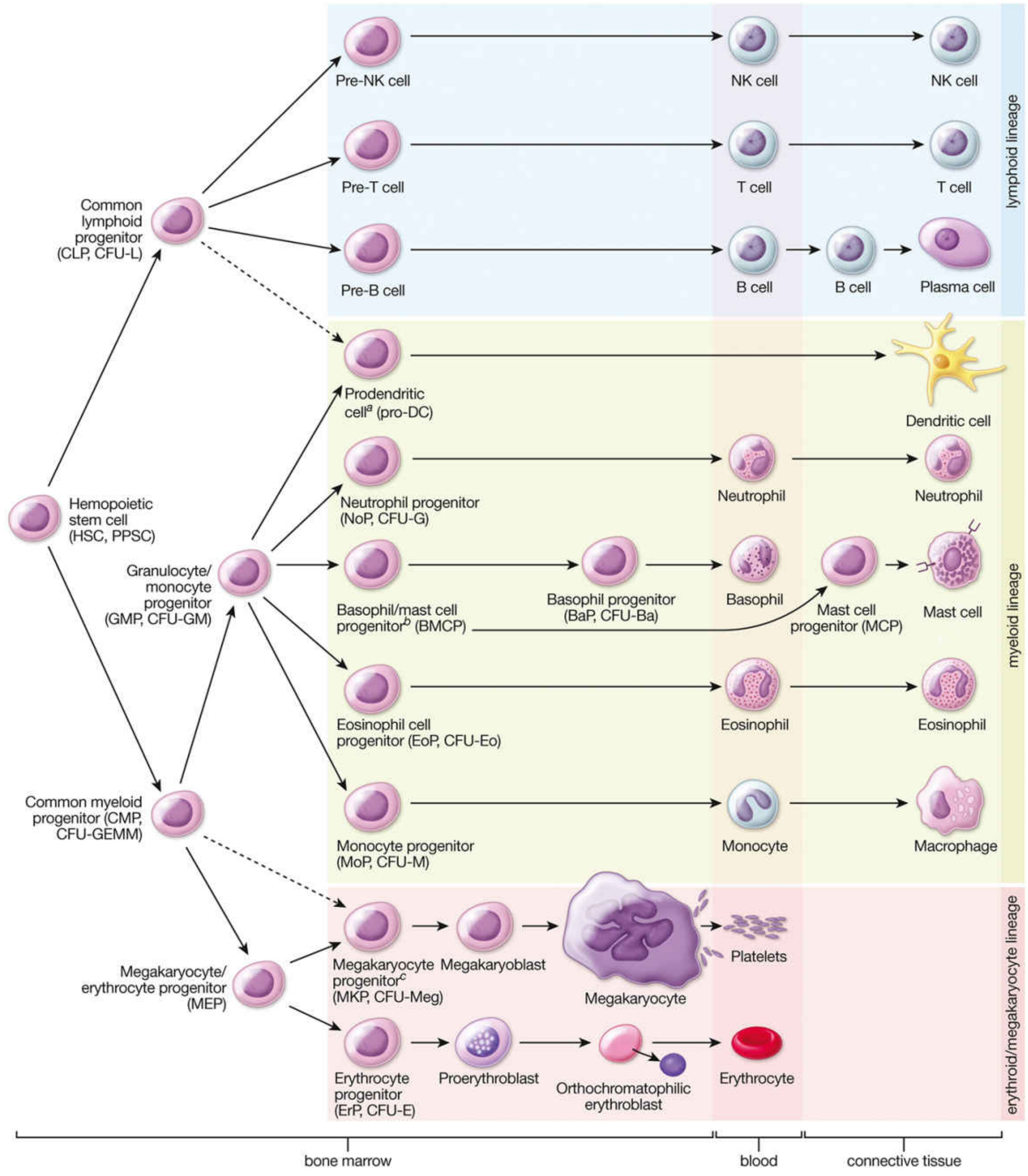
Granulopoiesis (Myeloid White Cells)
Granulocytes (neutrophils, eosinophils, basophils) originate from the CMP, which differentiates into the Granulocyte/Monocyte Progenitor (GMP, CFU-GM).
Neutrophil development passes through six morphologically identifiable stages:
- Myeloblast - earliest recognizable precursor
- Promyelocyte - large cell with primary (azurophilic) granules
- Myelocyte - first stage to exhibit specific (secondary) granules; cell still capable of division
- Metamyelocyte - kidney-shaped nucleus; no longer dividing
- Band (stab) cell - horseshoe-shaped nucleus; immature neutrophil
- Mature neutrophil (segmented) - multilobed nucleus
Eosinophil and basophil development follows a similar maturation pathway from their respective progenitors (EoP and BMCP).
Monopoiesis
Monocytes also arise from the GMP via the monocyte progenitor (MoP, CFU-M). On leaving the bloodstream, monocytes differentiate into tissue macrophages (e.g., Kupffer cells in liver, microglia in CNS, osteoclasts in bone).
Lymphopoiesis
Lymphocytes develop from the CLP and their fate (T vs. B vs. NK) depends on expression of specific transcription factors:
- T cells: mature in the thymus (thymus-dependent)
- B cells: mature in the bone marrow (bursa-equivalent)
- NK cells: develop from pre-NK cell precursors
- Plasma cells: terminal differentiation of activated B cells
Embryonic Leukopoiesis
Blood cell formation is initiated in early embryonic development. Some leukopoiesis occurs in the liver during embryogenesis, though the liver's main role is erythropoiesis in the second trimester fetal period. After birth, the bone marrow becomes the exclusive site for hemopoiesis in adults.
- Histology: A Text and Atlas, p. 731-732
Key Regulators (Cytokines / Growth Factors)
Cytokines act at all stages of leukopoiesis:
- G-CSF (Granulocyte Colony-Stimulating Factor) - promotes neutrophil production
- M-CSF - drives monocyte differentiation
- GM-CSF - stimulates both granulocyte and monocyte lines
- IL-5 - eosinophil maturation and survival
- SCF (stem cell factor) - acts on early HSCs
Clinically: Lithium therapy causes leukocytosis by a direct effect on leukopoiesis. Dopamine receptor antagonists (antipsychotics) can transiently depress leukopoiesis, rarely causing agranulocytosis.
Myeloid:Erythroid (M:E) Ratio
In bone marrow evaluation, the M:E ratio (myeloid to erythroid precursor ratio) is normally ~3:1. A decreased M:E ratio (<1.2:1) indicates either depression of leukopoiesis or erythroid hyperplasia (e.g., hemolytic anemia).
Leukocoria (Leucoria)
Definition
Leukocoria is a white pupillary reflex - the normally red fundal reflex is replaced by a white or yellowish-white glow seen through the pupil.
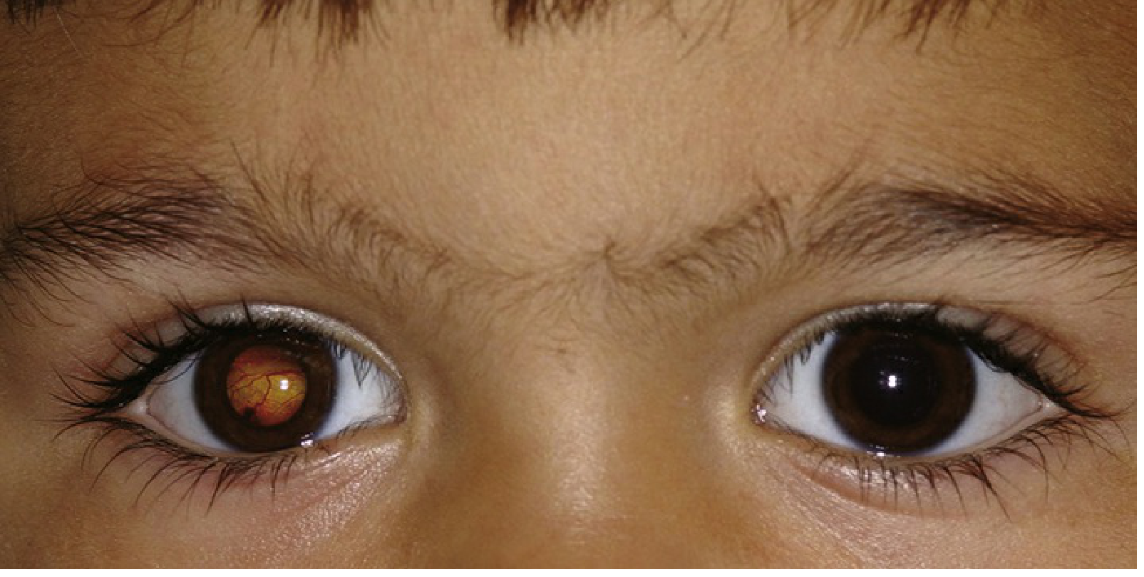
Note: The right eye shows the characteristic white/yellow reflex instead of the normal dark pupil. This is a red-flag sign requiring urgent ophthalmological evaluation.
- The Wills Eye Manual, Section 8.1
Clinical Importance
Leukocoria in a young child is critically important because it may be the presenting sign of retinoblastoma, a life-threatening intraocular malignancy. Despite routine pediatric screening, leukocoria is frequently first noticed by parents in flash photographs (where one eye shows a white reflection instead of red-eye).
Differential Diagnosis
The following conditions can all present with leukocoria:
| Cause | Key Features |
|---|---|
| Retinoblastoma | Most serious - malignant retinal tumor; mean age 18 months; may be bilateral; iris neovascularization in large tumors |
| Cataract (pediatric) | Lens opacity; may be isolated or associated with systemic disease |
| Persistent Fetal Vasculature (PFV) | Formerly "persistent hyperplastic primary vitreous" (PHPV); failure of regression of fetal hyaloid complex; eye usually smaller (microphthalmos) |
| Retinopathy of Prematurity (ROP) | Previously called retrolental fibroplasia; in premature infants receiving oxygen therapy |
| Coats Disease | Retinal vascular malformation with microaneurysms and macroaneurysms; exudative retinal detachment; almost exclusively males; first decade of life; rarely bilateral |
| Retinal Detachment | Various causes; white reflex from detached retina |
| Toxocariasis | Nematode infection (Toxocara); white elevated granuloma; age 6 months - 10 years; contact with puppies or pica history; serum ELISA positive |
| Familial Exudative Vitreoretinopathy (FEVR) | Hereditary vitreoretinal disorder mimicking ROP |
- Goldman-Cecil Medicine, Table 391-5; The Wills Eye Manual, 8.1
Workup
- Detailed fundoscopy (often under anesthesia / EUA in infants - "examination under anesthesia")
- Ultrasound of the globe - detects calcification (suggests retinoblastoma) and assesses retinal detachment
- MRI of orbit and brain - preferred over CT to avoid radiation; mandatory for bilateral retinoblastoma or family history
- Serum ELISA for Toxocara (positive at 1:8 in the majority of infected patients)
- Systemic pediatric evaluation if retinal astrocytic hamartoma, retinoblastoma, or systemic-disease-associated cataract is suspected
Caution: Anterior chamber paracentesis should be avoided if retinoblastoma is suspected - it risks tumor cell dissemination.
Treatment (by cause)
| Condition | Treatment |
|---|---|
| Retinoblastoma | Chemoreduction, intra-arterial chemotherapy, intravitreal chemotherapy, cryotherapy, thermotherapy, laser photocoagulation, or plaque radiotherapy (in combination). Enucleation for advanced/refractory cases. Systemic chemo for metastatic disease. |
| Toxocariasis | Topical/periocular/systemic steroids; vitrectomy if traction bands; consider laser to visible nematode; albendazole only for systemic disease |
| Coats Disease | FA-guided laser photocoagulation to leaking vessels; intravitreal anti-VEGF agents for posterior involvement; surgical drainage for severe retinal detachment |
| PFV | Cataract removal + retrolental stalk resection ± posterior vitrectomy; amblyopia treatment (visual outcome often poor) |
| Pediatric Cataract | Early cataract extraction, optical correction, amblyopia therapy |
Summary Comparison
| Feature | Leukopoiesis | Leukocoria |
|---|---|---|
| Type | Hematology / Histology | Ophthalmology |
| Meaning | Formation of white blood cells | White pupillary reflex |
| Site | Bone marrow (adults); liver/spleen (fetal) | Eye (pupil) |
| Key concern | Infections, leukemia, drug effects | Retinoblastoma until proven otherwise |
| Most important cause to exclude | Agranulocytosis (drug-induced) | Retinoblastoma |
Ovarian mass in a 17 year old female
Reading File
This is a broad clinical scenario with a special population (adolescent female). I'll run parallel searches across headings and paragraphs.
Finding Sources
Finding Sources
Finding Sources
Finding Sources
Finding Sources
Finding Sources
Finding Sources
Finding Sources
Good - Berek & Novak's Gynecology and Robbins Pathology are the key sources. Let me read those in parallel.
Reading File
Reading File
Reading File
Reading File
Reading File
Loading Image
Excellent content. Let me now read the Berek & Novak section further for management details on specific germ cell tumors, and get additional info on yolk sac tumor and immature teratoma.
Reading File
Reading File
I now have comprehensive content. Let me compile the full answer.
Ovarian Mass in a 17-Year-Old Female
Key Clinical Principle
In adolescent and young women (first two decades of life), ~70% of ovarian tumors are of germ cell origin, and approximately one-third of those are malignant. This is the inverse of the adult population, where epithelial tumors dominate. A 17-year-old with an ovarian mass must be evaluated with this age-specific differential in mind.
- Berek & Novak's Gynecology, p. 2377; Robbins & Kumar Basic Pathology, p. 7
Differential Diagnosis
1. Germ Cell Tumors (Most Likely in This Age Group)
The classification of ovarian germ cell tumors (WHO):
| Category | Examples |
|---|---|
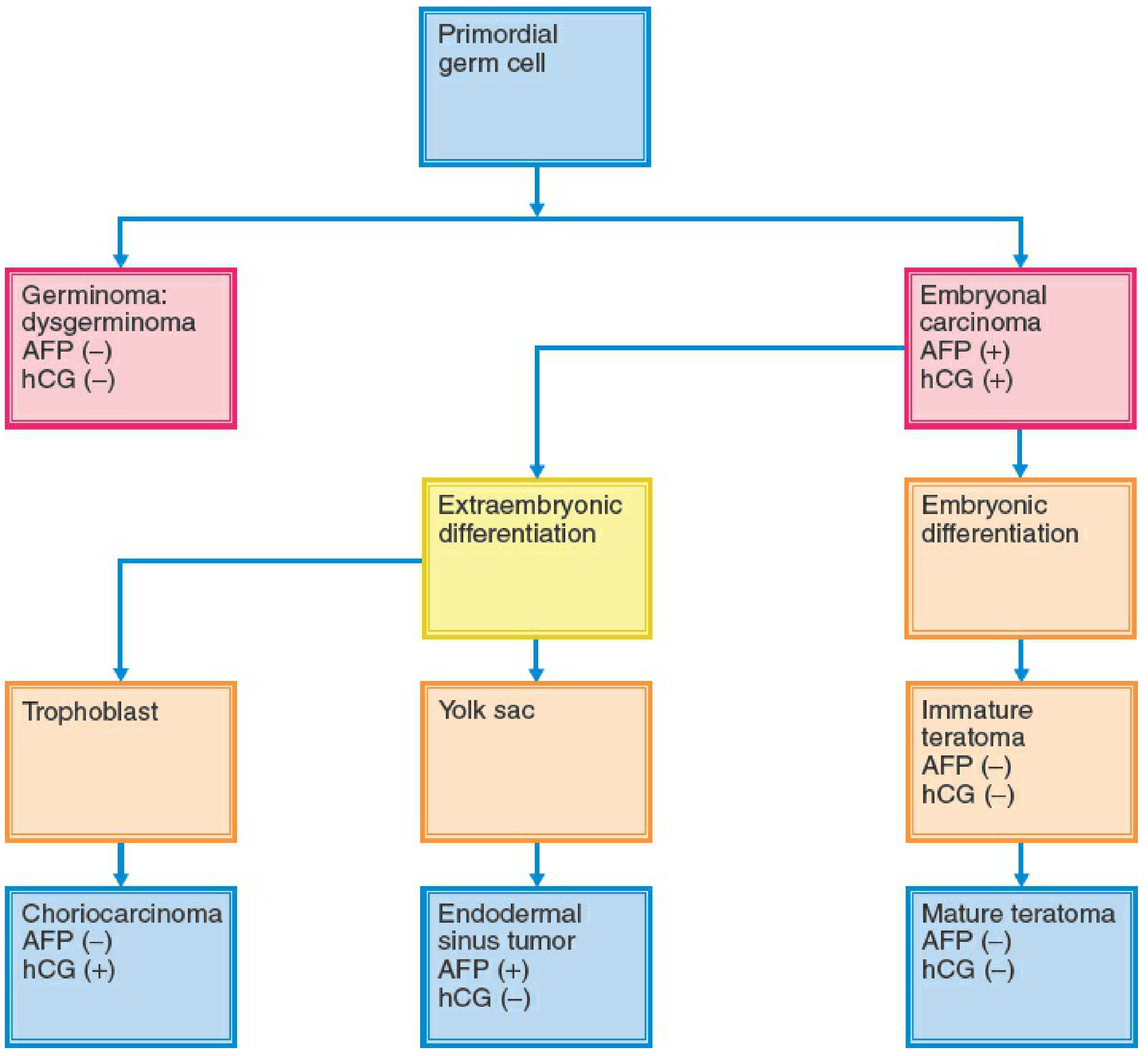
| Primitive germ cell tumors | Dysgerminoma, Yolk sac tumor (endodermal sinus tumor), Embryonal carcinoma, Choriocarcinoma, Polyembryoma, Mixed germ cell tumor |
| Biphasic/triphasic teratoma | Immature teratoma, Mature (dermoid) cyst |
| Monodermal teratoma | Struma ovarii, Carcinoid, Neuroectodermal tumor |
The tumor marker profile by cell type is shown below:
A. Mature Cystic Teratoma (Dermoid Cyst)
- Most common ovarian tumor in young women; vast majority are benign
- Contains derivatives of all three germ layers (teeth, hair, sebaceous material)
- Typically cystic; bilateral in ~10-15%
- AFP and hCG negative
- Complication: torsion (common due to size and mobility)
B. Dysgerminoma
- Most common malignant germ cell tumor
- Counterpart of testicular seminoma
- AFP negative, hCG negative; LDH and PLAP elevated in 95%
- May be bilateral (unique among germ cell tumors - ~10-15%)
- Associated with dysgenetic gonads (check karyotype - if 46,XY gonadal dysgenesis, both gonads must be removed)
- Highly radiosensitive and chemosensitive - excellent prognosis
C. Yolk Sac Tumor (Endodermal Sinus Tumor)
- Second most common malignant germ cell tumor
- AFP markedly elevated (key marker); hCG negative
- Aggressive; grows rapidly
- Schiller-Duval bodies on histology (pathognomonic)
D. Immature Teratoma
- Malignant; contains immature/embryonic neural tissue
- Graded 1-3 based on amount of immature neural elements
- AFP slightly elevated if hepatic/intestinal differentiation present; if markedly elevated, suggests mixed tumor
- Graded: Grade 1 (stage IA) can be managed with surgery alone
E. Embryonal Carcinoma
- Rare; AFP and hCG both positive
- Undifferentiated; precursor to other germ cell types
F. Nongestational Choriocarcinoma
- Very rare in pure form; hCG markedly elevated
- Isosexual precocious puberty due to hCG
G. Mixed Germ Cell Tumor
- Contains ≥2 germ cell elements
- Most common component: dysgerminoma (80%), then yolk sac tumor (70%), immature teratoma (53%)
2. Functional/Physiologic Cysts
- Follicular cyst: Most common; usually <8 cm; resolves spontaneously within 1-3 menstrual cycles; no intervention needed unless persistent or symptomatic
- Corpus luteum cyst: May rupture causing acute pain and hemoperitoneum
- Theca-lutein cyst: Associated with high hCG states
3. Sex Cord-Stromal Tumors (Less Common in Adolescents)
- Granulosa cell tumor (juvenile type): Produces estrogen → isosexual precocious puberty, irregular periods; inhibin B is the marker
- Sertoli-Leydig cell tumor: Produces androgens → virilization, amenorrhea, clitoromegaly
- Fibroma: Solid; associated with Meigs syndrome (ascites + pleural effusion)
4. Epithelial Tumors (Uncommon in This Age Group)
- Serous, mucinous, endometrioid - rare under 20; more common >40 years
- If present at this age, often borderline (low malignant potential)
5. Non-Neoplastic / Other
- Endometrioma ("chocolate cyst"): Endometriosis; associated with dysmenorrhea and dyspareunia
- Tubo-ovarian abscess (TOA): Pelvic inflammatory disease; fever, leukocytosis, cervical motion tenderness
- Paraovarian cyst: Adjacent to ovary, from Wolffian duct remnants
- Ectopic pregnancy: Must always be excluded - obtain serum beta-hCG
Immediate Complication to Rule Out: Ovarian Torsion
Any ovarian mass in an adolescent can precipitate torsion - a surgical emergency.
Features specific to adolescents:
- Torsion is more common at menarche and during adolescence
- In pre/peri-menarchal girls, torsion can occur even without an underlying mass (normal ovary, longer utero-ovarian ligament)
- Presents with sudden-onset severe unilateral pelvic pain, nausea/vomiting (70%)
- 50% of patients are initially misdiagnosed
- Nearly 70% of torsions occur on the right side
Imaging:
-
Transvaginal/transabdominal US with Doppler - primary modality
-
Ovary >4 cm is the most common US finding
-
Absent venous Doppler flow may be earliest sign (arterial flow can persist until late)
-
Normal Doppler does NOT exclude torsion
-
Tintinalli's Emergency Medicine, p. 670-672
Workup
History
- Menstrual history (LMP, cycle regularity)
- Sexual activity (exclude ectopic pregnancy)
- Pain: onset, character, radiation
- Symptoms: precocious puberty, virilization, menstrual irregularity
- Family history of ovarian/BRCA-related cancer
Physical Examination
- Abdominal/pelvic exam (mass characteristics: unilateral/bilateral, mobile/fixed, cystic/solid)
- Signs of virilization or feminization
- Ascites, lymphadenopathy
Investigations
| Test | Purpose |
|---|---|
| Serum beta-hCG | Exclude pregnancy; elevated in choriocarcinoma, embryonal CA |
| AFP | Yolk sac tumor, embryonal CA, mixed GCT |
| LDH | Dysgerminoma (elevated in 95%) |
| Inhibin B / estradiol | Granulosa cell tumor |
| Testosterone / DHEAS | Sertoli-Leydig, androgen-secreting tumors |
| CA-125 | Epithelial tumors (less useful in adolescents; non-specific) |
| Karyotype | Mandatory if dysgerminoma suspected (exclude 46,XY gonadal dysgenesis) |
| Pelvic US | First-line imaging - size, cystic vs solid, Doppler flow |
| MRI pelvis | Better soft tissue characterization than CT; preferred in adolescents (no radiation) |
| CT chest | Germ cell tumors can metastasize to lungs/mediastinum |
Adnexal masses ≥2 cm in premenarchal girls or ≥8 cm in other premenopausal patients generally require surgical exploration.
Surgical Management Principles
Fertility Preservation (Critical in Adolescents)
- Unilateral salpingo-oophorectomy is the standard approach for presumed malignant germ cell tumors - the contralateral ovary and uterus are preserved whenever possible
- Routine wedge biopsy of a normal-appearing contralateral ovary is not indicated (increases adhesion risk and may impair fertility)
- Exception: if karyotype reveals 46,XY, both gonads must be removed (malignant transformation risk)
By Tumor Type
| Tumor | Surgery | Adjuvant Therapy |
|---|---|---|
| Mature teratoma | Cystectomy (preserve ovarian tissue) or oophorectomy | None |
| Dysgerminoma stage IA | Unilateral SO + staging | Surveillance or BEP x3 cycles |
| Dysgerminoma advanced | Unilateral SO + cytoreduction | BEP chemotherapy |
| Yolk sac tumor | Unilateral SO + staging | BEP x3-4 cycles (mandatory) |
| Immature teratoma grade 1, stage IA | Unilateral SO + staging | Surveillance (no adjuvant needed) |
| Immature teratoma high grade or advanced | Unilateral SO + staging | BEP x3 cycles |
| Mixed GCT | Unilateral SO + staging | BEP x3-4 cycles |
Chemotherapy: BEP Regimen
BEP = Bleomycin + Etoposide + Cisplatin is the international standard of care for malignant ovarian germ cell tumors.
- 3 cycles for completely resected disease / adjuvant setting
- 4 cycles for macroscopic residual disease
- Must begin within 7-10 days of surgery (tumors can progress rapidly)
- BEP is superior to EP (bleomycin inclusion significantly improves relapse-free survival: 84% vs 69%)
- Cisplatin superior to carboplatin in metastatic GCT
Radiation therapy for dysgerminoma (2,500-3,500 cGy) is effective but rarely used as first-line due to loss of fertility.
Prognosis
Germ cell tumors respond dramatically to platinum-based chemotherapy. Even metastatic disease has cure rates >90% in many series. Fertility preservation is achievable in the majority of young patients.
Summary Algorithm
17-year-old with ovarian mass
↓
1. Is she pregnant? (beta-hCG)
2. Is this torsion? (acute pain → urgent US Doppler → surgical emergency)
↓
Tumor markers: AFP, beta-hCG, LDH, Inhibin B
Imaging: Pelvic US → MRI ± CT chest
↓
Predominantly CYSTIC Predominantly SOLID/MIXED
↓ ↓
Functional cyst? Likely neoplasm
Mature teratoma? Germ cell tumor most likely
Endometrioma? Send karyotype
↓
Surgical staging + Fertility-sparing surgery
↓
Histology → Adjuvant BEP if indicated
Sources: Berek & Novak's Gynecology, pp. 2375-2415; Robbins & Kumar Basic Pathology, p. 7; Tintinalli's Emergency Medicine, pp. 644-688
Looks neoplastic
Finding Sources
Finding Sources
Reading File
Reading File
Finding Sources
Reading File
Finding Sources
Reading File
Reading File
Loading Image
Reading File
I now have all the content needed. Here is the focused, clinically actionable answer for a neoplastic-appearing ovarian mass in a 17-year-old.
Neoplastic Ovarian Mass in a 17-Year-Old: Full Oncologic Approach
Given that the mass looks neoplastic, the framework shifts from "could this be benign/functional?" to "what type of ovarian neoplasm is this, and how do we stage and treat it while preserving fertility?"
Why This Age Changes Everything
In the first two decades of life, ~70% of ovarian tumors are germ cell origin, and one-third of those are malignant. Epithelial cancers (which dominate in adults >40 years) are rare here. This has major implications - germ cell tumors are highly chemosensitive, and fertility preservation is almost always achievable.
- Berek & Novak's Gynecology, p. 2377
Likely Neoplasm Types (Ranked by Probability at Age 17)
Germ Cell Tumors (Most Likely)
| Tumor | Key Features | Markers |
|---|---|---|
| Mature teratoma (dermoid) | Most common overall; usually benign; contains tooth/hair/sebum | AFP(-), hCG(-) |
| Dysgerminoma | Most common malignant GCT (30-40% of malignant GCTs); solid, bosselated surface; 75% occur age 10-30 | LDH↑, PLAP↑; AFP(-), hCG(-) |
| Immature teratoma | Malignant; graded 1-3 by neural tissue content | AFP mildly↑ only if hepatic differentiation; high AFP = mixed |
| Yolk sac tumor (endodermal sinus tumor) | Aggressive; second most common malignant GCT | AFP markedly↑; hCG(-) |
| Mixed germ cell tumor | ≥2 elements; dysgerminoma most common component (80%) | Variable - both AFP and hCG may be raised |
| Embryonal carcinoma | Rare; undifferentiated precursor | AFP(+), hCG(+) |
| Choriocarcinoma | Rare (non-gestational); hCG drives isosexual precocious puberty | hCG markedly↑; AFP(-) |
Sex Cord-Stromal Tumors (Less Common)
| Tumor | Key Features | Markers |
|---|---|---|
| Juvenile granulosa cell tumor | Occurs in women <30 (especially prepubertal/adolescent); produces estrogen → precocious puberty / irregular menses; low-grade malignancy; bilateral in only 2% | Inhibin B↑, estradiol↑ |
| Sertoli-Leydig cell tumor | Adolescents and young adults; produces androgens → virilization, amenorrhea, clitoromegaly | Testosterone↑, DHEAS↑ |
Epithelial Tumors (Rare at 17, but consider)
- Borderline (low malignant potential) serous or mucinous tumor - more likely than frankly invasive at this age
- CA-125 useful but non-specific
- If invasive epithelial cancer found, refer urgently to gynecologic oncologist
Macroscopic / Imaging Clues to Type
| Appearance | Likely Diagnosis |
|---|---|
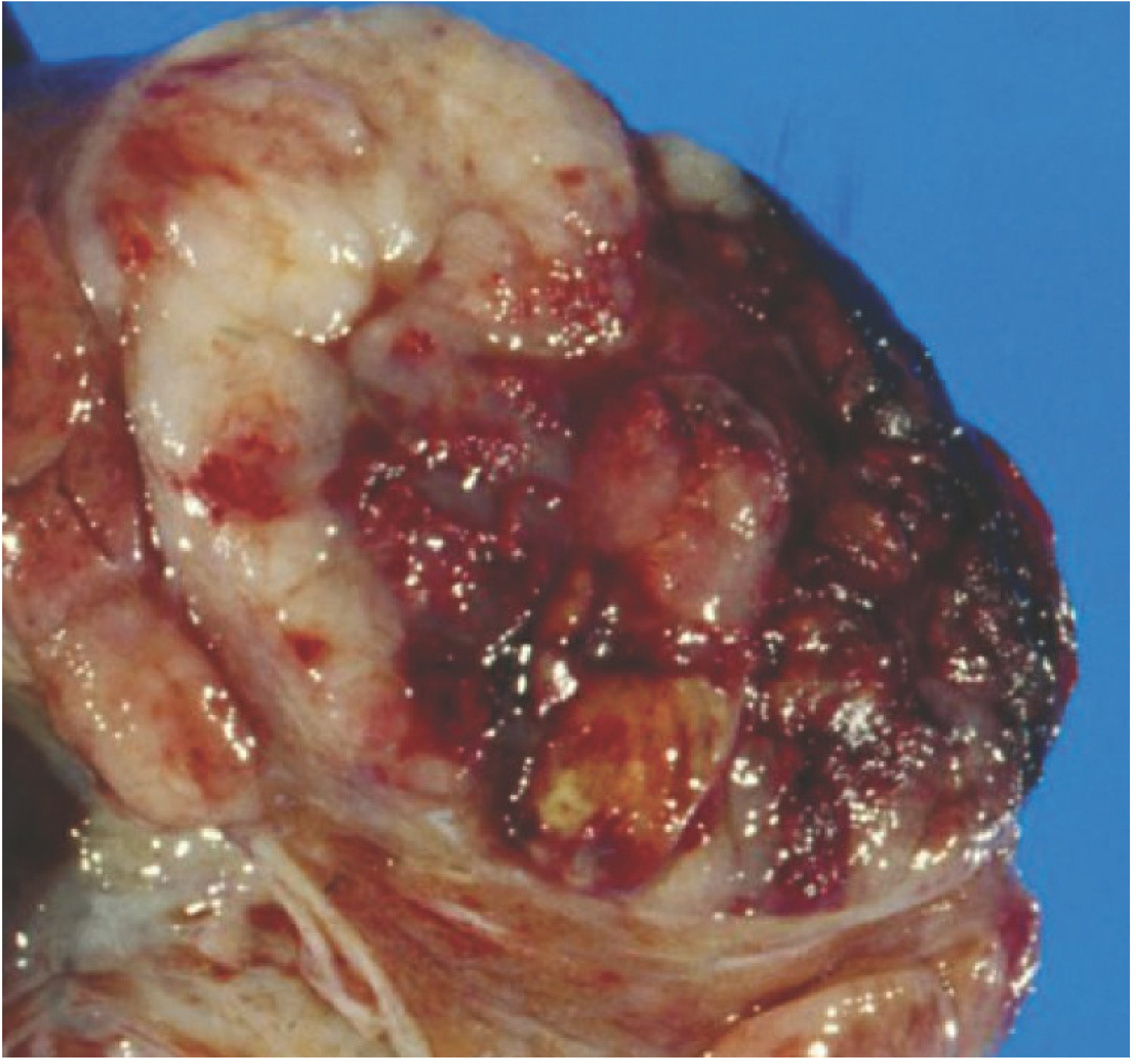
| Solid, fleshy, pale tan-grey, bosselated capsule | Dysgerminoma |
| Mixed solid-cystic with calcifications | Dysgerminoma with gonadoblastoma, or mixed GCT |
| Purely cystic with fat/calcium (teeth/hair on imaging) | Mature teratoma (dermoid) |
| Solid with areas of necrosis/hemorrhage, rapid growth | Yolk sac tumor, immature teratoma, embryonal CA |
| Solid, smooth, yellow-tinged | Granulosa cell tumor, fibroma |
Gross specimen of dysgerminoma - principally solid with cystic areas and necrosis:
Pre-operative Workup
Mandatory Blood Tests
| Test | What it detects |
|---|---|
| Serum AFP | Yolk sac tumor, embryonal CA, mixed GCT |
| Serum beta-hCG | Choriocarcinoma, embryonal CA; also excludes pregnancy |
| LDH | Dysgerminoma (elevated in 95%); also monitor disease |
| PLAP | Dysgerminoma |
| Inhibin B | Granulosa cell tumor |
| Testosterone / DHEAS | Sertoli-Leydig cell tumor |
| CA-125 | Less useful in young women but obtain as baseline |
| FBC, LFTs, renal function, coagulation | Pre-operative baseline |
Karyotype
- Mandatory in all premenopausal girls with a suspected malignancy, especially dysgerminoma
- If 46,XY gonadal dysgenesis (Swyer syndrome): malignant transformation risk in both gonads → both gonads must be removed (uterus may be preserved for future embryo transfer)
Imaging
- Pelvic MRI: best soft-tissue characterization; preferred over CT (no radiation in a young patient)
- CT chest: germ cell tumors metastasize to lungs and mediastinum
- CT abdomen/pelvis: assess retroperitoneal lymphadenopathy (dysgerminomas spread to para-aortic nodes around the renal vessels), liver metastases, ascites
FIGO Staging (Used at Surgery)
| Stage | Description |
|---|---|
| I | Tumor confined to ovaries |
| IA | One ovary; capsule intact; no surface involvement; no ascites |
| IB | Both ovaries; otherwise as IA |
| IC | Stage I + capsule rupture / surface involvement / malignant ascites or washings |
| II | Pelvic extension |
| III | Peritoneal metastasis outside pelvis ± retroperitoneal nodes |
| IV | Distant metastasis beyond peritoneal cavity |
Staging is based on surgical findings - a preoperative evaluation should exclude extraperitoneal metastases.
Surgical Management: Fertility-Sparing is the Default
Intra-operative Principles
- Unilateral salpingo-oophorectomy (USO) of the affected side - standard for germ cell and stromal tumors in young patients; uterus and contralateral ovary preserved
- Comprehensive staging at the same operation:
- Peritoneal washings / ascites for cytology
- Inspection and palpation of all peritoneal surfaces
- Biopsy of any suspicious lesion
- Unilateral pelvic lymphadenectomy + biopsy/palpation of para-aortic nodes (especially for dysgerminoma)
- Omental biopsy
- Do NOT do routine wedge biopsy of the contralateral ovary - increases adhesion risk and impairs fertility
- Exception: if contralateral ovary looks abnormal, excisional biopsy of the suspicious area is appropriate (dysgerminoma is the only GCT that tends to be bilateral)
- For immature teratomas: contralateral involvement is rare; do not resect a normal-appearing contralateral ovary
"Surgery should be fertility-sparing in the setting of a young patient with a germ cell tumor or low-grade epithelial tumor." - Sabiston Textbook of Surgery, p. 2804
Adjuvant Treatment by Tumor Type
| Tumor | Stage | Adjuvant Therapy |
|---|---|---|
| Dysgerminoma | IA (confined, unruptured) | Surveillance OR BEP x3 cycles; no chemotherapy needed if fully staged |
| Dysgerminoma | IC, II, III, IV | BEP x3-4 cycles |
| Yolk sac tumor | Any stage | BEP x3-4 cycles (mandatory - cannot observe) |
| Immature teratoma | IA, Grade 1 | Surveillance - no adjuvant needed; excellent prognosis |
| Immature teratoma | IA Grade 2-3 or advanced | BEP x3 cycles |
| Mixed GCT | Any stage | BEP x3-4 cycles |
| Granulosa cell (juvenile) | Stage I | USO alone (surgery curative in most stage I) |
| Granulosa cell (juvenile) | Advanced | Platinum-based chemo or BEP |
BEP Regimen Details
- Bleomycin + Etoposide + Platinum (cisplatin)
- International standard of care for malignant ovarian GCTs
- Start within 7-10 days of surgery (these tumors grow rapidly)
- 3 cycles for completely resected / adjuvant; 4 cycles for macroscopic residual disease
- Cisplatin is superior to carboplatin in metastatic GCT
- Bleomycin inclusion is important for non-dysgerminomatous tumors (BEP 84% RFS vs EP 69%)
- Second-line for relapse post-BEP: TIP (paclitaxel + ifosfamide + cisplatin) or VIP (vinblastine + ifosfamide + cisplatin)
Surveillance After Surgery
For stage IA tumors on surveillance (no adjuvant chemo):
- Regular clinical review + physical examination
- Tumor markers (AFP, hCG, LDH) - most frequent early on, as most relapses occur within 2 years (all relapses within 13 months in Charing Cross data)
- Interval pelvic/abdominal imaging
- Overall disease-specific survival with this approach: 94% (Charing Cross series)
- 75% of dysgerminoma recurrences occur within the first year; salvage with BEP is highly successful
Prognosis
Germ cell tumors in young women have outstanding outcomes with modern platinum-based chemotherapy. Even with metastatic disease, cure rates exceed 90%. The priority in a 17-year-old is cure with maximum fertility preservation - both goals are usually achievable.
Immediate Next Steps (Summary)
Neoplastic ovarian mass, 17-year-old
↓
1. Tumour markers: AFP, beta-hCG, LDH, PLAP, Inhibin B, Testosterone
2. Karyotype (mandatory)
3. MRI pelvis + CT chest + CT abdomen/pelvis
4. Refer to gynaecologic oncologist
↓
5. Fertility-sparing staging laparotomy / laparoscopy:
- Unilateral salpingo-oophorectomy
- Full surgical staging
- Frozen section if needed
↓
6. Histology result → adjuvant BEP if indicated
(start within 7-10 days of surgery)
↓
7. Surveillance with markers + imaging
- Berek & Novak's Gynecology, pp. 2375-2400; Sabiston Textbook of Surgery, p. 2804; Washington Manual of Medical Therapeutics, p. 857
Tubular, secretory changes in breast
Tubular carcinoma and secretory carcinoma are both special-type breast carcinomas with generally favorable prognoses. Tubular is characterized by well-formed tubules with low-grade nuclei; secretory (juvenile) carcinoma has intracellular and extracellular secretory material and is associated with the ETV6-NTRK3 fusion.
Searching sources...
Finding Sources
Finding Sources
Finding Sources
Finding Sources
Reading File
Reading File
Finding Sources
Finding Sources
Reading File
Loading Image
Finding Sources
Finding Sources
Reading File
Reading File
Excellent - I have comprehensive content from Robbins (big and basic), Schwartz's Surgery, and Berek & Novak. Let me now compose the full answer.
Tubular Carcinoma and Secretory Carcinoma of the Breast
These are two distinct special-type invasive breast carcinomas, both with characteristically favorable prognoses compared to the common invasive carcinoma of no special type (NST/IDC).
Overview: Special-Type Breast Carcinomas
Invasive breast carcinomas are classified as either no special type (NST) (formerly "invasive ductal carcinoma") or special type, when ≥90% of the tumor shows distinctive morphologic features. Special types are important because their histology often predicts molecular subtype, behavior, and prognosis.
The histologic panel of special types includes:
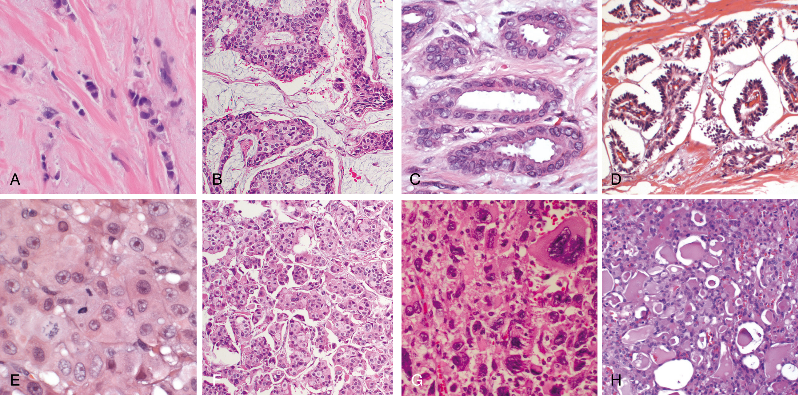
Panel C = Tubular carcinoma (elongated well-formed tubules with open lumens); Panel H = Secretory carcinoma (pale vacuolated cells with intracellular and extracellular secretory material)
Part 1: Tubular Carcinoma
Definition and Epidemiology
Tubular carcinoma is a special-type invasive breast carcinoma defined by the presence of well-formed open tubular structures comprising >90% of the tumor. It accounts for ~2% of all invasive breast cancers, but is detected in up to 20% of cancers found by mammographic screening (reflecting its small size and low-grade nature). It is most commonly diagnosed in the perimenopausal or early menopausal period.
- Schwartz's Principles of Surgery, p. 593-594; Robbins & Kumar Basic Pathology, p. 1286
Morphology
- Macroscopic: small, irregular, spiculated mass (almost always detected by mammography rather than palpation)
- Microscopic: haphazard array of small, randomly arranged, well-formed tubules infiltrating a desmoplastic stroma
- Tubules have open lumens lined by a single layer of epithelial cells
- Cells have low-grade nuclei - bland, uniform, similar to normal epithelium
- No myoepithelial cell layer (distinguishes from benign sclerosing adenosis - a key diagnostic pitfall)
- Sometimes mistaken for a benign sclerosing lesion on low power - hence >90% tubule formation requirement for the pure diagnosis
Molecular Subtype
Tubular carcinoma almost always falls within the Luminal (ER-positive/HER2-negative) subgroup:
- ER positive: 94% (SEER database)
- PR positive in the majority
- HER2 negative
- Low proliferation index (Ki-67 low)
- Genomic profile: quiet genome, 1q+, 16q- (typical luminal A)
Behavior and Prognosis
| Feature | Tubular Carcinoma |
|---|---|
| Lymph node metastasis | ~10% (rare) |
| Distant metastases | Rare |
| Long-term survival | Approaches 100% |
| Recurrence | Very low |
Even when 1-2 axillary lymph nodes are involved, this does not adversely affect survival - a unique feature of tubular carcinoma.
"Distant metastases are rare in tubular carcinoma and invasive cribriform carcinoma. Long-term survival approaches 100%." - Schwartz's Principles of Surgery, p. 594
Closely Related Tumor: Invasive Cribriform Carcinoma
Invasive cribriform carcinoma is closely related and has a similarly excellent prognosis. It forms invasive nests with a cribriform (sieve-like) architecture. Both tumors share the same favorable biology and are considered together in many staging and prognostic datasets.
Treatment Implications
- Due to its excellent prognosis, adjuvant systemic therapy may often be omitted or de-escalated
- Hormone receptor positivity means endocrine therapy (tamoxifen/aromatase inhibitor) is appropriate
- No role for HER2-directed therapy
- Sentinel lymph node biopsy is still standard for staging
Part 2: Secretory Carcinoma
Definition and Historical Context
Secretory carcinoma (previously called "juvenile carcinoma") is a rare special-type invasive breast carcinoma characterized by cells with abundant intracellular and extracellular secretory material (resembling lactational changes). It was originally described in children, giving it the older name "juvenile carcinoma," but it can occur at any age.
Molecular Hallmark: ETV6-NTRK3 Fusion
The defining molecular event is the t(12;15)(p13;q25) chromosomal translocation producing the ETV6-NTRK3 (TEL-TrkC) fusion gene. This is:
- Present in virtually all cases of secretory carcinoma
- The same translocation found in congenital fibrosarcoma and congenital mesoblastic nephroma (identical fusion gene, different tumor)
- Pathognomonic - its detection aids diagnosis
- Leads to constitutive activation of the TrkC tyrosine kinase → downstream PI3K and RAS/MAPK signaling
Molecular Subtype
Secretory carcinoma is one of the "salivary gland-like" breast carcinomas (along with adenoid cystic carcinoma and mucoepidermoid carcinoma). These are:
- Triple-negative (ER-, PR-, HER2-) in most cases - technically classified as TNBC
- However, this is a molecular exception within TNBC - they do NOT behave like typical high-grade TNBC
- Listed alongside adenoid cystic carcinoma as "rare TNBCs of special histologic type with a more favorable prognosis"
"Other rare special histologic types of TNBC with a relatively favorable prognosis are adenoid cystic carcinoma, mucoepidermoid carcinoma, and secretory carcinoma; these are collectively referred to as salivary gland-like carcinoma because they are identical to their more common counterparts in the salivary glands." - Robbins, Cotran & Kumar Pathologic Basis of Disease, p. 979
Note: The identical ETV6-NTRK3 translocation is also found in mammary analog secretory carcinoma (MASC) of the salivary glands - a recently recognized salivary tumor that mirrors breast secretory carcinoma histologically and molecularly.
Morphology
- Cells have abundant pale, vacuolated or granular cytoplasm with intracellular secretory vacuoles (Panel H in the image above)
- Extracellular secretory material in gland lumens (PAS-positive, diastase-resistant)
- Low-grade nuclei in most cases
- Resembles lactational changes (but occurs outside of pregnancy/lactation)
- Solid, microcystic, or tubular growth patterns
Behavior and Prognosis
- Generally favorable - indolent behavior, especially in younger/pediatric patients
- Low risk of nodal metastasis in pure low-grade cases
- However, some adult cases can behave more aggressively with nodal and even distant metastases
- Overall better prognosis than typical triple-negative breast cancer
Therapeutic Relevance: NTRK Inhibitors
Because secretory carcinoma harbors the NTRK3 fusion, it is potentially targetable with TRK inhibitors (larotrectinib, entrectinib) - FDA-approved for NTRK fusion-positive solid tumors. This is clinically relevant in recurrent or metastatic disease where conventional chemotherapy has failed.
Comparison Table
| Feature | Tubular Carcinoma | Secretory Carcinoma |
|---|---|---|
| Histology | Well-formed open tubules, single epithelial layer, low-grade nuclei | Pale vacuolated cells, intracellular + extracellular PAS+ secretory material |
| Molecular type | Luminal A (ER+/HER2-) | Triple-negative (TNBC) |
| Key marker | ER strongly positive (94%) | ETV6-NTRK3 fusion |
| Age group | Perimenopausal/early menopausal | Any age; originally described in children ("juvenile carcinoma") |
| Frequency | ~2% of invasive breast cancers | Very rare |
| Lymph node mets | ~10%; doesn't worsen survival | Low in pure cases; can occur in adults |
| Distant mets | Rare | Rare (but possible) |
| Prognosis | Excellent - survival ~100% | Favorable (much better than typical TNBC) |
| Diagnostic pitfall | Confused with sclerosing adenosis | Confused with lactational change or benign secretory adenosis |
| Treatment | Hormone therapy (ER+); no chemo usually | Surgery; NTRK inhibitors for advanced/metastatic |
| Related entities | Cribriform carcinoma | Adenoid cystic CA, mucoepidermoid CA; salivary gland MASC |
Grading Note
Both tumors are typically histologic grade 1 (low grade):
- Tubular: high tubule score (3 points) + low nuclear score (1) + low mitotic score (1) = grade 1 (Nottingham score 5/9)
- Secretory: low nuclear grade in most cases
Their low grade is a direct reflection of their excellent prognosis and luminal (or salivary gland-like) differentiation.
Sources: Robbins, Cotran & Kumar Pathologic Basis of Disease, pp. 978-980; Robbins & Kumar Basic Pathology, pp. 1286-1287; Schwartz's Principles of Surgery, p. 593-594
Role of PCV in Dengue
In dengue, the packed cell volume (PCV/hematocrit) is a key monitoring parameter. Rising PCV (≥20% increase from baseline) indicates plasma leakage - the hallmark of severe dengue - while a falling PCV with shock suggests internal hemorrhage. It guides fluid therapy decisions critically.
Searching sources...
Finding Sources
Finding Sources
Finding Sources
Finding Sources
Reading File
Finding Sources
Reading File
Reading File
Finding Sources
Loading Image
Loading Image
Reading File
I have all the essential content. Let me now deliver the complete, well-structured answer.
Role of PCV (Packed Cell Volume / Hematocrit) in Dengue
PCV (also called hematocrit, Hct) is the single most important laboratory parameter for monitoring dengue, particularly in dengue hemorrhagic fever (DHF) and dengue shock syndrome (DSS). It serves as a real-time mirror of plasma leakage - the central pathophysiological event in severe dengue.
Why PCV Matters: The Pathophysiology
In DHF/severe dengue, increased vascular permeability causes plasma (not red cells) to leak out of the intravascular compartment into the extravascular space (pleural cavity, peritoneum, tissues). Since red cells remain in the vessels while plasma leaks out:
- Blood becomes concentrated (haemoconcentration)
- PCV rises as the ratio of RBCs to total blood volume increases
- This rise is proportional to the degree of plasma leakage
This is why PCV is called a surrogate marker for plasma leakage - you cannot directly measure how much plasma has leaked, but a rising PCV tells you exactly that it is happening.
Key PCV Thresholds
| PCV Change | Interpretation |
|---|---|
| 5-10% rise above baseline | Early/mild plasma leakage - seen in uncomplicated dengue fever (DF) |
| ≥20% rise above baseline | Diagnostic of DHF (along with thrombocytopenia) - significant plasma leakage |
| Hct >45% in shock context | Ongoing plasma leakage; more fluid needed |
| Falling PCV despite persistent/worsening shock | Internal hemorrhage - NOT plasma leakage; blood transfusion indicated |
A haematocrit rise of ≥20% or more is a core criterion for DHF Grades I-IV (WHO classification).
- Park's Textbook of Preventive and Social Medicine, p. 295
WHO Classification: PCV as a Diagnostic Criterion
| Grade | Classification | PCV Finding |
|---|---|---|
| DF | Dengue Fever | Rising Hct 5-10% |
| DHF Grade I | Positive tourniquet test + plasma leakage | Hct rise ≥20%, platelets <100,000 |
| DHF Grade II | Spontaneous bleeding + Grade I | Hct rise ≥20%, platelets <100,000 |
| DHF Grade III | Circulatory failure (weak rapid pulse, pulse pressure ≤20 mmHg, hypotension) | Hct rise >20%, platelets <100,000 |
| DHF Grade IV / DSS | Profound shock, undetectable BP/pulse | Hct rise >20%, platelets <100,000 |
When and How to Monitor PCV
Baseline PCV (Febrile Phase)
- A full blood count including hematocrit at the first visit establishes the patient's own baseline
- This is essential because the ≥20% rise must be calculated relative to the individual's baseline, not a population average
- If baseline is unavailable, age-specific population hematocrit values can be used as a surrogate
Serial PCV Monitoring
- Daily from the 3rd day of illness until temperature has been normal for 1-2 days
- The critical period is the transition from febrile to afebrile phase (usually day 3-5 of illness)
- A rapidly falling platelet count in parallel with a rising hematocrit is the earliest warning of progression to the critical/plasma leakage phase
- If hematocrit measurement is unavailable, haemoglobin determination may be done as an alternative
- Patients in DHF should be monitored every hour for vital signs and urine output
PCV as a Guide to IV Fluid Therapy
This is the most critical clinical role of PCV - it drives every fluid management decision in dengue.
Indicator for Initiating IV Therapy
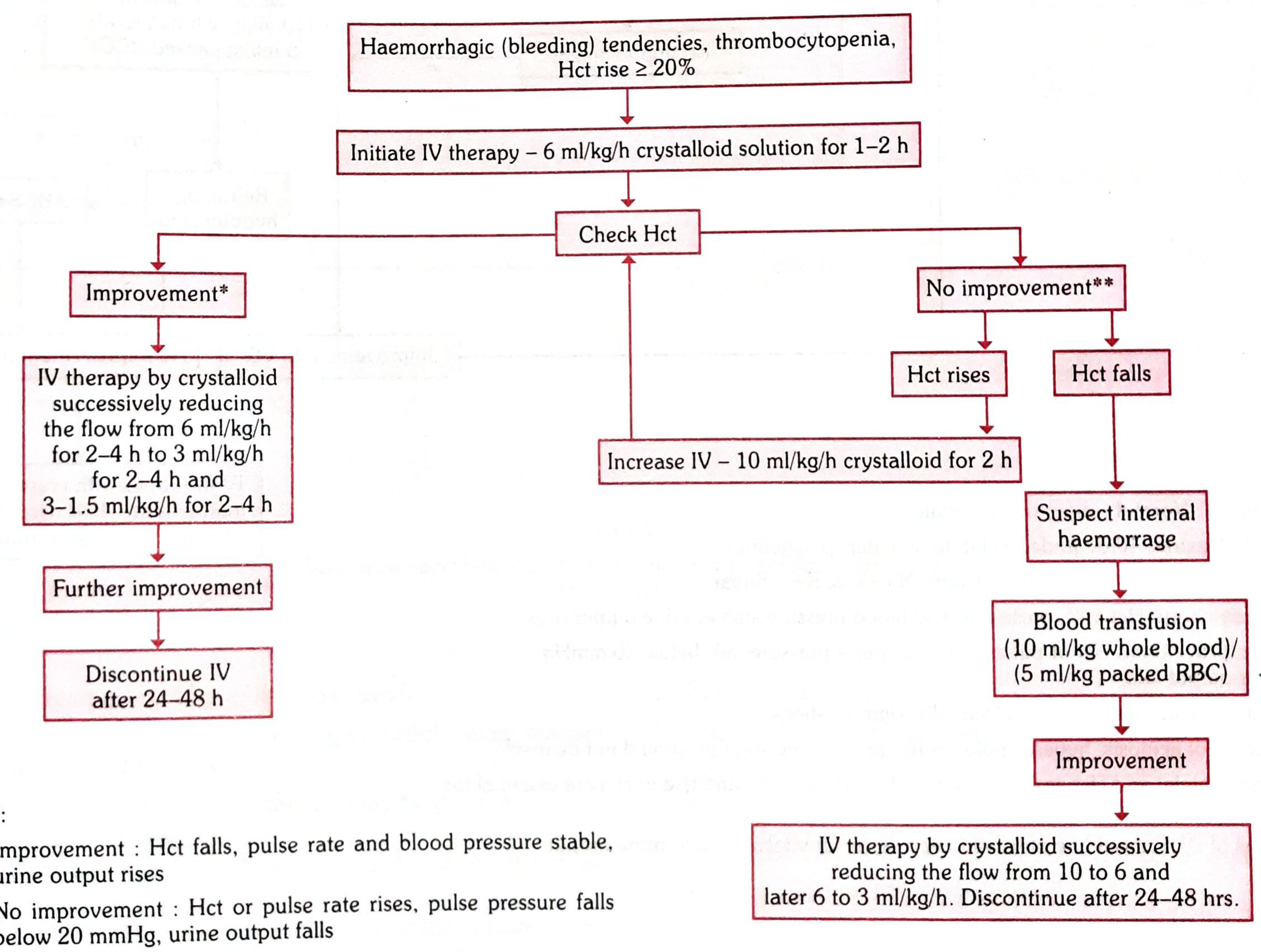
- Hct rise ≥20% = initiate IV therapy
- Start: 6 ml/kg/h crystalloid (normal saline or Ringer's lactate) for 1-2 hours
Indicator of Response to Treatment
- Improvement = Hct falls + pulse rate stable + BP stable + urine output rises → reduce IV rate step-wise (6 → 3 → 1.5 ml/kg/h) → discontinue after 24-48 hours
- No improvement = Hct continues to rise → escalate IV fluids to 10 ml/kg/h
The Critical PCV Decision: Rising vs. Falling in Shock
| Scenario | PCV Direction | Meaning | Action |
|---|---|---|---|
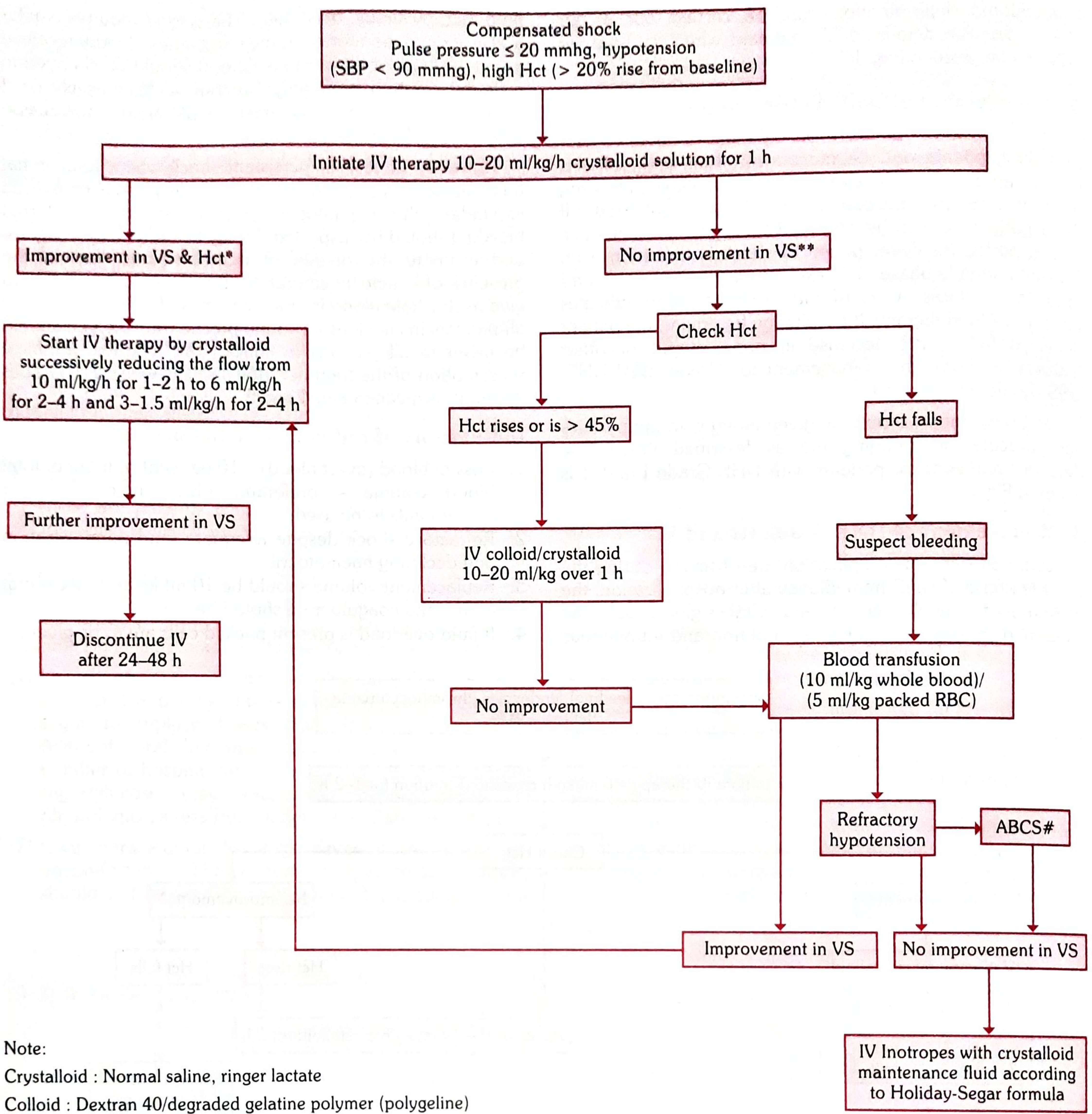
| Shock + Hct rising or >45% | ↑ | Ongoing plasma leakage | Escalate IV colloid/crystalloid 10-20 ml/kg/h |
| Shock + Hct falling | ↓ | Internal haemorrhage - NOT plasma leakage | Blood transfusion (10 ml/kg whole blood OR 5 ml/kg packed RBC) |
| Refractory shock despite fluids + Hct falling | ↓↓ | Severe haemorrhage | Blood transfusion + evaluate for coagulopathy (ABCS: acidosis, bleeding, calcium/electrolytes, sugar) |
"In cases of persistent shock when haematocrit continues to decline, internal bleeding should be suspected." - Park's Textbook of Preventive and Social Medicine, p. 296
This PCV decision point is the pivotal fork in dengue shock management - getting it wrong (giving more fluid to a bleeding patient) is dangerous.
Volume Replacement Algorithms (WHO / Park's)
For DHF Grade I & II (Moderate - Haemorrhagic tendencies + Hct rise ≥20%):
For DHF Grade III (Compensated shock - pulse pressure ≤20 mmHg, Hct >20% rise):
PCV and Blood / Platelet Transfusion Decisions
Red Cell Transfusion Indications (PCV-guided)
- Overt blood loss ≥10% of total blood volume
- Refractory shock with declining hematocrit (despite adequate fluids)
- Replacement: 10 ml/kg whole blood OR 5 ml/kg packed RBC at a time
- If fluid overload is present → packed cells preferred over whole blood
Platelet Transfusion
- PCV is NOT used to guide platelet transfusion - platelet count drives this decision
- No prophylactic platelet transfusion even at platelet count <20,000/cu.mm in general
- Give at <10,000/cu.mm, or in prolonged shock with coagulopathy, or systemic massive bleeding
Criteria for Discharge: PCV Stability Required
A patient can be discharged only when:
- Afebrile for ≥24 hours without antipyretics
- PCV/hematocrit is stable (no further rise or decline)
- Adequate urine output
- Improvement in clinical status (appetite returns, abdominal pain resolves)
Summary: PCV Roles at a Glance
| Role | Specific Use |
|---|---|
| Diagnosis | ≥20% rise = DHF criterion (with thrombocytopenia) |
| Monitoring | Daily from day 3; hourly in shock |
| Trigger for IV fluids | ≥20% rise from baseline |
| Response assessment | Falling Hct = responding; rising Hct = not responding |
| Differentiating plasma leakage vs. hemorrhage | Rising Hct = leakage (more fluids); Falling Hct = bleeding (blood transfusion) |
| Discharge criterion | Stable Hct confirms recovery phase |
Source: Park's Textbook of Preventive and Social Medicine, pp. 294-298 (WHO Dengue Management Guidelines)


image-1784972692531.jpg
IMG20260725151415.jpg
Peritoneal Fluid
analyse_media
analyse_media
Now let me query the library for peritoneal fluid cytology context to enrich the interpretation.
Finding Sources
Finding Sources
Finding Sources
Finding Sources
Finding Sources
Finding Sources
Finding Sources
Finding Sources
Reading File
Reading File
I now have all the information needed for a thorough interpretation. Here is the complete analysis.
Peritoneal Fluid Cytology - Microscopic Interpretation
Two Slides - Two Different Stains
You have provided two slides from the same peritoneal fluid specimen, prepared with different staining methods:
| Image 1 | Image 2 | |
|---|---|---|
| Stain | Papanicolaou (Pap) / H&E cytospin | Romanowsky-type (Diff-Quik / Wright-Giemsa) |
| Nuclear color | Dark blue-purple | Deep purple/royal blue |
| Cytoplasm | Pink-lavender-blue | Pale blue-grey |
| Background | Pink proteinaceous/fibrinous | Pale pink, lace-like, vacuolated/mucinous |
Cellular Findings (Both Slides)
1. Mesothelial Cells ✓
The dominant cellular component in both slides.
- Image 1: Present as both single cells and a cohesive 3D cluster with characteristic intercellular "windows" (gaps between adjacent cells) - the hallmark of reactive mesothelial cells
- Image 2: Medium-large round-to-oval single cells; some show a characteristic lighter "halo/fringe" at the outer border (another mesothelial feature)
- Nuclei: Round to oval, smooth nuclear membranes, evenly distributed benign chromatin, no significant atypia or hyperchromasia
- Cytoplasm: Moderate, pink-lavender (Pap) or pale blue-grey (Romanowsky)
- Assessment: Reactive/benign mesothelial cells - no malignant features
2. Macrophages / Histiocytes ✓
- Scattered individually throughout both smears
- Larger than lymphocytes; eccentric, kidney-shaped (reniform) or folded nuclei
- Abundant pale, vacuolated (foamy) cytoplasm containing phagocytosed debris
- Image 2: Some show large clear cytoplasmic vacuoles compressing the nucleus peripherally (vacuolated macrophages / "signet-ring-like" histiocytes - a benign reactive finding)
3. Lymphocytes ✓
- Small, mature mononuclear cells scattered individually
- High nuclear-to-cytoplasmic ratio
- Dense, dark chromatin; scant cytoplasm
4. Occasional Neutrophils ✓
- Segmented/multilobed nuclei
- Scattered, not dominant
5. Squamous Contaminant (Image 1 only)
- A single large, flat, polygonal superficial squamous epithelial cell in the upper-left quadrant
- Brightly eosinophilic cytoplasm with pyknotic nucleus
- Benign contaminant from skin or lower genital tract introduced during paracentesis - not clinically significant
Most Striking Feature: The Background
Both slides show a strikingly prominent vacuolated, bubbly, lace-like ("honeycomb") background that deserves special attention:
| Feature | Interpretation |
|---|---|
| Abundant pale-pink, finely vacuolated / "foamy" background | Mucin or lipid-rich proteinaceous material |
| Lace-like or honeycomb pattern (Image 2) | Highly characteristic of mucinous/gelatinous fluid |
| Dense pink amorphous clump (Image 2 right side) | Aggregated proteinaceous material, fibrin, or small clot |
| Overall low-to-moderate cellularity in a rich background | Pattern typical of mucinous effusion |
This pattern - abundant mucinous background with sparse, bland cells - is seen in:
- Mucinous ascites / Pseudomyxoma peritonei (PMP) - most important consideration
- Ruptured or leaking mucinous ovarian cyst
- Pancreatitis with lipid-rich exudate
- Highly proteinaceous reactive effusion
Overall Cytological Interpretation
Benign/Reactive Peritoneal Fluid with Prominent Mucinous/Proteinaceous BackgroundShowing: reactive mesothelial cells, macrophages, lymphocytes, and occasional neutrophils in a rich vacuolated mucinous background, with no malignant cells identified.
No evidence of:
- Malignant cells (no nuclear enlargement, hyperchromasia, prominent nucleoli, irregular nuclear membranes, high N:C ratio, or atypical mitoses)
- Carcinoma cells
- Lymphoma cells
- Malignant mesothelioma cells
The Critical Question: What Is Causing the Mucinous Background?
The prominent mucin background raises important differential diagnoses requiring clinical correlation:
Most Important - Pseudomyxoma Peritonei (PMP)
- A syndrome of mucinous ascites caused by accumulation of mucin in the peritoneal cavity
- Primarily arises from appendiceal mucinous neoplasms (low-grade: LAMN; high-grade: HAMN; mucinous adenocarcinoma)
- Can also arise from mucinous ovarian, pancreatic, colorectal, or gastric tumors
- Cytology typically shows acellular or paucicellular mucin with scattered bland epithelial cells or histiocytes - matching this appearance
- Clinical presentation: painless abdominal distention, "jelly belly"
- Definitive diagnosis: gelatinous material at laparotomy/laparoscopy
- Treatment: Cytoreductive surgery (CRS) + HIPEC (Hyperthermic Intraperitoneal Chemotherapy)
"Pseudomyxoma peritonei is a syndrome of mucinous ascites... primarily associated with appendiceal epithelial neoplasms... Presenting symptoms are usually painless abdominal distention." - Sleisenger and Fordtran's GI and Liver Disease, p. 645
Other Differentials
- Ruptured mucinous ovarian cyst/cystadenoma: mucin released into peritoneum; benign source
- Chronic inflammatory exudate: protein-rich but usually more cellular
- Pancreatitis: lipase-rich fluid may look vacuolated; clinical context distinguishes
Recommended Further Workup
Based on this cytology:
| Investigation | Rationale |
|---|---|
| CT abdomen/pelvis | Look for appendiceal lesion, ovarian mass, peritoneal implants |
| Tumour markers | CEA, CA 19-9, CA 125 (elevated in PMP/mucinous tumours) |
| Colonoscopy/appendix evaluation | Exclude appendiceal neoplasm |
| Gynaecological evaluation | Exclude mucinous ovarian tumour (especially if female) |
| Cell block from peritoneal fluid | May show epithelial cells not visible on smear; allows IHC |
| Fluid mucin staining | PAS, Alcian blue - confirm mucinous nature |
| Clinical correlation | Symptoms, physical exam (abdominal distension, mass) |
Summary Table
| Parameter | Finding |
|---|---|
| Stains used | Pap/H&E (Image 1) + Romanowsky/Diff-Quik (Image 2) |
| Cellularity | Low to moderate |
| Dominant cells | Reactive mesothelial cells |
| Other cells | Macrophages, lymphocytes, occasional neutrophils |
| Background | Prominent mucinous/vacuolated lace-like material |
| Malignant cells | Absent |
| Conclusion | Benign reactive peritoneal fluid with mucinous background |
| Key concern | Rule out pseudomyxoma peritonei / mucinous neoplasm |
Write a cytology report according to international society of reporting serous fluid
The International System for Reporting Serous Fluid Cytopathology (TIS) uses 5 diagnostic categories: Non-diagnostic, Negative for malignancy, Atypia of undetermined significance, Suspicious for malignancy, and Malignant.
Searching sources...
Finding Sources
Finding Sources
Searching the Web
International System Reporting Serous Fluid Cytopathology TIS 2020 categories report format
Your credits are exhausted. You can purchase a top-up or subscribe to a plan to continue using Orris. If you're already subscribed, your daily credits will reset tomorrow.
Lab Investigations of transfusion reaction and mismatched blood group
Your credits are exhausted. You can purchase a top-up or subscribe to a plan to continue using Orris. If you're already subscribed, your daily credits will reset tomorrow.
This is a shared conversation. Sign in to Orris to start your own chat.